


Services on Demand
Article
 English (pdf)
 Article in xml format
 Article references
Indicators

Related links
-
 Cited by Google
 -
 Similars in Google
Share

 Permalink
SAMJ: South African Medical Journal
On-line version ISSN 2078-5135
Print version ISSN 0256-9574
SAMJ, S. Afr. med. j. vol.108 n.12 Pretoria Dec. 2018
http://dx.doi.org/10.7196/samj.2018.v108i12.13453
CME
Preventing maternal deaths due to ectopic pregnancy
N F Moran
BM BCh, MA, FCOG (SA); Department of Obstetrics and Gynaecology, KwaZulu-Natal Department of Health, Durban, and Department of Obstetrics and Gynaecology, Nelson R Mandela School of Medicine, College of Health Sciences, University of KwaZulu-Natal, Durban, South Africa
ABSTRACT
The majority of maternal deaths due to ectopic pregnancy (EP) could be avoided if the health service renders better-quality care. There are two common clinical mistakes that lead to death from EP: (i) failure to make the diagnosis, because the attending healthcare worker does not consider this option; and (ii) failure to act urgently to stop the bleeding when there are signs of hypovolaemic shock. The only reliable way to stop the bleeding in a shocked patient with a ruptured EP is to urgently perform a laparotomy and clamp the bleeding vessels. Doctors at district hospitals must learn and maintain the skills required to provide anaesthesia for and operate on patients with a ruptured EP.
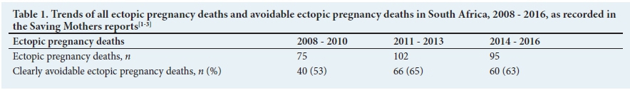
Ectopic pregnancy (EP), although a well-recognised cause of maternal death, is not one of the top five causes of maternal death in South Africa (SA). Nonetheless, it is an important topic to review for two reasons. Firstly, during the last three triennia, there has been no significant decrease in the number of maternal deaths due to EP (Table 1).[1-3] Secondly, in the Saving Mothers reports,[1-3] EP consistently features as one of the most avoidable causes of maternal death. This implies that there is a persistently poor quality of care in the management of EP, resulting in preventable maternal deaths. This situation should not be allowed to continue.
This article describes two cases that demonstrate common mistakes in the management of EP and offers advice on how to prevent unnecessary maternal deaths. In the Saving Mothers reports,[1-3] EP deaths are categorised into those before 20 weeks' gestation and those after 20 weeks' gestation (advanced extrauterine pregnancies). As >90% of EP deaths occurred before 20 weeks, this article is confined to dealing with this category rather than with advanced extrauterine pregnancies.
Case 1
An 18-year-old woman presented to the casualty department of a busy regional hospital at midnight. Her main complaint was epigastric pain associated with vomiting for the past 2 days. She was attended to by the medical officer (MO) on duty. On examination, her blood pressure (BP) was 96/61 mmHg, she looked pale and on palpation her abdomen was soft, with mild tenderness. There was a history of amenorrhoea for the past 6 weeks.
The MO's initial assessment was as follows: cystitis? anaemia? pregnancy?
Five hours later the following test results were reviewed:
-
Pregnancy test positive; haemoglobin (Hb) 10.7 g/dL; urea, electrolytes and amylase normal.
The patient was referred to the gynaecology outpatient department (GOPD), where she was seen by a sessional/part-time doctor later the same morning. This doctor's assessment and plan were as follows:
-
pregnant: 6 weeks' gestation; urinary tract infection and perineal warts
-
for antibiotics, review in 1 week.
At 15h00 the same day, the patient presented herself to the medical outpatient department at the same hospital, as she was experiencing severe abdominal pain. Her BP was 86/62 mmHg and she was noted to be extremely pale. She was immediately referred back to the GOPD.
She was attended to by the same doctor who had seen her earlier in the GOPD, and was admitted to the gynaecology ward with a diagnosis of urinary tract infection in early pregnancy. On admission at16h00, her BP was 110/60 mmHg and her pulse rate 82 bpm.
About 5 hours later, at 21h15, the ward nurses noted that the patient was groaning with pain and vomiting; her BP was 74/49 mmHg. They inserted an intravenous line and started an infusion of a litre of crystalloid fluid. Oxygen was administered by mask.
At 22h00, the MO on call for gynaecology was informed about the patient, but he was about to start an emergency caesarean delivery; he promised to review the patient after the procedure. He eventually assessed the patient at 00h10 the next day. She then had a BP of 82/40 mmHg, a pulse rate of 137 bpm and a temperature of 35oC. Her abdomen was distended with some guarding, but soft. The MO considered EP and pyelonephritis as possible diagnoses. His plan was: intravenous fluids, antibiotics, a repeat Hb test, and a formal scan to be arranged in the morning.
At 02h00, the repeat Hb of 7.1 g/dL was reported to the MO, who then thought that the diagnosis was most likely EP. He ordered 2 U blood to be cross-matched, and for the patient to be prepared for laparotomy, which could not be performed immediately, as the theatre was occupied. At 03h00, the patient arrested. Attempts at resuscitation failed.
Lesson 1: Diagnosing a ruptured ectopic pregnancy
The first common mistake that leads to a woman dying from EP is failure to make the diagnosis, usually because the attending doctor does not consider it. In some cases, it might not occur to the doctor that the patient is pregnant. She may be admitted to a medical or surgical ward for investigation of anaemia or ascites, or as a possible appendicitis patient who has to be resuscitated and considered for surgery. Even if pregnancy is diagnosed, as in case 1, the possibility that it is an EP sometimes evades the attending doctor. Instead, the patient may be admitted, or even sent home, with diagnoses such as anaemia, urinary tract infection or pelvic inflammatory disease. In cases where vaginal bleeding is one of the presenting complaints, an EP might be misdiagnosed as a miscarriage.
When the doctor in the GOPD in the case of patient 1 learnt about the outcome and realised his mistake in missing the correct diagnosis, he was distraught. He printed the following words in large font on paper and pinned it to the wall of the consulting room, so that he would never make the same mistake again: Think ectopic, every hour, every day.
This message is relevant, not only for those working in an obstetrics and gynaecology department, but also for anyone working in a casualty/emergency medicine department or a medical outpatient department, as well as for general practitioners. A diagnosis of EP must always be considered when a woman of reproductive age presents with lower abdominal pain or unexplained anaemia. Such patients should immediately be screened with an on-site urine pregnancy test. Pallor, dizziness, syncope and tachycardia are additional features consistent with a ruptured EP.
Further advice regarding the diagnosis of a ruptured EP:
-
Ruptured EPs do not always present with vaginal bleeding.
-
Pelvic inflammatory disease does not normally occur in early pregnancy; another diagnosis, such as an EP, should be considered.
-
If there is an incomplete miscarriage, the cervix will be open on examination. If the cervix is closed, think EP.
-
An ultrasound scan is a useful tool to aid diagnosis. If the patient has symptoms and signs in keeping with a ruptured EP, as well as a positive pregnancy test, the key features to look for on the scan are an empty uterus (without a clearly defined gestational sac) and free fluid in the abdomen. If these features are seen, the diagnosis is confirmed.
-
If a ruptured EP is suspected in a hospital setting, but no scanning facility is available, do not delay the diagnosis by referring the patient or letting her queue for a scan by a sonographer. The diagnosis can instead be confirmed by performing a colpocentesis/ culdocentesis. With the patient lying on the bed, the cervix is visualised through a speculum. Gently pull the posterior lip of the cervix forward and anteriorly, and insert a long needle (e.g. an 18-gauge spinal needle) attached to a 20 mL syringe in the midline through the posterior fornix of the vagina. Aspirate the contents of the pouch of Douglas. Alternatively, if the entire abdomen is distended with fluid, a paracentesis can be performed. Either way, if non-clotting blood is easily aspirated, this strongly suggests a diagnosis of a ruptured EP. Blood from the peritoneal cavity does not usually clot because of the fibrinolytic properties of the peritoneal fluid. Therefore, if the aspirated blood clots, it is more likely to be blood that has been aspirated from a blood vessel.
For more detail on the diagnosis and management of EP, the 2016 Guidelines for Maternity Care in South Africa[4]may be consulted.
Lesson 2: Stopping the bleeding in a ruptured ectopic pregnancy
The second common mistake that leads to women dying from EP is failure to act urgently to stop the bleeding when there are signs of hypovolaemic shock. A simple way to ascertain whether the patient is in shock or is no longer coping with blood loss is to measure the pulse rate and BP. If the pulse rate per minute is greater than the systolic BP (shock index >1), it suggests that the patient is decompensating. There may also be overt signs of shock, such as the inability to stand or a decreased consciousness level.
Further significant blood loss from this point is likely to lead to cardiac arrest and death. There is therefore an urgent need to stop the bleeding. In a patient with an EP, the only reliable way this can be done is to perform a laparotomy, identify the bleeding point, and clamp it off. It is not possible to effectively resuscitate the patient without surgical intervention, as the bleeding point cannot be accessed externally. While it is important to establish intravenous access, aggressive fluid resuscitation, by temporarily raising the BP, may increase the internal bleeding. Starting a blood transfusion with a view to raising the Hb level before taking the patient to theatre, is a misguided strategy, as the transfused blood will wastefully drain out through the uninhibited bleeding vessels, rather as if one is filling a leaking bucket.
In case 1, at 00h10, when the MO first considered the diagnosis of an EP, the patient was clearly already shocked. There was a missed opportunity at this stage. Rather than wait for a repeat Hb result and plan an ultrasound scan, immediate confirmation of the diagnosis could have been made by performing a paracentesis. At that stage, laparotomy without further delay was the only hope for this patient.
Lesson 2 implies that if a patient with a ruptured EP presents at a district hospital with signs of circulatory shock, a laparotomy should be performed without delay at that hospital. Planning a transfer to a higher level of care, especially where this may result in several hours' delay, is inappropriate. The following case illustrates this point.
Case 2
A 27-year-old woman was seen by an MO at 14h00 in the outpatient department of a district hospital. She complained of mild vaginal bleeding for the past 3 weeks, and collapsed at home that morning. Her BP was 93/44 mmHg, her pulse rate 140 bpm and her abdomen was distended and tender. The pregnancy test was positive and the Hb level was 3 g/dL. The MO made a diagnosis of EP.
At 15h00, the patient was admitted to the gynaecology ward and a blood transfusion was started. The regional hospital was contacted and transfer requested in view of the anaesthetic risk for the anticipated laparotomy that would be required.
The gynaecologist at the regional hospital advised urgent laparotomy at the district hospital, where an experienced surgical MO and an experienced anaesthetic MO were available. However, the anaesthetic MO refused to take the patient to theatre, stating that she was too 'unstable' for an anaesthetic at district hospital level. Eventually, at 15h30, the regional hospital accepted the patient for transfer. At 16h30, the ambulance arrived at the district hospital, and at 17h15 the patient was dead on arrival at the regional hospital.
Further points regarding surgical management of an ectopic pregnancy
-
Being haemodynamically unstable is not a contraindication for anaesthesia in the case of a ruptured EP; it is an urgent indication for anaesthesia to allow life-saving surgery and effective resuscitation. For a brief guide to administering anaesthesia for a ruptured EP, the 2016 Guidelines for Maternity Care in South Africa[4]may be consulted.
-
It is unacceptable and unethical for doctors to allow a patient to bleed to death without taking action to stop the bleeding.
-
Blood transfusion is sometimes indicated in cases of a ruptured EP. Where there is severe anaemia, with signs of shock, it is appropriate to have cross-matched or 'emergency' blood immediately accessible when starting a laparotomy. However, the transfusion should ideally only be started once the bleeding points have been clamped. In cases where blood for transfusion is not accessible, e.g. due to a national shortage of blood, this should not be a reason to delay surgery for a ruptured EP. The sooner the operation can be done in such circumstances, the less blood the woman will lose, and the better her chances of surviving.
-
A woman with a ruptured EP is the ideal candidate to benefit from autotransfusion through red cell-saving techniques, which reduce or eliminate the need for blood from a blood bank. Where cell-saving machines are available, these should always be used for a laparotomy for a ruptured EP. Where such machines are not available, and no other blood is accessible for transfusion, autotransfusion can be performed using low-technology methods, which might be life-saving.[5]
Conclusion
Two common mistakes that lead to maternal death from EP have been highlighted. To address this, there must be regular training of doctors and nurses regarding the recognition of EP and its management, particularly the need for immediate surgery if the patient is in shock. This applies especially to general practitioners, casualty doctors and nurses, generalist doctors working at district hospitals, as well as doctors working in gynaecology practice or departments. Casualty departments must have clear policies ensuring that gynaecological patients in shock are afforded equal priority and attention by casualty staff as any other category of shocked patient. Finally, managers of district hospitals must ensure that their doctors gain and/or maintain skills for providing safe general anaesthesia for laparotomy, as well as surgical skills for managing a ruptured EP.
Declaration. None.
Acknowledgements. None.
Author contributions. Sole author.
Funding. None.
Conflicts of interest. None.
References
1. Moran NF, Kunene B, Masasa M. Early pregnancy loss. In: Saving Mothers: Fifth Comprehensive Report on Confidential Enquiries into Maternal Deaths in South Africa 2008 - 2010. Pretoria: National Department of Health, 2012. [ Links ]
2. Moran NF, Kwet J. Early pregnancy loss. In: Saving Mothers: Sixth Comprehensive Report on Confidential Enquiries into Maternal Deaths in South Africa 2011 - 2013. Pretoria: National Department of Health, 2015. [ Links ]
3. National Department of Health. Saving Mothers 2014 - 2016: Short Report on Confidential Enquiries into Maternal Deaths in South Africa. Pretoria: NDoH, 2017. [ Links ]
4. National Maternity Guidelines Committee. Bleeding in early pregnancy. In: Guidelines for Maternity Care in South Africa: A Manual for Clinics, Community Health Centres and District Hospitals. 4th ed. Pretoria: National Department of Health, 2016. [ Links ]
5. Global Help. The impact of HIV on surgery. In: Cotton M, chief ed. Primary Surgery, vol. 1. 2nd ed. 2016. https://global-help.org/products/primary-surge (accessed 26 October 2018). [ Links ]
 Correspondence:
N F Moran
neil.moran@kznhealth.gov.za
Accepted 11 June 2018

