


Services on Demand
Article
 English (pdf)
 Article in xml format
 Article references
Indicators

Related links
-
 Cited by Google
 -
 Similars in Google
Share

 Permalink
SAMJ: South African Medical Journal
On-line version ISSN 2078-5135
Print version ISSN 0256-9574
SAMJ, S. Afr. med. j. vol.105 n.2 Pretoria Feb. 2015
http://dx.doi.org/10.7196/samj.9292
CONTINUING MEDICAL EDUCATION
CASE REPORT
M C Madua
MB ChB, FCP (SA); Chris Hani Baragwanath Academic Hospital, and Division of General Medicine, Department of Internal Medicine, Faculty of Health Sciences, University of the Witwatersrand, Johannesburg, South Africa
ABSTRACT
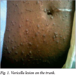
A 30-year-old woman presented to Tshepong Hospital, Klerksdorp, South Africa, with a history of rash with papules and pustules, which started on her face and spread to her entire body. There was a typical varicella lesion on the trunk. She was newly diagnosed as HIV-positive and was not yet on antiretroviral therapy.
A 30-year-old woman presented to Tshepong Hospital, Klerksdorp, South Africa, with a history of rash with papules and pustules, which started on her face and spread to her entire body. Fig. 1 depicts a typical varicella lesion on the trunk. She was newly diagnosed as HIV-positive, with a CD4 count of 232 cells μL, and was not yet on antiretroviral therapy.
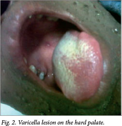
Examination showed that she was normotensive, but with a tachycardia of 138 beats per minute. She was afebrile, and had a tachypnoea of 28 breaths per minute, with 92% oxygen saturation on room air. Of note was a generalised varicella lesion involving the mucous membrane of the hard palate (Fig. 2).
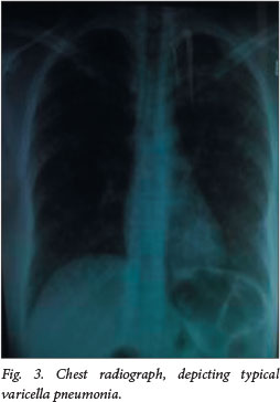
The rest of the examination revealed nothing abnormal. Arterial blood gases showed a pH of 7.5, PCO2 of 28.2 mmHg, PO2 of 92.7 mmHg and saturation of 98% on room air. Her HCO3- was 23 mmol/L. A chest radiograph revealed diffuse bilateral nodular interstitial infiltrates. Fig. 3 depicts typical varicella pneumonia.
The diagnosis was that of disseminated varicella in a woman newly diagnosed with HIV. We started treatment for varicella with intravenous acyclovir and instituted other supportive measures. The patient was sent to the intensive care unit for overnight observation.
Varicella in an immunocompromised patient often presents as severe disease. A full blood count showed a white cell count of 9 X 10⁹/L, haemoglobin of 14.7 g/dL, mean corpuscular volume of 78 fL, and platelet count of114 X 109/L, with a normal differential count. Urea and electrolytes were unremarkable, as was a liver function test. Serum crypotococcal antigen and blood culture were negative. A skin biopsy was sent for culture and histopathological examination. Tissue culture was negative and the histopathology was in keeping with varicella zoster infection.
Discussion
Disseminated zoster is associated with immunosuppression. In our setting, HIV is the most common association, although there are other causes for the immunosuppression. Dissemination of zoster in an immunocompromised patient may present mainly with cutaneous dissemination and visceral dissemination in the form of zoster pneumonitis, hepatitis and encephalitis.[1] The duration of the disease is generally longer in immunocompromised than immunocompetent patients.[1] The diagnosis is clinical. However, in our case a skin biopsy was taken to exclude cryptococcus and other opportunistic infections. Zoster in an immunocompromised patient does not follow a dermatomal pattern.[2] Other complications of varicella are myocarditis, corneal lesions, nephritis, arthritis, bleeding diatheses, acute glomerulonephritis and hepatitis. Acyclovir is still considered as a first-line agent. One should, however, note the potential for secondary bacterial infection and need for analgesia.
References
1. Cvjetkovic D, Jovanovic J, Hrnjakovic-Cvjetkovic I, Brkic S, Bogdanovic M. Reactivation of herpes-zoster virus infection by varicella-zoster virus. Medicinski Pregled 1999;52(3-5):125-128. [ Links ]
2. Gnann JW. Varicella-zoster virus: Atypical presentation and unusual complications. J Infect Dis 2002;186(Suppl 1):S91-S98. [ Links ]
 Correspondence:
M C Madua
chasneyza@yahoo.com

