


Services on Demand
Article
 English (pdf)
 Article in xml format
 Article references
Indicators

Related links
-
 Cited by Google
 -
 Similars in Google
Share

 Permalink
SAMJ: South African Medical Journal
On-line version ISSN 2078-5135
Print version ISSN 0256-9574
SAMJ, S. Afr. med. j. vol.105 n.2 Pretoria Feb. 2015
http://dx.doi.org/10.7196/samj.8654
RESEARCH
Prevalence of gastrointestinal pathogenic bacteria in patients with diarrhoea attending Groote Schuur Hospital, Cape Town, South Africa
B KullinI; R MeggerseeI; J D'AltonII; B GalvãoI; N RajaballyIII; A WhitelawIV; C BamfordV; S J ReidI; V R AbrattI
IPhD; Department of Molecular and Cell Biology, Faculty of Science, University of Cape Town, South Africa
IIBSc (Hons); Department of Molecular and Cell Biology, Faculty of Science, University of Cape Town, South Africa
IIIMB ChB, FCP (SA), Cert Gastroenterol; Division of Gastroenterology, Department of Medicine, Groote Schuur Hospital and Faculty of Health Sciences, University of Cape Town, South Africa
IVMB ChB, MSc, FCPath (Micro); National Health Laboratory Service, Groote Schuur Hospital and Division of Medical Microbiology, Faculty of Health Sciences, University of Cape Town, South Africa
VMB ChB, MMed (Med Micro), FCPath (Micro); National Health Laboratory Service, Groote Schuur Hospital and Division of Medical Microbiology, Faculty of Health Sciences, University of Cape Town, South Africa
ABSTRACT
BACKGROUND: Diarrhoea due to gastrointestinal infections is a significant problem facing the South African (SA) healthcare system. Infections can be acquired both from the community and from the hospital environment itself, the latter acting as a reservoir for potential pathogenic bacteria.
OBJECTIVES: To examine the prevalence of a panel of potential diarrhoea-causing bacteria in patients attending a tertiary healthcare facility in Cape Town, SA.
METHODS: Polymerase chain reaction (PCR) primers specific for Clostridium difficile, Shigella spp., Salmonella spp., Klebsiella oxytoca, enteropathogenic and enterohaemorrhagic Escherichia coli (EPEC/EHEC), Staphylococcus aureus, enterotoxigenic Bacteroides fragilis and Campylobacter spp. were used to screen total bacterial genomic DNA extracted from stool samples provided by 156 patients with diarrhoea attending Groote Schuur Hospital, Cape Town, SA.
RESULTS: C. difficile was the most frequently detected pathogen (16% of cases) in the 21 - 87-year-old patient range, but was not present in samples from the 16 - 20-year-old range. K. oxytoca (6%), EPEC/EHEC strains (9%) and S. aureus (6%) were also detected. The remaining pathogens were present at low frequencies (0 - 2.9%), and the occurrence of mixed infections was 5%. The majority of non-C. difficile-related diarrhoeas were community acquired.
CONCLUSION: C. difficile was the main cause of infectious diarrhoea in the sampled patients, while K. oxytoca and EPEC/EHEC strains were present as relatively minor but potentially significant pathogens.
Diarrhoea as a result of gastrointestinal tract infections is a significant problem facing much of Africa.[1] In 2000 alone, almost 4% of the deaths in South Africa (SA) were attributable to infectious diarrhoea, representing the fifth leading cause of years of life lost.[2] The causes of infectious diarrhoea are varied. Nosocomial infections are chiefly caused by Clostridium difficile and to a lesser extent by Klebsiella oxytoca, typically after antibiotic therapy, which allows the organisms to proliferate and cause disease.[3] Community-acquired diarrhoea resulting from person-to-person transmission or the consumption of contaminated and poorly prepared foodstuffs and water can be caused by a range of bacterial agents. These include several pathotypes of Escherichia coli, non-typhoidal Salmonella spp., Shigella spp., enterotoxigenic Bacteroides fragilis, Campylobacter spp. and Staphylococcus aureus, as well as several viruses (rotavirus, norovirus and adenovirus) and parasites (e.g. Giardia lamblia and Entamoeba spp.).[4]
Despite their potential to cause disease, there have been relatively few SA studies examining the prevalence of pathogenic microorganisms in 'non-outbreak' situations, particularly in the hospital environment. In addition, the majority of surveillance studies have examined paediatric populations that are at increased risk of developing diarrhoeal disease. There is little information regarding the prevalence of potential pathogens in adults. The aim of this study was therefore to identify the prevalence of a selected panel of potential pathogenic bacteria in routine stool samples provided by patients with diarrhoea attending Groote Schuur Hospital (GSH), Cape Town, SA.
Methods
Sample collection and study participants
Stool samples (N=139) were collected as part of a larger study from in- and outpatients presenting to GSH with diarrhoea between March 2012 and March 2013. Patients <16 years old were excluded from the study. Samples were transferred to the National Health Laboratory Service (NHLS) unit at GSH, where they were stored at -20°C until further processing. The presence of blood in stool samples was assessed visually. Ethics approval was obtained from the University of Cape Town, Human Research Ethics Committee (HREC Number 310/2008).
Genomic DNA extraction
Total faecal genomic DNA was extracted from stool samples using the GXT Stool Extraction Kit (Hain Lifesciences, SA) and the GenoXtract automated extraction instrument following the manufacturer's instructions. The quantity and purity of the DNA were assessed using a Nanodrop 1000 instrument (Nanodrop, USA). The quality of the genomic DNA and the absence of polymerase chain reaction (PCR) inhibitors were further assessed by PCR amplification of each sample using 50 ng template and the F27/R5 primer pair, which target the bacterial 16S rRNA gene.
Preparation of positive amplification controls
Bacterial-specific genomic DNA for use as a positive amplification control in the PCR screening amplifications was prepared from pure cultures of C. difficile (toxigenic isolate R20291), C. jejuni (a laboratory isolate provided by the NHLS, SA), S. aureus (a laboratory isolate) and S. enterica subsp. enterica serovar Typhimurium (ATCC 14028), using a genomic DNA extraction kit (ThermoScientific, USA) following the manufacturer's instructions. For the B. fragilis bft screening experiment, plasmids containing the cloned target region of the bft-1 subtype (prepared in this study) and the bft-2 subtype (kindly provided by Prof. Cynthia Sears), were used as positive amplification controls.!51 Plasmids containing cloned target regions of K. oxytoca (pehX), enteropathogenic and enterohaemorrhagic Escherichia coli (EPEC/EHEC) (eaeA) and the various pathogenic Shigella spp. (invC) were used as positive amplifications controls in the respective reactions.
Screening for potential pathogens
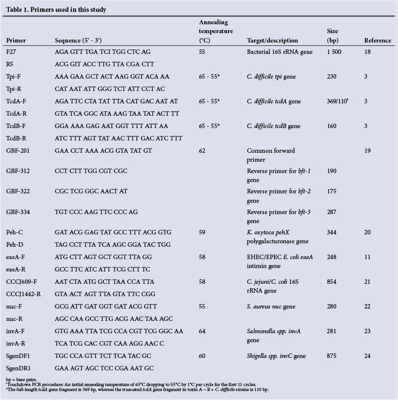
Total faecal genomic DNA (50 ng) prepared from each stool sample was used as template in a series of PCR amplifications using primers specific to each pathogen (Table 1). No template control reactions were included for each different primer set. PCR cycling parameters for the universal bacterial 16S rRNA gene, C. jejuni/C. coli, S. aureus, Salmonella spp., Shigella spp., K. oxytoca and EHEC/EPEC reactions were as follows: denaturation at 95°C for 5 minutes, followed by 35 cycles of denaturation at 94°C for 30 seconds, annealing at primer-specific temperature for 30 seconds, and extension at 72°C for 1 minute, and finally 72°C for 7 minutes.
Multiplex PCR reactions were used to screen for b_f-positive B. fragilis strains and toxigenic C. difficile. For the B. fragilis bft screening protocol, a common forward primer and three specific reverse primers that target the three different bft subtypes were used. The PCR cycling parameters were as follows: denaturation at 95°C for 5 minutes, followed by 35 cycles of denaturation at 94°C for 1 minute, annealing at 62°C for 30 seconds and extension at 72°C for 1 minute, and finally 72°C for 7 minutes. For the C. difficile screening protocol, primers targeting the species-specific tpi gene as well as the two toxin genes tcdA and tcdB were used to identify toxigenic strains. A touchdown PCR procedure was employed. An initial denaturation at 95°C for 5 minutes was performed, followed by 40 cycles of denaturation at 95°C for 30 seconds, annealing for 30 seconds at temperatures decreasing from 65°C to 55°C (decreasing by 1°C per cycle for the first 11 cycles) and extension at 72°C for 30 seconds. A final extension step was then carried out at 72°C for 7 minutes. All reaction products were analysed by electrophoresis through 2% (w/v) agarose gels and imaged using a ChemiDoc EC imager (Bio-rad, SA). Positive controls (50 ng of pure genomic DNA from target strains or 50 ng of plasmid DNA containing the relevant target fragment) and negative controls containing no template were included in each PCR experiment.
Results
Basic demographic data
A total of 139 stool samples were analysed, of which 80 (57.6%) were from female patients. Patient ages ranged from 16 to 87 years, with the majority of the patients (73%) between 20 and 60 years of age.
Prevalence of selected pathogenic bacteria in diarrhoea samples
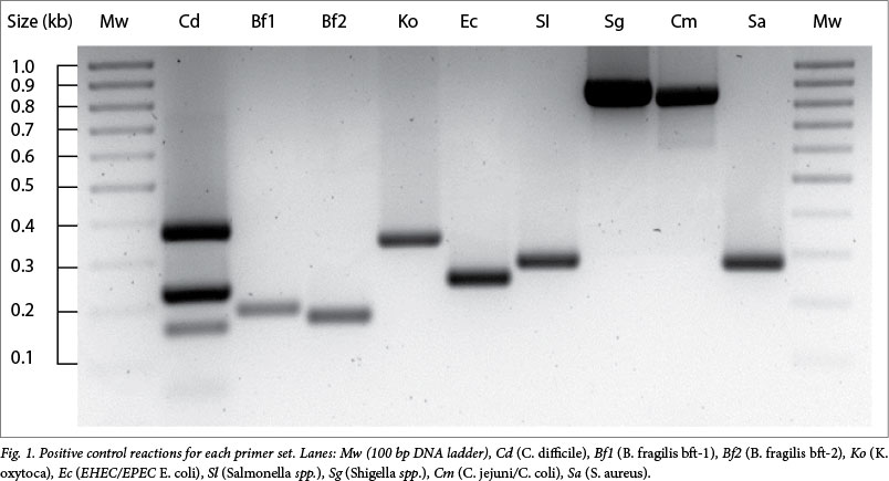
All samples showed the predicted 1.5 kb product when screened using the universal bacterial 16S rRNA gene primers (result not shown). Each of the primer sets gave the specific, expected product when used to amplify control target DNA under the study conditions (Fig. 1). The results of screening the study samples are summarised in Fig. 2. Of the 139 samples screened, 53 (approximately 38%) contained one or more of the target organisms possibly linked to the patient symptoms, while the remainder gave a negative result reflecting a diarrhoea of unknown origin. Toxigenic C. difficile was the most prevalent potential pathogen. It was found in approximately 16% of samples overall and in all age groups except the 16 - 20 years group. EPEC/EHEC E. coli were present in approximately 9% of cases and occurred across all age groups. K. oxytoca and S. aureus both occurred in 6% of samples, Salmonella spp., Shigella spp., and bft-positive B. fragilis together were present in approximately 8% of the samples, and Campylobacter spp. were not detected in any of them (Fig. 2). Mixed populations of potential pathogens were found in approximately 5% of the samples. Of the patient cohort, ten individuals (7%) showed evidence of blood in their stool. Two of these stools came from patients colonised by Shigella spp. and one from a patient colonised by S. aureus, while the remaining seven bloody stools did not contain any of the pathogens included in the screening procedures.
Discussion
Routine surveillance of bacteria that are known agents of infectious diarrhoea is very seldom carried out for organisms other than Shigella spp. and non-typhoidal Salmonella spp. In this study, we sought to examine the prevalence of other potential bacterial pathogens present in diarrhoea samples from patients attending a hospital in Cape Town. The results suggested that C. difficile was the most prevalent pathogen among patients in the 21 - 87-year-old range, with the rest of the selected potential pathogens making up a relatively small percentage of the cases.
C. difficile is a frequent cause of nosocomial diarrhoea, accounting for up to 20% of such cases worldwide.[3] There have also been reports suggesting that the prevalence of community-acquired cases of C. difficile is increasing.[6] Importantly, there are few data on the prevalence of C. difficile in SA. A previous study, also carried out at GSH but employing an enzyme-immunoassay (EIA)-based technique, determined a prevalence of 9.2% for toxigenic C. difficile in patients with diarrhoea.[7] It is possible, however, that some C. difficile cases were overlooked in this earlier study, as EIA-based tests have been reported to suffer from inferior diagnostic sensitivity.[8]
The only other PCR-based analysis reported a prevalence of 11.4% of toxigenic C. difficile in individuals with diarrhoea in the Vhembe district, suggesting that C. difficile-related infections are an important but possibly under-reported cause of diarrhoea in SA.[9] A more detailed analysis of the prevalence and epidemiology of C. difficile at GSH as well as a comparison of various diagnostic testing modalities is currently being prepared for publication.
Pathogenic E. coli are traditionally divided into several different pathotypes. While enteroaggregative E. coli are increasingly recognised as an important cause of diarrhoea in both Africa and the rest of the world, their broad genetic diversity means that in order to detect them using a PCR-based screening method, several primer combinations detecting different targets need to be employed.[10] We therefore limited our screening procedure to detect EPEC and EHEC strains, both of which have been known to cause outbreaks of diarrhoea in Africa and can be detected by the presence of the eaeA gene.[11] In the current study, EPEC and EHEC strains were present in just under 9% of the diarrhoea cases. Of these cases, 8/12 (67%) developed prior to hospital admission, suggesting that the majority of the E. coli infections were community acquired. Blood in the stool was not evident in any of the patients colonised by EPEC/EHEC strains. A previous study by Bisi-Johnson et al.[12] identified EPEC and EHEC strains in approximately 13% of patients with diarrhoea attending a tertiary hospital in the Eastern Cape Province, SA. However, that study had a large proportion of young patients (30% of the patients were between the ages of 7 and 13), who are at increased risk of developing E. coli-related diarrhoea.
K. oxytoca has been implicated as a cause of antibiotic-associated haemorrhagic colitis. Clinical isolates have been shown to constitutively produce β-lactamases, which confer resistance to both amino- and carboxypenicillins and allow the organism to survive antibiotic therapy and initiate infection.[13] A small proportion of healthy individuals (1.6%) are asymptomatic carriers of K. oxytoca, although the carriage rate in non-symptomatic patients attending GSH is currently unknown.[14] None of the samples positive for K. oxytoca showed evidence of blood in the stool, suggesting that K. oxytoca was not a major cause of diarrhoea among the patients examined.
Non-typhoidal Salmonella spp. and Shigella spp. are responsible for a significant number of cases of diarrhoea in Africa and are regarded by the World Health Organization as organisms of global significance.[1,15] Shigella spp. in particular have been known to cause outbreaks of bloody diarrhoea among adults in much of the developing world.[4] Of the four patients colonised by pathogenic Shigella spp., two showed strong evidence of blood in the stool. Although occasional nosocomial outbreaks have been reported in SA, both pathogens are predominantly acquired through the ingestion of contaminated food and water or from person-to-person transmission via the faecal-oral route. In the current study, all the diarrhoea cases in which either pathogen was present were community acquired. However, the combined prevalence of approximately 5% suggests that neither pathogen was a major cause of diarrhoea in the sample group.
B. fragilis is a human gut commensal that is also able to cause opportunistic invasive infections. In addition, certain strains also produce a metalloprotease enterotoxin encoded by the bft gene that enables the bacterium to cause diarrhoea.[5] Screening of the samples in the current study using primers that target all three subtypes of the bft gene revealed a low prevalence of bft-positive B. fragilis strains.
Enterotoxin-producing S. aureus is a fairly rare cause of diarrhoea, but is of particular significance in the hospital environment owing to its role in postoperative infections. There have also been reports that many samples from patients with antibiotic-associated diarrhoea that were positive for enterotoxin-producing S. aureus were also positive for C. difficile.[16] In the current study, 5/8 (62.5%) of the samples positive for S. aureus also harboured at least one of the other potential pathogens that were included in the screen. C. difficile was co-present in three of these samples and is presumed to be the main cause of diarrhoea in these patients. It is not clear from these results whether the presence of S. aureus promoted colonisation by other potential pathogens or vice versa.
In developing countries, Campylobacter spp., predominantly C. jejuni and C. coli, are the most common bacterial cause of diarrhoea in babies in the first year of life.[4] A previous study by Samie et al.[17] reported prevalences of 12.5% and 7% for C. jejuni and C. coli, respectively, among patients between the ages of 0 and 88 attending hospitals in the Venda region of SA. However, in the patient group examined in our study, it was not possible to detect either species in the stool samples using the described primer set, the specificity of which was validated using the C. jejuni-positive control DNA.
An initial screening of the purified DNA using universal primers targeted to the bacterial 16S rRNA gene yielded positive products for each of the samples, indicating that the quality of the extracted DNA was suitable for PCR analysis. However, it is possible that in cases where targets were not abundant in the samples, these may not have been detected by the individual PCR screening experiments. In addition, other bacterial strains, parasites and viruses as well as non-infectious factors (e.g. direct gut toxicity of administered antibiotics) were not included in this pilot study and are reflected as being of unknown origin (Fig. 2). These might be contributing to the diarrhoea cases observed and should be included in further studies. In particular, protocols to detect the prevalence of rotavirus (which has an RNA not a DNA genome) should be used during stool analysis. Nevertheless, the results presented here suggest that, in addition to C. difficile, other bacterial pathogens such as EPEC/EHEC strains that are not routinely screened for in the hospital setting may be responsible for a number of episodes of diarrhoea among adults in SA, and this warrants further investigation.
Acknowledgements. The authors acknowledge funding from the National Research Foundation of South Africa. BK acknowledges bursary funding from the Carnegie Corporation and the Claude Leon Foundation. The authors are grateful to Hain Lifescience for the loan of the GenoXtract automated extraction instrument and extraction kits for use in the study, and to the staff of the NHLS at GSH for assistance in the collection of samples.
References
1. Lamberti LM, Bourgeois AL, Fischer Walker CL, Black RE, Sack D. Estimating diarrheal illness and deaths attributable to Shigellae and enterotoxigenic Escherichia coli among older children, adolescents, and adults in South Asia and Africa. PLoS Negl Trop Dis 2014;8(2):e2705. [http://dx.doi.org/10.1371/journal.pntd.0002705] [ Links ]
2. Bradshaw D, Groenewald P, Laubscher R, et al. Initial burden of disease estimates for South Africa, 2000. S Afr Med J 2003;93(9):682-688. [ Links ]
3. Lemee L, Dhalluin A, Testelin S, et al. Multiplex PCR targeting tpi (triose phosphate isomerase), tcdA (toxin A), and tcdB (toxin B) genes for toxigenic culture of Clostridium difficile. J Clin Microbiol 2004;42(12):5710-5714. [http://dx.doi.org/10.1128/JCM.42.12.5710-5714.2004] [ Links ]
4. Pfeiffer ML, DuPont HL, Ochoa TJ. The patient presenting with acute dysentery - a systematic review. J Infect 2012;64(4):374-386. [http://dx.doi.org/10.1016/j.jinf.2012.01.006] [ Links ]
5. Franco AA, Cheng RK, Goodman A, Sears CL. Modulation of bft expression by the Bacteroides fragilis pathogenicity island and its flanking region. Mol Microbiol 2002;45(4):1067-1077. [http://dx.doi.org/10.1046/j.1365-2958.2002.03077.x] [ Links ]
6. Eyre DW, Cule ML, Wilson DJ, et al. Diverse sources of C. difficile infection identified on whole-genome sequencing. N Engl J Med 2013;369(13):1195-1205. [http://dx.doi.org/10.1056/NEJMoa1216064] [ Links ]
7. Rajabally NM, Pentecost M, Pretorius G, Whitelaw A, Mendelson M, Watermeyer G. The Clostridium difficile problem: A South African tertiary institution's prospective perspective. S Afr Med J 2013;103(3):168-172. [http://dx.doi.org/10.7196/SAMJ.6012] [ Links ]
8. Humphries RM, Uslan DZ, Rubin Z. Performance of Clostridium difficile toxin enzyme immunoassay and nucleic acid amplification tests stratified by patient disease severity. J Clin Microbiol 2013;51(3):869-873. [http://dx.doi.org/10.1128/JCM.02970-12] [ Links ]
9. Samie A, Obi CL, Franasiak J, et al. PCR detection of Clostridium difficile triose phosphate isomerase (ipi), toxin A (tcdA), toxin B (tcdB), binary toxin (cdtA, cdtB), and tcdC genes in Vhembe District, South Africa. Am J Trop Med Hyg 2008;78(4):577-585. [ Links ]
10. Okeke IN. Diarrheagenic Escherichia coli in sub-Saharan Africa: Status, uncertainties and necessities. J Infect Dev Ctries 2009;3(11):817-842. [http://dx.doi.org/10.3855/jidc.586] [ Links ]
11. Wang G, Clark CG, Rodgers FG. Detection in Escherichia coli of the genes encoding the major virulence factors, the genes defining the O157:H7 serotype, and components ofthe type 2 Shiga toxin family by multiplex PCR. J Clin Microbiol 2002;40(10):3613-3619. [http://dx.doi.org/10.1128/JCM.40.10.3613-3619.2002] [ Links ]
12. Bisi-Johnson MA, Obi CL, Vasaikar SD, Baba KA, Hattori T. Molecular basis of virulence in clinical isolates of Escherichia coli and Salmonella species from a tertiary hospital in the Eastern Cape, South Africa. Gut Pathog 2011;3(1):9. [http://dx.doi.org/10.1186/1757-4749-3-9] [ Links ]
13. Decré D, Burghoffer B, Gautier V, Petit J-C, Arlet G. Outbreak of multi-resistant Klebsiella oxytoca involving strains with extended-spectrum beta-lactamases and strains with extended-spectrum activity of the chromosomal beta-lactamase. J Antimicrob Chemother 2004;54(5):881-888. [http://dx.doi.org/10.1093/jac/dkh440] [ Links ]
14. Högenauer C, Langner C, Beubler E, et al Klebsiella oxytoca as a causative organism of antibiotic-associated hemorrhagic colitis. N Engl J Med 2006;355(23):2418-2426. [http://dx.doi.org/10.1056/NEJMoa054765] [ Links ]
15. Morpeth SC, Ramadhani HO, Crump JA. Invasive non-typhi Salmonella disease in Africa. Clin Infect Dis 2009;49(4):606-611. [http://dx.doi.org/10.1086/603553] [ Links ]
16. Ackermann G, Thomalla S, Ackermann F, Schaumann R, Rodloff AC, Ruf BR. Prevalence and characteristics of bacteria and host factors in an outbreak situation of antibiotic-associated diarrhoea. J Med Microbiol 2005;54(2):149-153. [http://dx.doi.org/10.1099/jmm.0.45812-0] [ Links ]
17. Samie A, Obi CL, Barrett LJ, Powell SM, Guerrant RL. Prevalence of Campylobacter species, Helicobacter pylori and Arcobacter species in stool samples from the Venda region, Limpopo, South Africa: Studies using molecular diagnostic methods. J Infect 2007;54(6):558-566. [http://dx.doi.org/10.1016/j.jinf.2006.10.047] [ Links ]
18. Weisburg WG, Barns SM, Pelletier DA, Lane DJ. 16S ribosomal DNA amplification for phylogenetic study. J Bacteriol 1991;173(2):697-703. [ Links ]
19. Kato N, Liu CX, Kato H, et al. A new subtype of the metalloprotease toxin gene and the incidence of the three bft subtypes among Bacteroides fragilis isolates in Japan. FEMS Microbiol Lett 2000;182(1):171-176. [http://dx.doi.org/10.1016/S0378-1097(99)00585-6] [ Links ]
20. Kovtunovych G, Lytvynenko T, Negrutska V, Lar O, Brisse S, Kozyrovska N. Identification of Klebsiella oxytoca using a specific PCR assay targeting the polygalacturonasepehXgene. Res Microbiol 2003;154(8):587-592. [http://dx.doi.org/10.1016/S0923-2508(03)00148-7] [ Links ]
21. Linton D, Lawson AJ, Owen RJ, Stanley J. PCR detection, identification to species level, and fingerprinting of Campylobacter jejuni and Campylobacter coli direct from diarrheic samples. J Clin Microbiol 1997;35(10):2568-2572. [ Links ]
22. Brakstad OG, Aasbakk K, Maeland JA. Detection of Staphylococcus aureus by polymerase chain reaction amplification of the nuc gene. J Clin Microbiol 1992;30(7):1654-1660. [ Links ]
23. Rahn K, de Grandis SA, Clarke RC, et al. Amplification of an invA gene sequence of Salmonella typhimurium by polymerase chain reaction as a specific method of detection of Salmonella. Mol Cell Probes 1992;6(4):271-279. [http://dx.doi.org/10.1016/0890-8508(92)90002-F] [ Links ]
24. Ojha SC, Yean Yean C, Ismail A, Singh K-KB. A pentaplex PCR assay for the detection and differentiation of Shigella species. Biomed Res Int 2013;2013:412370. [http://dx.doi.org/10.1155/2013/412370] [ Links ]
 Correspondence:
V R Abratt
valerie.abratt@uct.ac.za
Accepted 9 December 2014

