


Servicios Personalizados
Articulo
 Inglés (pdf)
 Articulo en XML
 Referencias del artículo
Indicadores

Links relacionados
-
 Citado por Google
 -
 Similares en Google
Compartir

 Permalink
SAMJ: South African Medical Journal
versión On-line ISSN 2078-5135
versión impresa ISSN 0256-9574
SAMJ, S. Afr. med. j. vol.103 no.6 Pretoria jun. 2013
RESEARCH
AIDS-related progressive multifocal leukoencephalopathy (PML): A retrospective study from Pretoria, South Africa
C-M SchutteI; N RanchhodII; M KakazaIII; M PillayIII
IMMed (Neurology), MD. Department of Neurology, Steve Biko Academic Hospital and University of Pretoria, Pretoria, South Africa
IIMB ChB. Department of Neurology, Steve Biko Academic Hospital and University of Pretoria, Pretoria, South Africa
IIIMMed (Neurology). Department of Neurology, Steve Biko Academic Hospital and University of Pretoria, Pretoria, South Africa
ABSTRACT
INTRODUCTION AND OBJECTIVES: Progressive multifocal leukoencephalopathy (PML), caused by the John Cunningham (JC) virus, results from lytic infection of predominantly oligodendrocytes. Following the HIV pandemic, the incidence of PML has risen sharply, but has rarely been reported in Africa. An increasing number of PML cases were seen recently in a tertiary South African hospital, and this study describes their clinical and radiological features.
METHODS: Patients with positive cerebrospinal fluid (CSF) JC virus confirmed by real-time polymerase chain reaction (PCR) were retrospectively identified from January 2008 to June 2012. Adults seen at Neurology with PML were identified, and clinical features, laboratory findings and imaging studies were analysed.
RESULTS: Of 121 specimens, 19 were positive; records of 17 patients were available (ages 27 - 64; CD4 counts 11 - 32 8 x10⁶/µl); clinical manifestations included focal weakness (47%), impaired co-ordination (41%), and speech disturbances (12%), and CSF analysis showed high protein in 76%, and pleocytosis in 35%. Fifteen patients had CT brain scans, showing white matter involvement in 12; MRI studies in 13 patients showed typical PML lesions.
CONCLUSION: This report is the first case series of patients with PML from a South African neurology unit, emphasising the fact that PML occurs commonly in South African patients with HIV infection.
Progressive multifocal leukoencephalopathy (PML) is caused by the John Cunningham (JC) virus, a human polyoma virus that is widespread around the globe. Initial subclinical infection probably occurs in childhood; however, in severe immunosuppression, reactivation of the virus leads to a lytic infection of oligodendrocytes in the brain.[1] In HIV-positive patients during the pre-HAART (highly active antiretroviral therapy) era, infection by the JC virus in the brain with PML was rapidly fatal, with a median survival of 3 - 6 months; up to 5% of HIV-positive patients eventually succumbed to PML. Of fatal central nervous system (CNS) diseases, PML occurs in up to 18% of patients.[2]
In the USA and Europe, seroprevalence studies have shown that 60 - 80% of adults have antibodies against the JC virus; a minimum of 7 viral genotypes may be identified according to the sequence analysis of the major capsid protein (VP1). It has been shown that the major type of JC virus in the USA is JCV1, while type 4 is seen in Europe and the USA, type 2 in Asia, and types 3 and 6 appear to be prevalent in western Africa.[3]
Reports of JC virus-confirmed PML from Africa have been rare. Two cases of PML have been reported from Gambia, and 4 additional cases have been described in African patients living in Europe.[4] In South Africa, a study that examined the JC virus in the cerebrospinal fluid (CSF) of patients with suspected PML showed JC virus type 3 in 3 patients, and type 7 and 2 in 1 patient each, confirming that the virus occurs in South African patients.[5] However, the study only analysed the types of JC virus, and no clinical data of the affected patients were reported.
With the high burden of HIV infection in sub-Saharan Africa, the reasons for the lack of reports of patients with PML have been debated. HIV clade C infection predominates in Africa, and the possibility exists that the clade C virus interacts differently to the clade B virus with other pathogens, including the JC virus. The paucity of reports may also reflect the absence of sophisticated technology necessary to confirm a diagnosis of PML in Africa.[6] A recent study from South Africa showed that JC virus detection in CSF was improved when a T-antigen-specific fluorescence resonance energy transfer hybridisation probe real-time PCR method was used, compared with the VP1 nested PCR previously employed.[7]
During the last 2 years, an increasing number of patients with PML were seen at the Neurology Department of the Steve Biko Academic Hospital in Pretoria. Since there are no clinical reports of PML from South Africa, and the absence of studies from Africa has been the cause of speculation in the literature, the objective of this study is to describe the clinical and radiological features of these patients.
Methods
The study was performed retrospectively from January 2008 to the end of June 2012. All patients with a positive PCR for JC virus in CSF were identified from the National Health Laboratory Service Database. All adult patients (age >13 years) who were seen by the Department of Neurology of the University of Pretoria with PML were included in the study. The discharge reports from the Department of Neurology's database, as well as all hospital record files of the patients, were retrieved. The data were then analysed regarding presenting complaints, clinical features, laboratory findings and imaging studies. The JC virus was confirmed with T-antigen-specific fluorescence energy transfer hybridisation probe real-time PCR.[7]
Results
Of 121 CSF specimens identified from the database, 19 were positive for the JC virus. Of these, the records of 17 patients (8 males and 9 females) with PML were available at Steve Biko Academic Hospital, University of Pretoria, while 2 were from other hospitals in Gauteng.
Twelve patients were seen during 2011 and the first 6 months of 2012.
The ages of patients ranged from 27 to 64 years (mean 38); 13 were HAART-naïve at admission. The most common clinical manifestations were focal weakness (47%), impaired co-ordination (41%) and speech disturbances (12%); cognitive dysfunction and status epilepticus were seen in 1 patient each. CD4 counts were very low, ranging from 11 to 32 8 x 10⁶/µl with a mean of 76 x 106/µl. CSF analysis showed a high protein in 13/17 (76%) of patients, and a pleocytosis in 6/17 (35%); the data are shown in Table 1.
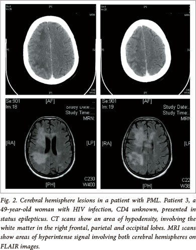
Radiological imaging was available for all patients: 15 had computerised tomography (CT) of the brain, and 13 had magnetic resonance imaging (MRI) studies done. Of the CT scans, 12 showed white matter involvement, 1 showed cerebral oedema, and 2 were normal. The MRIs revealed typical areas of hyperintensity on T2-weighted imaging and FLAIR images, with hypointensity on T1-weighted imaging, and no contrast enhancement or mass effect. Multiple lesions were seen in 10 patients; in 2, a single lesion was seen in the centrum semi-ovale, and bilateral cerebellar lesions were seen in 1. The 2 normal CT scans both revealed a single lesion in the centrum semi-ovale on MR imaging and, in 4 of the MRI scans, multiple lesions were visible, which had appeared as single lesions on the CT scans. Two sets of CT and MRI brain scans of 2 patients are shown in Figs 1 and 2 respectively.

Two patients were considered to have had PML immune reconstitution inflammatory syndrome (IRIS). The first patient (patient no. 13) had numerous white matter lesions in the brainstem and cerebellum, with some of the lesions in the cerebellum showing ring enhancement post gadolinium. This patient had been started on HAART about 6 months before the current admission. The other patient (patient no. 17) deteriorated while in the ward after HAART had been started, but an MRI was not repeated at that stage. She improved slightly after steroid treatment.
In 6 patients, electroencephalography (EEG) was available. All patients showed diffuse abnormalities with excessive slow wave activity; the patient who presented in status epilepticus had periodic lateralising epileptiform discharges (PLEDs), and 1 patient showed triphasic complexes and episodic sharp waves in the fronto-central regions.
Discussion
Despite the high prevalence of HIV infection in South Africa, very few reports of PML have been published and only a small number of JCV strains been characterised. In one report, a child with ataxia and a hemiparesis with radiological features of PML IRIS was described;[8] in another, a patient with new-onset seizures was found to have a positive PCR for JCV with radiological signs of PML in the setting of profound immunosuppression.[9] Reasons for this low prevalence of PML in Africa and India have been debated, and range from possible underreporting or underdiagnosing the condition to possible reduced neurovirulence of circulating JCV genotypes and host genetic differences in the development of PML.([6] In our study, most patients were seen in the last 2 years, suggesting that the disorder is becoming more frequent or that the diagnosis is now more readily made. The real-time PCR method used in this study employs a T-antigen-specific fluorescence energy transfer hybridisation probe that may be able to detect a lower concentration of JCV DNA. The probe targets the large T-antigen that is 100% conserved in all known JCV genotypes as opposed to the nested PCR which targets only the variable VP1 region. It will have to be evaluated over time whether this test will also be able to detect viral DNA in the setting of HIV-infected patients who have been taking HAART in South Africa, since reports have shown that the demonstration of the JCV in CSF is becoming more difficult in patients on HAART, with sensitivities as low as 58%.[10]
The patients in this study presented with typical clinical findings of PML as described in the HIV-related literature. Focal weakness was seen in 47% of patients, which is similar to the 43% found in a study of PML from Denmark[11] and lower than in a study from Brazil[12] where this clinical sign occurred in 75% of 12 patients. Impaired co-ordination occurred in 41% of our patients, compared with the 68% and 42% in Engsig and Vidal's studies.[11,12] Surprisingly, cognitive decline was a complaint in only 1 (6%) of our patients, which is very low compared with the 57% and 42% in the above studies. The mean CD4 count in our study (76 x 106/µl) was comparable to that in other studies.[11,12]
CSF findings usually only serve to rule out other conditions in PML and are typically normal, or may show a mild lymphocytic pleocytosis in 15% of patients and a mildly elevated protein in 20 - 30% of cases.[13] In our patients, pleocytosis occurred slightly more commonly (6/17 (35%)), and elevated protein levels much more commonly (13/17 (76%)). CSF abnormalities are often interpreted as part of the nonspecific findings of patients who are HIV positive.
The typical lesions of PML on imaging are subcortical hypodensities, often in the parieto-occipital areas, confined to the white matter of the brain. The most common findings are bilateral confluent multifocal supratentorial white matter lesions, but single lesions restricted to the subcortical white matter may also be found. [13,14] Lesions may also appear in the corpus callosum, thalamus, basal ganglia and cerebellum.[13,15] Our patients had typical findings on CT and MR imaging, with a large proportion showing lesions in the posterior fossa. MRI remains the imaging of choice when evaluating patients with PML,[14,16,17] and it was also clear in our study that MRI yielded better results, since two CT brain scans that were reported as normal, showed clear single white matter lesions on MRI. Two patients had PML IRIS - both had had HAART initiated recently, and all received steroid treatment in the ward, with limited improvement.
In conclusion: This is the first report of a series of patients with PML from South Africa, emphasising that the condition is seen with increasing frequency in Gauteng Province, South Africa.
References
1. Koralnik IJ. Progressive multifocal leukoencephalopathy revisited: Has the disease outgrown its name: Ann Neurol 2006;60:162-173. [ Links ]
2. Cinque P, Koralnik, IJ, Gerevini S, et al. PML in HIV-1 infection. Lancet Infect Dis 2009;9(10):625-636. [http://dx.doi.org/10.1016/S1473-3099(09)70226-9] [ Links ]
3. Egli A, Infanti L, Dumoulin A, et al. Prevalence of Polyoma BK and JC infection and replication in 400 healthy blood donors. J Infect Dis 2009;199(6):837-846. [ Links ]
4. Stoner GL, Agostini HT, Ryschkewitsch CF, et al. Detection of JC virus in two African cases of progressive multifocal leukoencephalopathy including identification of JCV type 3 in a Gambian AIDS patient. J Med Microbiol 1998;47(8):733-742. [ Links ]
5. Venter M, Smit SB, Leman P, Swanepoel R. Phylogenetic evidence of widespread distribution of genotype 3 JC virus in Africa and identification of a type 7 isolate in an African AIDS patient. J Gen Virol 2004;85:2215-2219. [ Links ]
6. Shankar SK, Satishchandra P, Mahadevan A, et al. Low prevalence of progressive multifocal leukoencephalopathy in India and Africa: Is there a biological explanation? J NeuroVirol 2003;9(suppl 1):59-67. [ Links ]
7. Glass AJ, Venter M. Improved detection of JC virus in AIDS patients with progressive multifocal leukoencephalopathy by T-antigen specific fluorescence energy transfer hybridization probe real-time PCR: Evidence of diverse JC virus genotypes associated with progressive multifocal leukoenceohalopathy in Southern Africa. J Med Virol 2009;81(11):1929-1937. [http://dx.doi.org/10.1002/jmv.21618] [ Links ]
8. Nuttall JJ, Wilmshurst JM, Ndondo AP, et al. Progressive multifocal leukoencephalopathy after initiation of highly active antiretroviral therapy in a child with advanced human immunodeficiency virus infection: A case of immune reconstitution inflammatory syndrome. Pediatr Infect Dis J 2004;23(7):683-685. [ Links ]
9. Modi M. Progressive multifocal leukoencephalopathy: A case report. S Afr Med J 2008;98(4):263-264. [ Links ]
10. Marzocchetti A, Di Giambenedetto S, Cingolani A, Ammassari A, Cauda R, De Luca A. Reduced rate of diagnostic positive detection of JC virus DNA in cerebrospinal fluid in cases of suspected progressive multifocal leukoencephalopathy in the era of potent antiretroviral therapy. J Clin Microbiol 2005;43(8):4175-4177. [ Links ]
11. Ensig FN, Hansen A. Incidence, clinical presentation, and outcome of PML HIV-positive patients during the highly active antiretroviral therapy era: A nationwide cohort study. J Infect Dis 2009;199(1):77-83. [http://dx.doi.org/10.1086/595299] [ Links ]
12. Vidal JE, Penalva de Oliveira AC, Fink MC, Pannuti CS, Trujillo JR. AIDS-related progressive multifocal leukoencephalopathy: A retrospective study in a referral center in Sao Paulo, Brazil. Rev Inst Med Trop S Paulo 2008;50(4):209-212. [ Links ]
13. Berger JR. The clinical features of PML. Cleveland Clin J Med 2011;78 (suppl 2):S8-S12. [http://dx.doi.org/10.3949/ccjm.78.s2.03] [ Links ]
14. Bag AK, Cure JK, Chapman PR, Roberson GH, Shah R. JC virus infection of the brain. Am J Neuroradiol 2010;31(9):1564-1576.[http://dx.doi.org/10.3174/ajnr.A2035] [ Links ]
15. Whiteman ML, Post MJ, Berger JR, Tate LG, Bell MD, Limonte LP. Progressive multifocal leukoencephalopathy in 47 HIV-seropositive patients: neuroimaging with clinical and pathological correlation. Radiology 1993;187(1):233-240. [ Links ]
16. Thurnher MM, Thurnher SA, Muhlbauer B, et al. Progressive multifocal leukoencephalopathy in AIDS: Initial and follow-up CT and MRI. Neuroradiology 1997;39(9):611-618. [ Links ]
17. Mark AS, Atlas SW. Progressive multifocal leukoencephalopathy in patients with AIDS: Appearance on MR images. Radiology 1989;173(2):517-521. [ Links ]
Accepted 18 October 2012.
 Correspondence: C-M Schutte (cschutte@medic.up.ac.za)

