


Servicios Personalizados
Articulo
 Inglés (pdf)
 Articulo en XML
 Referencias del artículo
Indicadores

Links relacionados
-
 Citado por Google
 -
 Similares en Google
Compartir

 Permalink
SAMJ: South African Medical Journal
versión On-line ISSN 2078-5135
versión impresa ISSN 0256-9574
SAMJ, S. Afr. med. j. vol.102 no.6 Pretoria jun. 2012
CORRESPONDENCE
Hydatid cysts of the breast and parotid gland
To the Editor: We report 2 interesting cases of hydatid cysts in unusual sites: in the breast and the deep lobe of the parotid gland.
Hydatid cysts are an infectious disease caused by the larval stage of the cestode Echinococcus. Humans are mostly affected by E. granulosus as incidental intermediate hosts. The liver (70%) and lungs (25%) are most commonly affected, with the spleen, heart, kidneys, bone, nervous system and soft tissue less frequently affected.1 Even in endemic areas, hydatid disease of the head, neck and breast is extremely rare. The incidence of hydatid cyst in the breast has been reported as 0.27%.2 No parotid gland incidence figures are available.
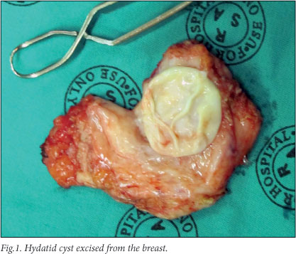
A 24-year-old woman from Van Wyksvlei was referred to the surgical clinic with a lump in the superolateral quadrant of the left breast. Fine-needle aspiration was performed at a local clinic before referral to the surgical clinic; parasitic hooklets were observed, diagnosing an Echinococcus cyst. Chest X-ray (CXR) and abdominal sonar showed no other cysts. Pre-operative albendazole was administered and the cyst was removed by excision biopsy (Fig. 1). There were no postoperative complications.
In the second case, an HIV-negative 20-year-old man from Britstown was referred with a cystic mass in the right parotid area. The cyst, present for about 2 years, had fluctuated in size. It appeared superficial on examination. In the absence of a radiologist at the facility, informed consent was obtained for excision of the cyst with or without superficial parotidectomy. On excision, the cyst appeared to extend into the deep lobe of the parotid gland. Superficial parotidectomy and excision of the cyst was performed without injury to the facial nerve. Histology confirmed normal superficial parotid tissue and an Echinococcus cyst. CXR and abdominal sonar showed no other cysts. The patient was given albendazole and discharged; he returned once for follow-up.
Hydatid disease is a prevalent parasitic infection in sheep-rearing areas such as the Northern Cape. Although hydatid cysts in the head, neck and breast are extremely rare, Echinococcus infection should be considered as a differential diagnosis in patients from endemic areas.
Johlene du Plessis
Central Karoo Hospital De Aar
Northern Cape
johlene_dup@yahoo.com
REFERENCES
1. Ahmad S, Jalil S, Saleem Y, Suleman BA, Chughtai N. Hydatid cysts at unusual sites: reports of two cases in the neck and breast. J Pak Med Assoc 2010;60(3):232-234. [ Links ]
2. Epstein NA. Hydatid cyst of the breast: diagnosis using cytological techniques. Acta Cytol 1969:13(7):420-421. [ Links ]

