


Serviços Personalizados
Artigo
 Inglês (pdf)
 Artigo em XML
 Referências do artigo
Indicadores

Links relacionados
-
 Citado por Google
 -
 Similares em Google
Compartilhar

 Permalink
SAMJ: South African Medical Journal
versão On-line ISSN 2078-5135
versão impressa ISSN 0256-9574
SAMJ, S. Afr. med. j. vol.100 no.9 Pretoria Set. 2010
SCIENTIFIC LETTERS
New fly species causing human myiasis identified in Eastern Cape, South Africa
S K KuriaI; H J C KinguII; S D VasaikarIII; J N MkhizeIV; J M IisaV; A DhaffalaVI
IPhD. Department of Biological Sciences, Walter Sisulu University, Mthatha, E Cape
IIMD, MMed (Surg). Department of Surgery, Walter Sisulu University
IIIMD (Med Microbiol), MB BS. Department of Medical Microbiology, Walter Sisulu University
IVPhD. Department of Biological Sciences, Walter Sisulu University, Mthatha, E Cape
VBVetMed. Department of Biological Sciences, Walter Sisulu University, Mthatha, E Cape
VIMB ChB, MMed (Surg). Department of Surgery, Walter Sisulu University
To the Editor: Myiasis is the infestation of tissues of live vertebrates (humans and/or animals) by dipterous larvae. Human myiasis is classified according to the type of larva producing the lesion, location and clinical signs.1 Myiases in humans are thought to have originated from the close association between humans and domestic animals in ancient times.2 Flies that cause myiasis are grouped into three families: Calliphoridae (blowflies), Sarcophagidae (flesh flies) and Oestridae (bot flies).2 For a long period species of Sarcophaga have been implicated in myiasis in man and animals.3 Other dipterans have also been implicated with causing myiasis in humans, including Cochliomyia hominivorax (Coquerel),1 C. macellaria Fab., Phormia regina Meig., Lucilia sericata Meig., L. illustris Meig., Calliphora erythrocephala Meig. and Cynomyia cadaverina Desv.4 Nevertheless, several species of flies have been used for maggot debridement therapy (MDT), the commonest being L. sericata Meigen, a greenbottle blowfly which is closely related to the greenbottle L. cuprina Wiedmann; however, L. cuprina feeds on live as well as necrotic tissue, which is undesirable in MDT.5
We aimed to elucidate the fly species that cause myiasis in humans in the Eastern Cape province, South Africa. We collected maggots from necrotic human wounds in patients attending Nelson Mandela Academic Hospital (NMAH) and other clinics within the O R Tambo municipality. These maggots were allowed to grow and pupate, and the emerging flies were subsequently identified.6
Materials, methods and results
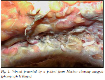
NMAH is one of the three provincial and tertiary Eastern Cape academic hospitals. It undertakes teaching and research for undergraduate and postgraduate students in the Faculty of Health Sciences at Walter Sisulu University. It offers secondary and tertiary medical services in the former Transkei region of the Eastern Cape province. Maggots were harvested from a full-thickness burn wound in a patient (aged 22 years) from Maclear (Fig. 1) and a dry-gangrene foot of a 57-year-old patient from Slovo Park suburb, Mthatha. The maggots were bred in plastic containers in the laboratory using chicken liver until they pupated. The emerging flies were later identified to species level. The fly species that emerged from maggots harvested from the patient from Maclear were identified as Sarcophaga (Liosarcophaga) nodosa Engel (Fig. 2), while those harvested from the other patient belonged to L. cuprina (Fig. 3).


Discussion
Myiasis is the infestation of human or animal tissue by larvae of dipteran flies. It is commonly reported in Mexico and South and Central America,7 but there is minimal information on myiasis in South Africa. Many species of Calliphoridae are saprophages that feed on animal carcasses, whereas others are obligate parasites.2 S. (Liosarcophaga) nodosa Engel is a widespread and common species that breeds in carrion and dead arthropods, and in 1972 there was no clear answer about whether it could cause myiasis, either in humans or even in other animals.6 This is the first time this species has been recorded as causing myiasis in humans. It is possible that there may be many more fly species that can cause myiasis in humans that are still not known. Flies that emerged from maggots collected from the second patient from Slovo Park, Mthatha belong to L. cuprina (Fig. 3). The research project to determine other fly species associated with human wounds in this region is still ongoing.
We thank M Villet from Rhodes University for identifying the fly specimens, and A Gova who worked tirelessly to ensure that the maggots survived in the laboratory. We also appreciate the assistance of S Swanepoel in editing the photos. This project was supported by a research grant from Walter Sisulu University through the Research Directorate.
References
1. Duro EA, Mariluis JC, Mulieri PR. Perinatal/neonatal case presentation: umbilical myiasis in a human newborn. J Perinatol 2007; 27: 250-251. [ Links ]
2. Stevens JR, Wallman JF, Otranto D, Wall R, Papes T. The evolution of myiasis in humans and other animals in the Old and New Worlds (part 11): biological and life-history studies. Trends Parasitol 2006; 22: 181-188. [ Links ]
3. Knipling EF. A comparative study of the first-instar larvae of the genus Sarcophaga (Calliphoridae, Diptera), with notes on the biology. J Parasitol 1936; 22: 417-454. [ Links ]
4. Knipling EF, Rainwater HT. Species and incidence of dipterous larvae concerned in wound myiasis. J Parasitol 1937; 23: 451-455. [ Links ]
5. Williams KA, Cronje FJ, Avenant L, Villet MH. Identifying flies used for maggot debridement therapy. S Afr Med J 2008; 98: 630-631. [ Links ]
6. Zumpt F. Calliphoridae (Diptera, Cyclorrhapha). Part IV. Sarcophaginae. Exploration du Parc National des Virunga, Mission de Witte 1933-1935. 1972; 101: 1-264. [ Links ]
7. Çetinkaya M, Özkan H, Köksal N, Coşkun ŞZ, Hacımustafaoğlu M, Girişgin O. Neonatal myiasis: a case report. Turk J Pediatr 2008; 50: 581-584. [ Links ]
Accepted 14 June 2010.
Corresponding author: S K Kuria (kkuria@wsu.ac.za)

