


Services on Demand
Article
 English (pdf)
 Article in xml format
 Article references
Indicators

Related links
-
 Cited by Google
 -
 Similars in Google
Share

 Permalink
SAMJ: South African Medical Journal
On-line version ISSN 2078-5135
Print version ISSN 0256-9574
SAMJ, S. Afr. med. j. vol.100 n.6 Pretoria Jun. 2010
ORIGINAL ARTICLES
Warfarin-induced skin necrosis in HIV-1-infected patients with tuberculosis and venous thrombosis
F BhaijeeI; H WainwrightII; G MeintjesIII; R J WilkinsonIV; G ToddV; E de VriesVI; D J PepperVII
IMB ChB. Groote Schuur Hospital, Cape Town; and Institute of Infectious Diseases and Molecular Medicine and Department of Medicine, University of Cape Town
IIMB ChB, FCPath (Anat) (SA). Groote Schuur Hospital, Cape Town; and Department of Pathology, Division of Anatomical Pathology, University of Cape Town
IIIMB ChB, MRCP, FCP (SA), DipHIVMan (SA). Institute of Infectious Diseases and Molecular Medicine and Department of Medicine, University of Cape Town; and Infectious Diseases Unit, G F Jooste Hospital, Cape Town
IVFRCP. Institute of Infectious Diseases and Molecular Medicine and Department of Medicine, University of Cape Town; Infectious Diseases Unit, G F Jooste Hospital; Division of Medicine, Imperial College, London, UK; and National Institute for Medical Research, Mill Hill, London
VBSc, MB ChB, FFDerm (SA), PhD. Department of Dermatology, Groote Schuur Hospital and University of Cape Town
VIMB ChB, MFamMed. School of Public Health and Family Medicine, University of Cape Town
VIIMB ChB, MD. Institute of Infectious Diseases and Molecular Medicine and Department of Medicine, University of Cape Town; and Infectious Diseases Unit, G F Jooste Hospital, Cape Town
ABSTRACT
BACKGROUND: At the turn of the century, only 300 cases of warfarin-induced skin necrosis (WISN) had been reported. WISN is a rare but potentially fatal complication of warfarin therapy. There are no published reports of WISN occurring in patients with HIV-1 infection or tuberculosis (TB).
METHODS: We retrospectively reviewed cases of WISN presenting from April 2005 to July 2008 at a referral hospital in Cape Town, South Africa.
RESULTS: Six cases of WISN occurred in 973 patients receiving warfarin therapy for venous thrombosis (0.62%, 95% CI 0.25 - 1.37%). All 6 cases occurred in HIV-1-infected women (median age 30 years, range 27 - 42) with microbiologically confirmed TB and venous thrombosis. All were profoundly immunosuppressed (median CD4+ count at TB diagnosis 49 cells/µl, interquartile range 23 - 170). Of the 3 patients receiving combination antiretroviral therapy, 2 had TB-IRIS (immune reconstitution inflammatory syndrome). The median interval from initiation of antituberculosis treatment to venous thrombosis was 37 days (range 0 - 150). The median duration of parallel heparin and warfarin therapy was 2 days (range 1 - 6). WISN manifested 6 days (range 4 - 8) after initiation of warfarin therapy. The international normalised ratio (INR) at WISN onset was supra-therapeutic, median 5.6 (range 3.8 - 6.6). Sites of WISN included breasts, buttocks and thighs. Four of 6 WISN sites were secondarily infected with drug-resistant nosocomial bacteria (methicillin-resistant Staphylococcus aureus (MRSA), Acinetobacter, extended-spectrum β-lactamase (ESBL)-producing Escherichia coli and Klebsiella pneumoniae) 17 - 37 days after WISN onset. In 4 patients, the median interval from WISN onset to death was 43 days (range 25 - 45). One of the 2 patients who survived underwent bilateral mastectomies and extensive skin grafting at a specialist centre.
CONCLUSION: This is one of the largest case series of WISN. We report a novel clinical entity: WISN in HIV-1 infected patients with TB and venous thrombosis. The occurrence of 6 WISN cases in a 40-month period may be attributed to (i) hypercoagulability, secondary to HIV-1 and TB; (ii) short concurrent heparin and warfarin therapy; and (iii) high loading doses of warfarin. Active prevention and appropriate management of WISN are likely to improve the dire morbidity and mortality of this unusual condition.
Warfarin-induced skin necrosis (WISN) is a rare complication of warfarin therapy, with an estimated prevalence of 0.01 - 0.1% in individuals receiving warfarin.1,2 WISN is associated with high morbidity, often necessitating aggressive surgical intervention, and may be fatal in the absence of early accurate diagnosis and treatment. Originally described in 1943, WISN was first associated with oral anticoagulants in 1954.3 By 2000, only 300 cases had been reported internationally.4 Most cases occur in patients receiving treatment for venous thrombo-embolism (VTE); 25% of WISN occurs in patients with cardiac indications for therapy (e.g. atrial fibrillation, valve replacement) or cerebrovascular insufficiency.2 To date, published reports do not associate WISN with HIV-1 infection or tuberculosis (TB). We describe 6 cases of WISN with poor outcome occurring in HIV-1-infected patients receiving treatment for TB.
Methods
Setting
We retrospectively reviewed 6 cases of WISN seen at G F Jooste Hospital (Cape Town, South Africa) from April 2005 through July 2008. G F Jooste Hospital is a 200-bed adult (>15 years) public hospital that receives referrals from primary care clinics serving a catchment population of 1.3 million high-density, low-income people. We have previously described national guidelines for antituberculosis treatment and antiretroviral therapy in South Africa.5,6 The Research Ethics Committee of the University of Cape Town approved the study (REF: 182/2009).
Definitions
We defined venous thrombosis as either visualisation of a noncompressible thrombus with Doppler ultrasound (popliteal or femoral venous thrombosis) or a venous filling defect with radio-contrast during computed tomography (CT) (inferior vena cava or superior sagittal sinus thrombosis). A radiologist performed sonography and interpreted the CT findings. WISN was defined as a characteristic drug eruption on the skin, occurring shortly after starting warfarin therapy for a venous thrombosis and progressing to skin and subcutaneous tissue loss. We defined the following: microbiologically confirmed TB as Mycobacterium tuberculosis cultured or acid-fast bacilli (AFB) seen in sputum or a lymph node aspirate; TB-IRIS (immune reconstitution inflammatory syndrome) using the consensus clinical case definition of paradoxical TB-IRIS for resource-limited settings;7 extended-spectrum beta-lactamase (ESBL)-producing bacteria as bacteria having clavulanateinhibited transferable enzymes able to hydrolyse third- and fourth-generation cephalosporins as tested by the disc diffusion (fishtail) method; and methicillin-resistant Staphylococcus aureus (MRSA) as having an oxacillin minimum inhibitory concentration of >4 mg/l.
Materials
We obtained clinical information from hospital notes, laboratory reports and communication with attending physicians. The following data were reviewed: patient demographics, HIV-1 status, CD4+ counts (nadir and post-ART where available), TB episode (microbiological confirmation, drug susceptibility testing), site of venous thrombosis, anticoagulation therapy, site of WISN, international normalised ratio (INR) at WISN onset, antibiotic treatment, and outcome (e.g. death, surgical intervention). All patients admitted to G F Jooste Hospital from 2005 through 2008 were managed using a standardised venous thrombosis protocol. Following diagnosis of venous thrombosis, low-molecular-weight (LMW) heparin (enoxaparin 1 mg/kg twice daily by deep subcutaneous injection) was prescribed for a maximum of 5 days. Warfarin was started 2 days after heparin initiation to minimise the risk of warfarin-induced skin necrosis. If the patient was receiving TB treatment for 10 days or longer, the loading dose of warfarin was adjusted from 5 mg to 10 mg.
Results
Baseline characteristics
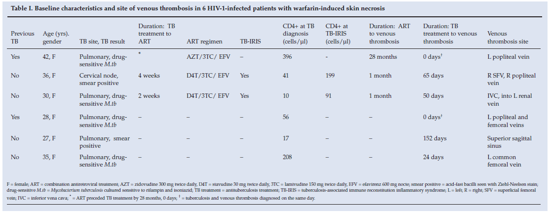
Nine hundred and seventy-three patients were diagnosed with venous thromboses and received warfarin therapy at G F Jooste Hospital over the 40-month study period. WISN occurred in 6 HIV-1-infected women receiving treatment for microbiologically confirmed TB (Table I). The prevalence of WISN in our study population was 0.62% (6/973) (95% CI 0.25 - 1.37%). The median age was 33 years (range 27 - 42). The median CD4+ count at TB diagnosis was 49 cells/µl (interquartile range 23 - 170). Three patients received ART (regimens specified in Table I). Two patients (cases 2 and 3) were diagnosed with TB-IRIS. The median interval from initiation of antituberculosis therapy to venous thrombosis was 37 days (range 0 - 150). Venous thrombosis sites included popliteal and femoral veins, the inferior vena cava, and the superior sagittal sinus. Only patient 3 was an inpatient at the time of venous thrombosis (and received LMW heparin prophylaxis); the remaining patients were admitted to hospital as a result of venous thrombosis. No patient had a personal or family history of previous venous thrombosis.
Clinical features at WISN and outcomes
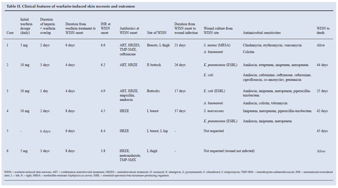
The warfarin loading dose was 5 mg or 10 mg (Table II). The median duration of parallel heparin and warfarin therapy was 2 days (range 1 - 6) and the median interval from initiation of warfarin therapy to WISN was 6 days (range 4 - 8). The INR at WISN onset was supra-therapeutic, median 5.6 (range 3.8 - 6.6). Activated partial thromboplastin times (aPTT) were not measured. Sites of WISN included the breasts, buttocks and thighs (Fig. 1). Skin biopsy was performed in 1 patient (Fig. 1). After WISN diagnosis, warfarin was stopped and LMW heparin was used to manage anticoagulation. Wound cultures from infected WISN sites produced the following drug-resistant nosocomial organisms: Escherichia coli (ESBL), Klebsiella pneumoniae (ESBL), S. aureus (MRSA), Acinetobacter baumannii and Serratia marcescens. Antimicrobial sensitivities of each organism are listed in Table II.
Four patients died, and no autopsies were performed. All 4 patients were profoundly immunosuppressed at TB diagnosis. The median interval from WISN onset to death was 43 days (range 25 - 45). The two surviving patients' CD4+ counts at TB diagnosis exceeded 200 cells/µl. Patient 1 was referred to a specialist centre for aggressive surgical management (Fig. 1, D - F). Patient 6 recovered with appropriate wound care and prophylactic broad-spectrum intravenous antibiotics (a thirdgeneration cephalosporin).
Discussion
This is one of the largest case series of WISN. We report a novel clinical entity: WISN occurring in HIV-1-infected patients with TB and venous thrombosis.
All 6 patients were chronically ill women of reproductive age with venous thromboses. WISN typically occurs in obese, perimenopausal women who are receiving anticoagulant therapy for a deep-vein thrombosis or pulmonary embolism.2 Women are affected more frequently than men (4:1)2 - the reason for this predilection is unclear. In women, the breast is most commonly affected, followed by the buttocks and thighs;8 our patients were similarly affected. It is postulated that local tissue factors contribute to the development of WISN at these sites, and that such factors include trauma and variation in local temperature and perfusion.9,10
About 90% of affected patients develop symptoms between the 3rd and 6th day of warfarin therapy,2,4,11 which is similar to our experience. The clinical presentation of WISN is characteristic (Fig. 1, A - C); all our patients demonstrated these clinical features. Widespread disease may result in deep tissue necrosis, secondary infection and multi-organ failure.2
Mortality within 3 months of WISN onset is substantial (15%), even with appropriate treatment.2 Four of our 6 patients died within 45 days of onset. All deaths were probably due to a sepsis syndrome complicating wound infection. All wound cultures were taken from infected wounds. These were not surveillance cultures. Prior rifampicin, trimethoprimsulfamethoxazole and cephalosporin use in our patients may have favoured the selection of highly antibiotic-resistant organisms. In South Africa, more than 50% of S. aureus isolates from public hospitals are resistant to rifampicin and/ or trimethoprim sulfamethoxazole.12 Failure of effective infection control measures, lack of appropriate antimicrobial chemotherapy,6 delayed referral to a specialist centre for surgical debridement, and profound immunosuppression at TB diagnosis probably contributed to the 4 deaths. In contrast, the superior immune function at TB diagnosis of patients 1 and 6 may account for their survival. Pulmonary emboli may have also complicated warfarin cessation and contributed to death. We were unable to determine the exact cause of death, as the patients' relatives declined consent to perform autopsies. It is important to note that despite prompt referral to a specialist centre, patient 1 had considerable morbidity including bilateral mastectomies, an impaired gait due to a contracture of her left thigh, and the associated psychosocial stigma.
Classic histological features of WISN include full-thickness epidermal necrosis and thrombosed vessels in the dermis. While the underlying pathophysiological mechanisms remain unclear, it is postulated that WISN results from an imbalance between intrinsic pro- and anticoagulant factors during the first few days of warfarin therapy.2,11 Warfarin is a vitamin K antagonist and reduces serum levels of vitamin K-dependent factors, which include factors II, VII, IX and X, protein C and protein S. Serum levels of factor VII (a procoagulant factor), and proteins C and S (anticoagulant factors) decline more rapidly than serum levels of factors II, IX and X (procoagulant factors) on warfarin therapy.2,11 This results in an initial hypercoagulable state, which, especially in the presence of additional risk factors such as protein C and/or S deficiency, may predispose to WISN.4 The INR is factor VII-dependent, so patients will have a raised INR, but a relative protein C deficiency will nonetheless result in a hypercoagulable state.13 Screening for these conditions before warfarin initiation is not recommended, however, as they lack the necessary sensitivity and specificity to accurately predict the risk of developing WISN.2,4 Owing to the retrospective nature of our study, serum levels of protein C, protein S and antithrombin III were not measured. The lack of genetic testing and coagulation work-up is a limitation of our study.
The prevalence of WISN in our study population is 0.62% (6/973), which is six times higher than that reported in HIVuninfected patients.1,2 The occurrence of 6 WISN cases in a 40-month period at one centre is unusual. It may be a result of the short duration of parallel heparin and warfarin therapy (median 2 days) observed in our patients. Parallel heparin and warfarin therapy is postulated to prevent the development of WISN, and should be continued until the vitamin K-dependent clotting factors have been consumed (72 - 96 hours).2,4,8,11 In our patients, premature cessation of heparin during the initial hypercoagulable period of warfarin therapy may have exacerbated an underlying hypercoagulable disorder (such as a protein C or S deficiency) and culminated in WISN. In our setting, we routinely prescribe a loading dose of 5 or 10 mg of warfarin in TB patients with venous thromboses, as rifampicin induces the rate of warfarin clearance by cytochrome p450 (CYP) 2C9.14 This dose of warfarin with a short window of parallel heparin and warfarin therapy may have contributed to the high prevalence of WISN (0.62%).
HIV infection is a widely acknowledged risk factor for VTE.15-17 Some reports cite a tenfold increase in the incidence of deep-vein thrombosis (DVT) in HIV/AIDS as opposed to the general population.15 The following independent risk factors have been identified for VTE in HIV-positive patients: low CD4 count, high viral load, advanced stage of immunocompromise, opportunistic infections, AIDS-related neoplasms, HIVassociated auto-immune disorders (e.g. auto-immune haemolytic anaemia), hospitalisation in the past 3 months, and central venous catheter use in the past 3 months.16-18 Exposure to antiretroviral therapy (ART) has not been associated with VTE.16,17 HIV-positive patients are also more likely to demonstrate multiple acquired and persistent thrombophilic abnormalities; the frequency of these abnormalities increases with progression to AIDS, and their presence may contribute to the high prevalence of venous and arterial thrombosis in patients with HIV infection.19 These abnormalities include antiphospholipid antibodies, lupus anticoagulant, anticardiolipin antibodies, increased von Willebrand factor, increased d-dimers, and deficiencies of protein C, protein S, antithrombin and heparin cofactor II.20 The acquired protein S and protein C deficiencies seen in acutely ill patients may be reversible following treatment for opportunistic infections and/or ART.18
M. tuberculosis infection may present clinically as DVT; 2 of our patients (patients 1 and 4) were diagnosed with TB and DVT simultaneously. DVT usually occurs shortly after initiating antituberculosis therapy (about 2 weeks).21 Rifampicin-based regimens have a fivefold increased risk of DVT (relative risk = 5), so DVT prevention is recommended in patients on rifampicin.21 DVT is also associated with advanced HIV infection and PTB. The following thrombogenic factors probably contribute to this association: acquired protein C and protein S deficiencies, elevated plasma fibrinogen, impaired fibrinolysis, depressed ATIII, reactive thrombocytosis, increased platelet aggregation, and antiphospholipid antibodies.22 These parameters may improve with antituberculosis treatment.22
It is not known whether IRIS predisposes to venous thrombosis. A single case is reported of IRIS manifesting as disseminated TB, myelopathy, encephalopathy and DVT;23 with appropriate treatment, IRIS resolved and no adverse drug effects occurred. We report the first 2 cases of TB-IRIS and WISN occurring simultaneously. The 2 patients diagnosed with TB-IRIS were profoundly immunosuppressed, had a short duration from starting antituberculosis treatment to initiation of ART, and presented with recurrence of TB symptoms soon after initiating ART.7
Active prevention and appropriate management of venous thromboses are likely to alleviate the dire morbidity and mortality associated with WISN. Prophylactic heparinisation of acutely ill hospital patients with HIV-1 infection and/ or TB will reduce the incidence of venous thrombosis. In patients with venous thrombosis, parallel heparin therapy for at least the first 4 days of warfarinisation2,4,8,11 may limit the occurrence of WISN. WISN should be considered in all newly warfarinised patients with new skin lesions. Effective infection control measures and expedited referral to specialist centres for surgical review may reduce mortality.
We thank the dedicated medical and nursing staff at G F Jooste Hospital for the care administered to their patients.
Graeme Meintjes and Robert J Wilkinson are supported by Wellcome Trust fellowships. Dominique J Pepper is supported by funding from the US Agency for International Development and PEPFAR via the Perinatal HIV Research Unit, and received SATBAT research training funded by the Fogarty International Center and the National Institutes of Health (NIH/FIC 1U2RTW007373-01A1). The content is solely the responsibility of the authors and does not necessarily represent the official views of the Fogarty International Center, the NIH, USAID or the US government.
References
1. Warkentin TE, Sikov WM, Lillicrap DP. Multicentric warfarin-induced skin necrosis complicating heparin-induced thrombocytopenia. Am J Hematol 1999; 62(1): 44-48. [ Links ]
2. Irwin RS, Rippe JM. Warfarin induced skin necrosis. In: Irwin and Rippe's Intensive Care Medicine. 6th ed. Philadelphia: Lippincott Williams & Wilkins, 2007: 2267. [ Links ]
3. Verhagen H. Local haemorrhage and necrosis of the skin and underlying tissues, during anticoagulant therapy with dicumarol or dicumacyl. Acta Med Scand 1954; 148(6): 453-467. [ Links ]
4. Chan YC, Valenti D, Mansfield AO, Stansby G. Warfarin induced skin necrosis. Br J Surg 2000; 87(3): 266-272. [ Links ]
5. Pepper DJ, Maartens G, Rebe K, et al. Neurological manifestations of paradoxical tuberculosis-associated immune reconstitution inflammatory syndrome: a case series. Clin Infect Dis 2009; 48(11): e96-107. [ Links ]
6. Pepper DJ, Rebe K, Morroni C, Wilkinson RJ, Meintjes G. Clinical deterioration during antitubercular treatment at a district hospital in South Africa: the importance of drug resistance and AIDS defining illnesses. PLoS ONE 2009; 4(2): e4520. [ Links ]
7. Meintjes G, Lawn SD, Scano F, et al. Tuberculosis-associated immune reconstitution inflammatory syndrome: case definitions for use in resource-limited settings. Lancet Infect Dis 2008; 8(8): 516-523. [ Links ]
8. Cole MS, Minifee PK, Wolma FJ. Coumarin necrosis - a review of the literature. Surgery 1988; 103(3): 271-277. [ Links ]
9. Broekmans AW, Bertina RM, Loeliger EA, Hofmann V, Klingemann HG. Protein C and the development of skin necrosis during anticoagulant therapy. Thromb Haemost 1983; 49(3): 251. [ Links ]
10. Comp PC, Elrod JP, Karzenski S. Warfarin-induced skin necrosis. Semin Thromb Hemost 1990; 16(4): 293-298. [ Links ]
11. Essex DW, Wynn SS, Jin DK. Late-onset warfarin-induced skin necrosis: case report and review of the literature. Am J Hematol 1998; 57(3): 233-237. [ Links ]
12. Marais E, Aithma N, Perovic O, et al. Antimicrobial susceptibility of methicillin-resistant Staphylococcus aureus isolates from South Africa. S Afr Med J 2009; 99: 170-173. [ Links ]
13. McKnight JT, Maxwell AJ, Anderson RL. Warfarin necrosis. Archives of Family Medicine 1992; 1: 105-108. [ Links ]
14. Niemi M, Backman JT, Fromm MF, Neuvonen PJ, Kivisto KT. Pharmacokinetic interactions with rifampicin: clinical relevance. Clin Pharmacokinet 2003; 42(9): 819-50. [ Links ]
15. Saber AA, Aboolian A, LaRaja RD, Baron H, Hanna K. HIV/AIDS and the risk of deep vein thrombosis: a study of 45 patients with lower extremity involvement. Am Surg 2001; 67(7): 645-647. [ Links ]
16. Ahonkhai AA, Gebo KA, Streiff MB, Moore RD, Segal JB. Venous thromboembolism in patients with HIV/AIDS: a case-control study. J Acquir Immune Defic Syndr 2008; 48(3): 310-304. [ Links ]
17. Crum-Cianflone NF, Weekes J, Bavaro M. Review: thromboses among HIV-infected patients during the highly active antiretroviral therapy era. AIDS Patient Care STDS 2008; 22(10): 771-778. [ Links ]
18. Saif MW, Bona R, Greenberg B. AIDS and thrombosis: retrospective study of 131 HIV-infected patients. AIDS Patient Care STDS 2001; 15(6): 311-320. [ Links ]
19. Lijfering WM, Sprenger HG, Georg RR, van der Meulen PA, van der Meer J. Relationship between progression to AIDS and thrombophilic abnormalities in HIV infection. Clin Chem 2008l; 54(7): 1226-1233. [ Links ]
20. Saif MW, Greenberg B. HIV and thrombosis: a review. AIDS Patient Care STDS 2001; 15(1): 15-24. [ Links ]
21. White NW. Venous thrombosis and rifampicin. Lancet 1989; 334: 434-435. [ Links ]
22. Turken O, Kunter E, Sezer M, et al. Hemostatic changes in active pulmonary tuberculosis. Int J Tuberc Lung Dis 2002; 6(10): 927-932. [ Links ]
23. Tahir M, Sinha S, Sharma SK, Mitsuyasu RT. Immune reconstitution inflammatory syndrome manifesting as disseminated tuberculosis, deep venous thrombosis, encephalopathy and myelopathy. Indian J Chest Dis Allied Sci 2008; 50(4): 363-364. [ Links ]
Accepted 3 November 2009.
Corresponding author: F Bhaijee (fbhaijee@gmail.com)

