


Serviços Personalizados
Artigo
 Inglês (pdf)
 Artigo em XML
 Referências do artigo
Indicadores

Links relacionados
-
 Citado por Google
 -
 Similares em Google
Compartilhar

 Permalink
SAMJ: South African Medical Journal
versão On-line ISSN 2078-5135
versão impressa ISSN 0256-9574
SAMJ, S. Afr. med. j. vol.99 no.11 Pretoria Nov. 2009
ORIGINAL ARTICLES
Peripheral arterial disease and intermittent claudication: efficacy of short-term upper body strength training, dynamic exercise training, and advice to exercise at home
B M ParrI; T D NoakesII; E W DermanIII
IMSc (Med) Exercise Science. Cape Peninsula University of Technology, Cape Town
IIMB ChB, MD, DSc, FACSM. University of Cape Town MRC/UCT Research Unit for Exercise Science and Sports Medicine, Cape Town
IIIMB ChB, PhD, FACSM. University of Cape Town MRC/UCT Research Unit for Exercise Science and Sports Medicine, Cape Town
ABSTRACT
OBJECTIVE: To compare the effect of two training programmes and advice to exercise at home on physiological adaptations in patients with peripheral arterial disease (PAD).
DESIGN: 30 patients with a typical history of PAD and intermittent claudication were randomised to either an upper body strength training programme (UBST), a dynamic (walking, cycling, circuit) conventional exercise rehabilitation programme (CER), or advice to 'walk as much as possible at home' (CONT). Before and after intervention groups performed a standard graded treadmill exercise test (GTET) and a 6-minute walk test (SMWT) to determine peak physiological parameters and walking distances. Maximal walking distance (MWD), pain-free walking distance (PFWD), peak oxygen uptake (VO2) , heart rate and perceived pain were measured.
RESULTS: MWD on the GTET increased significantly in the CER group compared with the CONT and UBST groups (93.9±79% v. 7.0±19.8% v. 7.3±46%; CER v. UBST v. CONT p=0.003). Similarly, peak VO2 increased with CER compared with the CONT and UBST groups (28.4±20 v. -6.2±15 v. -1.0±21%; CER v. UBST v. CONT p=0.004). During the SMWT the CER and UBST groups improved in PFWD compared with the CONT group (37±47% v. 27±71% v. -30±29%; CER v. UBST v. CONT p=0.03), and perceived pain decreased in the CER group compared with the UBST group (-24±39% v. 27±48%; CER v. UBST p=0.01).
CONCLUSION: CER improves physiological parameters and walking distances more than UBST does. CER is effective within 6 weeks. Verbal encouragement to exercise is an ineffective form of management.
Exercise training improves walking tolerance in patients with peripheral arterial disease (PAD).1-8 Typically this includes walking as the principal mode of training.4,5 Patients with PAD who walk with pain have greater improvements in walking distances than those who walk only to the point at which pain begins.6,7 However, forcing patients with PAD to walk with increasing pain is challenging as because of their personality type they tend to experience negative emotions more readily than people without PAD.9 They may therefore not tolerate pain well, which could lead to poor attendance at training sessions. However, no studies have specifically examined the relationship between prescription of exercise likely to cause pain and compliance with exercise programmes.
Walker et al. found that a 6-week pain-free upper limb cycle ergometry exercise training programme and lower limb cycling training produced equal improvements in maximal walking distance (MWD) and pain-free walking distance (PFWD).1 Increases in MWD and PFWD are therefore independent of mode of training in patients with PAD. Developing an effective pain-free exercise training programme is important for patients with PAD.
Arm ergometers are not freely available at many gymnasia in South Africa, whereas most have upper body strength training equipment. A study on the effects of lower limb strength training in patients with PAD reported no improvement in treadmill exercise duration or peak oxygen uptake (VO2) in the strength training group.10
We therefore aimed to compare the effects of a structured upper body strength training programme (UBST) with those of a structured dynamic conventional exercise rehabilitation programme (CER) as typically prescribed for patients with PAD. The control group were patients with PAD who were told to 'walk as much as possible at home'.
We hypothesised that if the effects of exercise training are not specific to the limbs that are used, upper body training could produce changes equivalent to exercising the diseased limbs in patients with PAD.
Although the study by Walker et al. was over 6 weeks,1 the duration of training in other randomised controlled trials was 3 or 6 months.2-5 Since patients may find commitment to prolonged treatment unacceptable, we aimed to determine whether a short-duration (6-week) CER programme could improve walking tolerance in patients with PAD.
We also aimed to determine whether conventional medical advice to 'walk as much as possible at home' without the support of a structured programme can produce equivalent benefits.
Methods
Study design
Thirty patients with PAD were recruited from the Department of Vascular Surgery at Groote Schuur Hospital, Cape Town, and Claremont Hospital, Cape Town. All had a medical history of PAD with intermittent claudication and were referred for duplex flow Doppler to confirm the diagnosis. Patients with rest pain, exercise tolerance limited by medical conditions other than PAD, or significant chronic obstructive pulmonary disease (COPD) were excluded. A detailed medical history was obtained from the patient's vascular surgeon and/or cardiologist.
The study was approved by the Research and Ethics Committee of the Faculty of Health Sciences of the University of Cape Town.
Patients gave signed informed consent to participate. Randomisation was decided by patients drawing an intervention group name. One who was randomised to the control group refused and was excluded. An anthropometric analysis was performed; patients were familiarised with the treadmill and visited the laboratory the following day for determination of ankle and brachial blood pressure, blood analysis, an ECG, a 6-minute walk test and a graded treadmill exercise test to maximal claudication pain.
The three treatment groups were CER, UBST and CONT (the control group). The duration of intervention was 6 weeks. In the CER group blood pressure, walking distance and time and in the UBST group blood pressure, weight lifted and repetitions were recorded at each session.
Anthropometric analysis. Body mass and height, and skinfold thickness to the closest 0.1 mm at the triceps, biceps, supra-iliac and subscapular sites on the right side of the body using a Harpenden skinfold calliper (British Indicator HSC Body 918760, UK), were recorded. Using a formula, skinfolds predicted the body fat percentage.11
Ankle and arm blood pressure. Resting brachial blood pressure measured by audible sphygmomanometry (Korotkoff phase I and IV) was recorded with the patient supine. Simultaneously, left and right ankle pressures were measured with a portable office Doppler (Huntleigh Technology PLC 1997 Dopplex Advanced Pocket Dopplers, UK) and blood pressure cuffs were applied just above the ankles with the probe placed over the posterior tibial or dorsalis pedis artery. The ankle brachial index (ABI) was calculated by dividing the ankle pressure by the brachial pressure. Ankle pressure gradient was also measured as the percentage fall in ankle pressure from the resting value [(pre-post)/pre]×100. The postexercise ABI was measured as for the resting ABI immediately after the patients had walked to their maximum walking distance.
Graded treadmill exercise test (GTET). During the treadmill protocol12 the speed was held constant at 3.2 km/h. The gradient increased by 2% every 2 minutes, when heart rate, brachial blood pressure and perceived pain were recorded. Using the perception of pain scale, pain-free walking distance (PFWD) was recorded when patients first noticed calf pain. Maximum walking distance (MWD) was defined as the distance that had been walked on the treadmill when patients were unable to continue because of severe claudication pain.
During exercise total inspiratory ventilation (VE), VO2 and respiratory exchange ratio (RER) were measured over 15second intervals (Oxycon Alpha, Erich Jaeger, Wuerzburg, Germany). Before each test, the gas meter was calibrated and the analysers were calibrated with room air and a 4% CO2-16% O2 and the balance N2 gas. Heart rate during the graded treadmill exercise test was recorded using an electrocardiogram.13
Six-minute walk test (SMWT). After resting for 20 minutes, patients completed a 6-minute walk around an indoor track. They were instructed to cover as much ground as they could in 6 minutes with the tester walking behind them. PFWD and MWD, heart rate every minute, rating of perceived pain every minute and recovery heart rate 1 minute after exercise were recorded.
Strength testing. Patients performed 15 repetitions of 10 upper body weight plated machines. Since they were elderly and had not exercised before, the weight at which they could comfortably complete 15 repetitions was recorded as the starting strength.
Intervention. Patients in the CER and UBST groups attended structured exercise rehabilitation 3 times per week for a 45minute period for 6 weeks.
The unstructured exercise training group were advised to 'walk as much as possible at home' and told that they could join the exercise programme at completion of the study.
The CER training consisted of repetitive walking on a treadmill at a speed that produced claudication pain within 5 - 10 minutes, followed by rest. Further exercise/rest was continued until a total of at least 10 minutes of walking initially, and 20 minutes after 2 weeks, was completed. Once patients could complete 20 minutes with ease, the intensity of the treadmill-training programme was increased weekly by increasing the walking speed (by 0.3 km/h) or the gradient (by 1%), which is equivalent to a quarter of a MET per week14 (a MET is one metabolic unit which is equal to an oxygen consumption of 3.5 ml O2/kg/min13). After walking, patients cycled for 5 minutes followed by 15 minutes of a circuit training programme in which they completed 6 different exercises of 15 repetitions on the upper and lower body weight plated machines once a week, floor exercises (consisting of either step, aerobics or core stabilisation exercises) once a week, and a 15-minute spin class once a week. Training sessions ended with 5 minutes of stretching.
The UBST group initially completed 15 repetitions of exercises on 10 upper body weight plated machines and 30 repetitions of 4 upper body dumbbell exercises (weight of 1.5 kg). The initial weight was set so the patient could comfortably complete 15 repetitions. Weights were increased between 4 and 16 pounds (1.8 and 7.3 kg) per week. The UBST group received no cardiovascular training.
Statistical analysis
Results were analysed using Statistica (StatSoft Inc., Tulsa, OK, USA). Data are expressed as mean ± SD. A between-subjects analysis of variance was used to detect differences between groups on entry to the study and differences in length of the intervention period between groups. Changes in each group over time were expressed as a percentage and an analysis of variance was used to detect differences between groups over time. An LSD post hoc analysis was used to detect differences between groups from pre-treatment to post-treatment. Pain ratings for each patient were converted to a single variable by plotting an area under the curve for them. Data were normalised by expressing the pain measure (y axis) relative to 100% of elapsed time (x axis).
The Kruskal-Wallis ANOVA was used to detect differences between groups in Δ (delta) pain-free walking distance on the treadmill exercise test (pre-treatment to post-treatment) and Δ pain-free walking distance on the 6-minute walk test (pretreatment to post-treatment), as these data were not normally distributed.
Fisher's exact test was used to assess differences between groups in morbidity, gender, race and medications taken by each group.
A dependent t-test assessed changes in strength in the UBST group before and after treatment. An independent t-test was used to assess differences in compliance with training sessions between the UBST group and the CER group.
As the sample size of the groups are small, the standardised difference in the means or Cohen's d effect size (ES) statistic was used to detect the relative magnitude of the experimental treatment.
The ES values were calculated using an Excel statistical program.15 The qualitative terms below are used to discuss the results: negligible effect = -0.15 and <0.15; small effect = 0.15 and <0.40; medium effect = 0.40 and <0.75; large effect = 0.75 and <1.10; very large effect = 1.10 and <1.45; huge effect >1.45.
Results
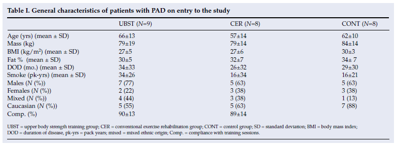
Thirty patients with PAD and intermittent claudication were recruited; 5 dropped out, 1 suffered a stroke, 2 underwent peripheral vascular angioplasty before re-testing, 1 suffered a muscle tear and 1 was diagnosed with cancer. Their general characteristics are listed in Table I. There was no significant difference in duration of disease, pack-years of smoking, body mass index, mass, fat percentage or age between groups on entry to the study.
After 6 weeks of training there was a statistically significant improvement in strength in all the muscle movements trained in the UBST group except for rotary shoulder movement (data not shown). The average improvement across all pieces of equipment was 53.9±24.8%.
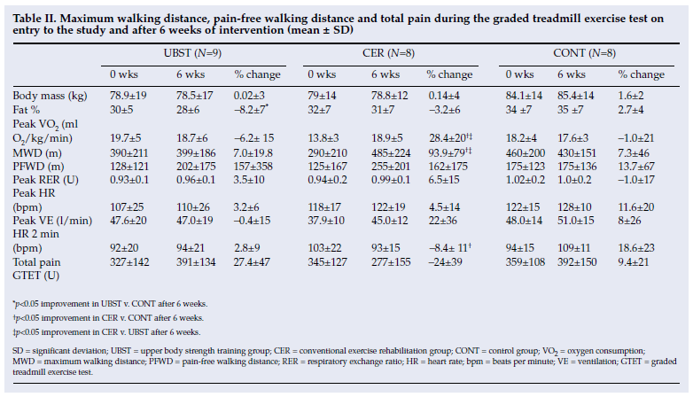
Tables II and III list the variables measured during the GTET and SMWT, respectively. There were no significant differences measured in the variables on entry to the study between the different groups except for peak VO2. The CER group had significantly lower peak VO2 values compared with the UBST and CONT groups (CER and CONT p=0.04 and CER v. UBST p=0.006).
MWD on the GTET increased significantly in the CER group compared with the CONT and UBST groups ((93.9±79% v. 7.0±19.8% v. 7.3±46%; CER v. UBST v. CONT p=0.003; ES=1.56 (CER v. UBST) and ES=1.43 (CER v. CONT)).
Similarly, peak VO2 increased with CER compared with the CONT and UBST groups (28.4±20% v. -6.2±15% v. -1.0±21%; CER v. UBST v. CONT p=0.004; ES=2.1 (CER v. UBST) and ES=1.53 (CER v. CONT)), indicating a training response in the CER group.
Furthermore, heart rate at minute 2 during the GTET was significantly lower after 6 weeks in the CER group compared with the CONT group (-8.4±11% v. 18.6±23%; CER v. CONT p=0.002; ES=1.6 (CER v. CONT)), indicating a training response in the CER group.
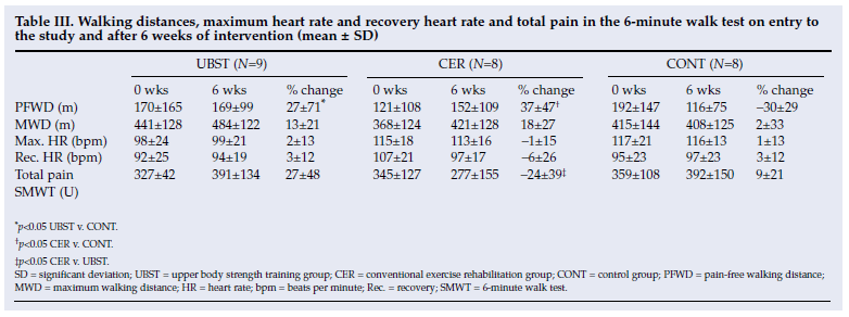
Pain-free walking distance during the SMWT increased significantly in the CER and UBST groups compared with the CONT group after training (37±47% v. 27±71% v. -30±29%; CER v. UBST v. CONT p=0.03; ES=1.83 (CER v. CONT) and ES=1.12 (UBST v. CONT)), and total pain perceived during the 6-minute walk decreased in the CER group compared with the UBST group (-24±39% v. 27±48%; CER v. UBST p=0.01; ES=1.25).
Recovery heart rate after the 6-minute walk tended to be reduced in the CER group after training, but this was not significant.
After training the fat percentage was found to have decreased in the UBST group compared with the control group (-8.2±7% v. 2.7±4%; UBST v. CONT p=0.0008; ES=2).
There was no significant difference between groups in ankle or brachial systolic blood pressure on entry to the trial. No significant change occurred in any of the groups after intervention (data not shown).
Discussion
We are not aware of assessments of the effects of a structured short-duration (6-week) conventional exercise rehabilitation programme and an upper body strength training programme in patients with PAD and intermittent claudication. Using this training model we draw several conclusions.
The CER group displayed marked improvements in functional capacity compared with the other groups. A study weakness is that despite randomisation this group had a significantly lower peak VO2 at entry, and improvements in functional capacity may have been easier to demonstrate in them. The CER group increased peak VO2 and MWD and also had lower sub-maximal heart rates than the UBST group and the CONT group during the treadmill exercise test. Although the UBST improved significantly more than the CONT group in PFWD in the SMWT, only the CER group improved significantly in PFWD and total pain perceived during the 6minute walk test.
In contrast, the control group showed no significant changes in walking distances, VO2 or sub-maximal heart rates. Since physicians do not conventionally ask patients to keep a log book of their walking when giving advice to walk at home, we did not do so in this study. We therefore do not know whether this group did not improve because they did not walk. However, we assume that the advice to exercise at home was not followed as these patients' sub-maximal and maximal heart rates increased and their peak VO2 was lower after the intervention period.
Finally, although upper body strength training increased strength and reduced the percentage of body fat, it did not cause any change in peak VO2 or sub-maximal heart rate values.
Owing to the small sample size in this pilot study, further studies should be conducted to see whether these exercise regimens elicit similar results.
Conclusion
Although 6 weeks of structured UBST produces improvements in fat percentage and strength and tends to improve PFWD, it is not as effective as a structured CER programme in producing functional improvements in patients with PVD.
Those advised to 'walk at home as much as possible' showed no improvement in functional capacity or walking distance, and such standard advice is therefore likely to be ineffective. A 6-week programme is sufficient to improve walking distances and cardiovascular fitness in patients with PVD.
References
1. Walker RD, Nawaz S, Wilkinson CH, Saxton JM, Pockley AG, Wood RFM. Influence of upper- and lower-limb exercise training on cardiovascular function and walking distances in patients with intermittent claudication. J Vasc Surg 2000; 31: 662-669. [ Links ]
2. Creasy TA, McMillan PJ, Fletcher EW, Collin J, Morris PJ. Is percutaneous transluminal angioplasty better than exercise for claudication? Preliminary results from a prospective randomised trial. Eur J Vasc Surg 1990; 4: 135-139. [ Links ]
3. Mannarino E, Pasqualini L, Menna M, Maragoni G, Orlandi U. Effects of training on peripheral vascular disease: A controlled study. Angiology 1989; 40(1): 5-10. [ Links ]
4. Hiatt WR, Regensteiner JG, Hargarten ME, Wolfel EE, Brass EP. Benefit of exercise conditioning for patients with peripheral vascular disease. Circulation 1990; 81: 602-609. [ Links ]
5. Parker-Jones P, Skinner JS, Kent Smith L, John FM, Bryant CX. Functional improvements following stairmaster vs treadmill exercise training for patients with intermittent claudication. J Cardiopulm Rehabil 1996; 16: 47-55. [ Links ]
6. Gardner AW, Poehlman ET. Exercise rehabilitation programs for the treatment of claudication pain. JAMA 1995; 274: 975-980. [ Links ]
7. Gardner AW, Montgomery PS, Flinn WR, Katzel LI. The effect of exercise intensity on the response to exercise rehabilitation in patients with intermittent claudication. J Vasc Surg 1995; 42: 702-709. [ Links ]
8. Regensteiner JG, Meyer TJ, Krupski WC, Cranford LS, Hiatt WR. Hospital vs home-based exercise rehabilitation for patients with peripheral arterial occlusive disease. Angiology 1997; 48(4): 291-300. [ Links ]
9. Aquarius AE, Denollet J, Hanning JF, De Vines J. Role of disease status and type D personality in outcomes in patients with peripheral arterial disease. Am J Cardiol 2005; 96(7): 996-1001. [ Links ]
10. Hiatt WR, Wolfel EE, Meier RH, Regensteiner JG. Superiority of treadmill walking exercise vs strength training for patients with peripheral arterial disease. Implications for mechanism of the training response. Circulation 1994; 90: 602-609. [ Links ]
11. Brozek J, Grande F, Anderson J, Keys A. Densitometric analysis of body composition: Revision of some quantitative assumptions. Ann NY Acad Sci 1963; 110: 113-140. [ Links ]
12. Perakyla T, Tikkanen H, Von Knorring J, Lepantalo M. Poor reproducibility of exercise test in assessment of claudication. Clin Physiol 1998; 18: 187-193. [ Links ]
13. American College of Sports Medicine. Guidelines for Exercise Testing and Prescription. London: Williams & Wilkins, 1995. [ Links ]
14. Gordon N, Gibbons L. The Complete Heart Recovery Guide. Cape Town: Oxford University Press, 1991: 371-372. [ Links ]
15. Thalheimer W, Cook S. How to calculate effect sizes from published research articles: A simplified methodology. http://work-learning.com/effect_sizes.htm (accessed 31 November 2002). [ Links ]
Accepted 17 June 2009.
Corresponding author: B M Parr (parrb@cput.ac.za)

