


Services on Demand
Article
 English (pdf)
 Article in xml format
 Article references
Indicators

Related links
-
 Cited by Google
 -
 Similars in Google
Share

 Permalink
SAMJ: South African Medical Journal
On-line version ISSN 2078-5135
Print version ISSN 0256-9574
SAMJ, S. Afr. med. j. vol.99 n.10 Pretoria Oct. 2009
SAMJ FORUM
CLINICAL IMAGES
Malignant otitis externa with ecthyma gangrenosum and pneumonia in an infant
Suhail Amin Patigaroo; Nazia Mehfooz; S F Hashmi
Our patient, a previously healthy 5-month-old infant, presented with malignant otitis externa, ecthyma gangrenosum and pneumonia, but without bacteraemia.
Background
Necrotising (malignant) external otitis (MEO), an infection involving the temporal and adjacent bones, is a rare complication of external otitis.1-3 It is characterised by unremitting pain and purulent discharge, and tends to invade cartilage, bone, nerve and adjacent soft tissues. It primarily affects elderly diabetics. The causative agent is almost uniformly Pseudomonas aeruginosa and may progress to involve the base of the skull, with multiple cranial nerve palsies and meningitis, leading to death in 30 - 80% of cases.4 Blood cultures can be sterile.5 Ecthyma gangrenosum (EG) is a rare and invasive cutaneous infection caused by Pseudomonas in the majority of cases.6-8 Pseudomonal pneumonia typically occurs in a patient who is immunocompromised; it is the most common cause of hospital-acquired pneumonia, sometimes in the absence of bacteraemia.9,10
Case report
An infant who is immunocompromised can develop all three of these conditions, as in our patient, a 5-month-old previously healthy infant, who developed transient neutropenia and presented with these manifestations of P. aeruginosa infection and without bacteraemia. His ear commenced a slight, yellowish discharge which became profuse, greenish and mixed with blood. There was blackening and ulceration of the skin of the right ear (Fig. 1) and facial deviation; one black spot over the neck was followed by two other spots on the scalp and forehead which progressed. The infant had a cough, difficulty in breathing and refused to feed. He was well-nourished, pale, restless and irritable. His heart rate was 156, respiratory rate 60, perfusion <3 seconds, temperature 102ºF, and SpO2 89% without oxygen. There was facial weakness and deviation of the mouth to the left on crying (Fig. 2). There was intercostal and subcostal recession, accessory muscles were working, and bilateral decreased air entry and bilateral crepitus were present.
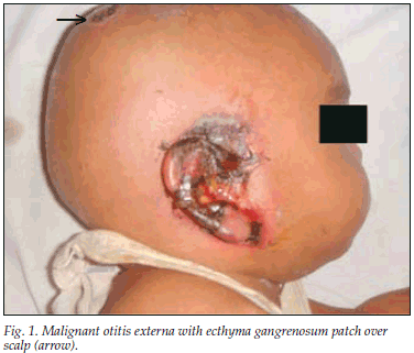
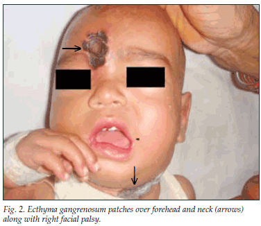
There was ulceration, necrosis and blackening of the right external auditory canal and part of the pinna (Fig. 1). The external auditory canal was filled with reddish soft tissue and greenish pus along with three blackish eschars over the scalp (Fig. 1), neck and forehead (Fig. 2).
Haemoglobin was 10 gm/dl, neutrophils 8%, absolute neutrophil count 0.5 x 10⁹/l; ESR 110 mm/h; HIV ELISA and VDRL were negative; and serum immunoglobulin levels were normal.
Pus from the external auditory canal revealed P. aeruginosa sensitive to ceftazidime, amikacin and piperacillin and resistant to cefeprazone, ceftriaxone and amoxicillin. Blood cultures on admission 12 hours apart revealed no growth. The eschar revealed Gram-negative rods and on culture revealed Pseudomonas growth. Histology of the mass in the auditory canal showed granulation tissue. Chest X-ray showed bilateral pneumonic patches. Contrast-enhanced computed tomography (CECT) showed severe swelling of soft tissues in the right external canal with a soft-tissue mass in the middle ear and partial erosion of the posterior wall of the bony canal.
The patient responded well to treatment with piperacillin and amikacin, local debridement of EG lesions and necrotic lesions of the ear, and regular dressings. A deformed right ear with loss of tympanic membrane and ossicles and partial facial paralysis remained.
Discussion
Few cases of MEO have been described in the paediatric age group and in infants.11 We have not found another case in the literature with this combination of MEO with EG and pneumonia.
Paediatric case reports stress anaemia, poor general condition, and concurrent systemic disease as predisposing factors. All paediatric patients with MEO are immunocompromised, which permits this opportunistic infection to be progressive and virulent. The most frequently cultured pathogen is P. aeruginosa; others include Staphylococcus epidermidis, Gram-negative bacteria, and fungi. Diagnosis requires culture of ear secretions and pathological examination of granulation tissue from the infection site to exclude malignancy. The characteristic lesions of EG are haemorrhagic pustules or infarcted-appearing areas with surrounding erythema that evolve into necrotic ulcers surrounded by erythematic halo. EG may appear in any location on the body; however, it predominantly affects the anogenital and axillary areas. Our patient had lesions over the scalp, forehead, and neck.
Patients at high risk for EG include those with malignancies, hypogammaglobulinaemia, patients on steroid therapy and those who are immunodeficient. Our patient also had pneumonia which, although not proved to be of pseudomonal origin, responded to antipseudomonal antibiotics.
1. Chandler JR. Malignant external otitis. Laryngoscope 1968; 78: 1257-1293. [ Links ]
2. Ophir H, Doron H. Necrotizing (malignant) external otitis. Am Fam Physician 2003; 68: 309-312. [ Links ]
3. Meltzer PE, Kelemen G. Pyocyaneous osteomyelitis of the temporal bone, mandible and zygoma. Laryngoscope 1959; 169: 1300-1316. [ Links ]
4. Chandler JR. Malignant external otitis, further considerations. Ann Otol, Rhinol Laryngol 1977; 86: 417-428. [ Links ]
5. Dan NIR, Tsila NIR. Clinical records malignant external otitis in an infant. J Laryngol Otol 1990; 104: 488-490. [ Links ]
6. Song WK, Kim YC. Ecthyma gangrenosum without bacteraemia in a leukaemic patient. Clin Exp Dermatol 2001; 26: 395-397. [ Links ]
7. Mull CC, Scarfone RJ, Conway D. Ecthyma gangrenosum as a manifestation of Pseudomonas sepsis in a previously healthy child. Ann Emerg Med 2000; 36: 383-387. [ Links ]
8. Huminer D, Siegman-Igra Y, Morduchowicz G, Pitlik SD. Ecthyma gangrenosum without bacteraemia. Report of six cases and review of the literature. Arch Intern Med 1987; 147: 299-301. [ Links ]
9. Iannini PB, Claffey T. Bacteraemia pseudomonas pneumonia. JAMA 1974; 230; 558. [ Links ]
10. Tillotson JR, Lerner AM. Characteristics of nonbacteremic pseudomonas pneumonia. Ann Intern Med 1968; 68: 295. [ Links ]
11. Rubinstein E, Ostfeld E, Ben-Zaray S, Schiby G. Necrotizing external otitis. Paediatrics 1980; 66: 618-620. [ Links ]
Accepted 1 April 2009.
Dr Patigaroo is a junior resident in the Department of ENT at Jawaharlal Nehru Medical College, AMU, Aligarh, India. Dr Mehfooz is a junior resident in the Department of Tuberculosis and Respiratory Diseases, AMU, Aligarh. Dr Hashmi is a reader in the Department of ENT, Jawaharlal Nehru Medical College, AMU, Aligarh.
Corresponding author: S A Patigaroo (Dr_suhail_jnmc@yahoo.co.in)

