


Serviços Personalizados
Artigo
 Inglês (pdf)
 Artigo em XML
 Referências do artigo
Indicadores

Links relacionados
-
 Citado por Google
 -
 Similares em Google
Compartilhar

 Permalink
SAMJ: South African Medical Journal
versão On-line ISSN 2078-5135
versão impressa ISSN 0256-9574
SAMJ, S. Afr. med. j. vol.98 no.4 Pretoria Abr. 2008
SAMJ FORUM
CLINICAL IMAGES
Auto-amputation of a breast due to ductal carcinoma
P van der Bijl
Pieter van der Bijl qualified MB ChB (cum laude) at Stellenbosch University in 2005. He performed his internship at Dihlabeng Regional Hospital, Bethlehem, Free State, in 2006, and was a community service medical officer at Kimberley Hospital Complex, Kimberley, Northern Cape, in 2007. He received the Diploma in Anaesthetics (SA) during that year. He is currently a registrar in internal medicine at Stellenbosch University
Auto-amputation of a breast as the result of a malignant process has been reported twice.1,2 In one case, a malignant melanoma of the breast originating in the nipple was responsible for the gross tissue destruction,1 the breast progressively atrophying and disintegrating to the point of disappearance. Another auto-amputation was described as one of tissue 'decay', denoting a somewhat different pathological process.2 No other references to a breast auto-amputating in toto as a consequence of mamma carcinoma could be found.
Case report
The patient, a 58-year-old woman, presented to the outpatient clinic at the Department of Oncology, Kimberley Hospital, in 2007. Her history was of a bloody discharge from her left nipple, with an accompanying ulcer on the left breast that had been developing more than 4 months before she presented to us. Approximately 4 months prior to the date of presentation, the left breast auto-amputated as an entity. It was discarded by the patient, and hence could not be subjected to histopathological investigation.
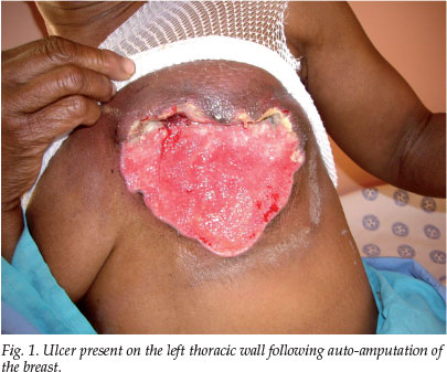
An ulcer (15x12 cm), with a clean base and a small amount of granulation tissue, was present on the left thoracic wall (Fig. 1). A fixed, hard lymph node (1x1 cm) was found in the right axilla, and another (2x1.5 cm) in the left axilla. A biopsy of the ulcer base demonstrated an infiltrating ductal carcinoma. No signs of pulmonary, hepatic or skeletal metastases were detected. A full blood count demonstrated thrombocytosis that was considered secondary to the neoplastic process.
On clinical and histological grounds, the diagnosis of an infiltrating ductal mamma carcinoma staged T4cN2aM0 with subsequent auto-amputation was made and chemotherapy was initiated.
Discussion
One can only speculate on the mechanism of auto-amputation of breast tissue as it has not been described. Owing to the late presentation of our patient, the clinical evolution of the process was not witnessed, and we therefore cannot add to knowledge of the process. From the history, it is only known that nipple discharge and an ulcer preceded the auto-amputation.
Necrosis can occur in malignancies (ductal carcinoma in situ as well as invasive carcinomas) of the female breast.3 Ultrastructural studies have indicated that the observed necrotic processes are a combination of apoptosis (active/ programmed cell death) and oncosis (passive/accidental cell death).4 It is reasonable to assume that auto-amputation of breast tissue is related to necrosis. Auto-amputation presumably starts with necrosis of the skin and supporting tissues of the breast, superimposed on the tumour necrosis. This extensive destruction of deep and superficial structures is then followed by eventual detachment of the breast from the thoracic wall. The auto-amputation in toto described in this case, as opposed to gradual auto-amputation,1,2 might, however, have been the result of differing pathological processes.
Late presentation of mamma carcinoma is common at our clinic and in South Africa, which has no national mammographic screening programme. Diagnostic and treatment delays are also associated with socio-cultural beliefs causing misconceptions about disease and its management.
Education of patients in regular self-examination of their breasts cannot be overemphasised. Furthermore, doctors should be strongly encouraged to include an examination of the breasts as part of a general medical examination.
1. Kuten A, Cohen Y, Kerner V. Malignant melanoma of the nipple with auto-amputation of the breast. Harefuah 1976; 90: 321-322. [ Links ]
2. Mintz U, Keinan Z. Mammary auto-amputation in inoperable carcinoma of the female breast. Harefuah 1975; 88: 531-532. [ Links ]
3. Jaffer S, Bleiweiss IJ. Intraductal papilloma with 'comedo-like' necrosis, a diagnostic pitfall. Ann Diag Path 2004; 8: 276-279. [ Links ]
4. Moinfar F, Mannion C, Man Y, et al. Mammary 'comedo' DCIS: apoptosis, oncosis and necrosis: an electron microscopic examination of 8 cases. Ultrastruct Pathol 2000; 24: 135-144. [ Links ]
 Correspondence:
P van der Bijl
(pieter.vanderbijl@gmail.com)

