


Services on Demand
Article
 English (pdf)
 Article in xml format
 Article references
Indicators

Related links
-
 Cited by Google
 -
 Similars in Google
Share

 Permalink
South African Journal of Surgery
On-line version ISSN 2078-5151
Print version ISSN 0038-2361
S. Afr. j. surg. vol.61 n.4 Cape Town 2023
http://dx.doi.org/10.36303/sajs.4183
OPINION PIECE
Tutankhamun - Africa's first reported road traffic crash victim?
AB van AsI; R BrownII
IDepartment of Surgery, School of Medicine, University of Limpopo, South Africa
IIDivision of Paediatric Surgery, University of Cape Town, South Africa
Keywords: Tutankhamun, cause of death, chest injury, clubfoot, femur fracture, mechanism of injury, post-mortem
Introduction
The most famous of all the Egyptian pharaohs was Tutankhamun, also known as King Tut, who died aged 18 in January 1343 BC. King Tut is famous because his tomb was left in almost perfect condition and contained wonderful treasures upon its discovery. The tomb permitted discovering the world of ancient Egypt. However, the cause of King Tut's death remains wrapped in a mystery.
We critically review the circumstantial findings in the tomb together with the various international examinations that were performed on the corpse of King Tut over the last 100 years. They include post-mortems, radiographic examination, CT-scanning, and DNA testing, and propose a hypothesis as to the cause of his death.
Background
Tutankhamun is possibly the most famous pharaoh in modern times, not necessarily because of his place in history (a minor pharaoh of the 18th dynasty of Egypt), but because of the findings of his nearly intact tomb in 1922 by Howard Carter. His famous forefathers were Thutmose IV, Amenhotep III and Amenhotep IV (Akhenaton).1 Akhenaton, his father, notoriously caused a revolution in Egypt by shifting the focus of religion from henotheistic worship to the monotheistic worship of Aten, the Sun Disk. In order to achieve this, he had many of the images and statues of the old gods destroyed and is likely to have undermined the class of conservative priests by closing the temples of Amun and depriving them of their enormous wealth. As part of his novel ideas he also moved the religious capital from Thebes (the contemporary Luxor) to a newly designed and built city, Akhetaten (the Horizon of the Sun Disk), hundreds of kilometres north. Upon Akhenaton's death in 1332, Tutankhamun ascended the throne as an 8-year-old boy, under the name of Tut-Ankh-Aten (Living Image of Aten). He was married to his half-sister Ankhesenamun (who at that time was known as Ankhesenpaaten). As a minor he was most likely educated and controlled by Ay and Horemheb, the non-royals who succeeded him as he left no heir. Ay performed the most important 'opening of the mouth ceremony' at the burial of Tutankhamun, thereby ensuring he would become the next pharaoh.
Tomb evidence
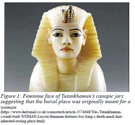
Tutankhamun was buried in a surprisingly small, hastily adapted tomb in the Valley of the Kings. Contrary to common belief, the tomb was not intact; yet, upon discovery, it was still loaded with marvellous treasures. Carter knew that the tomb had been compromised because of a re-plastered and sealed hole in the outer doorway. In addition, the findings of chaotically placed artefacts and damaged jewellery chests were an early indication that much of value had been taken. Of particular interest were a number of dismantled chariots found in the tomb. Pictures on a painted box also show Tutankhamun driving a chariot during a hunt. Of all the artefacts identified, it is estimated that approximately 80% were meant for someone else - the name of Tutankhamun being superimposed on the names of Akhenaton and his wife Nefertiti (Figure 1).
The major coffin itself was a yellow quartzite sarcophagus with a mismatching red granite lid and contained three smaller coffins all of which appeared with a face unlike that of Tutankhamun. Even the famous golden mask and the four canopic jars which held the pharaoh's viscera were probably not made for Tutankhamun. In summary, both the size of the tomb as well as the contents indicate that the death was unexpected, and the funeral rushed. The tomb was probably intended for a senior official, most likely Ay or perhaps Horemheb.
Mummy extraction
The mummy of Tutankhamun was very poorly prepared when compared to other pharaohs of the 18th dynasty. Excess resin was used that charred the skin and converted the mummy wrappings to mere soot. The copious amounts of resin also caused the mummy to stick to the inner coffin and face mask. As a result, Howard Carter was not able to remove the body intact. It was removed piecemeal causing great damage to the head and limbs. Considering the over-utilisation of resin, it is possible that, as part of a hasty funeral, the process of embalming was also adversely affected.
Tutankhamun's 'medical conditions'
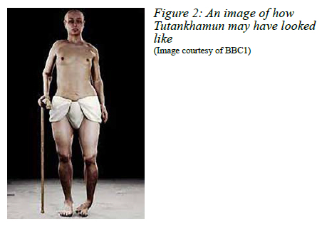
Several morphological 'idiosyncrasies' have been described, including a long narrow skull, a small cleft palate and extra occipital bones, like his father Akhenaten, and buck teeth, long hands and feet similar to other members of the family. This led to the postulates that he had one or more of the following medical diagnoses - Klinefelter's syndrome, feminising adrenal tumour, testicular feminisation syndrome and Wilson's disease. He was 165 cm tall, of slight build with wide hips and appeared well-nourished. Clothing found in the tomb suggested chest-waist-hip measurements of 31-29-43 cm. He was noted to have an abnormal left clubfoot but there was no sign of tuberculosis or any other disease. Figure 2 depicts his most likely body habitus.
1925 Post-mortem
From 11 November 1925, Dr Douglas Deny, an anatomical pathologist from the American University in Cairo, and Dr Saleh Bey Hamdi, along with Carter and other members of the expedition team, started the post-mortem study of the mummified pharaoh. It was extremely complicated to unwrap the mummy since the anointing oils and resins utilised in the process of mummification caused the fine linen wrappings to adhere to the body. As each layer was removed, the team began to discover many protective amulets, including gold jewellery, daggers, and pieces of armour wrapped between the layers all over Tutankhamun's body. Once the layers had been removed, they could finally begin to examine the actual corpse and make detailed anatomical notes on the body.
1968 Radiological examination
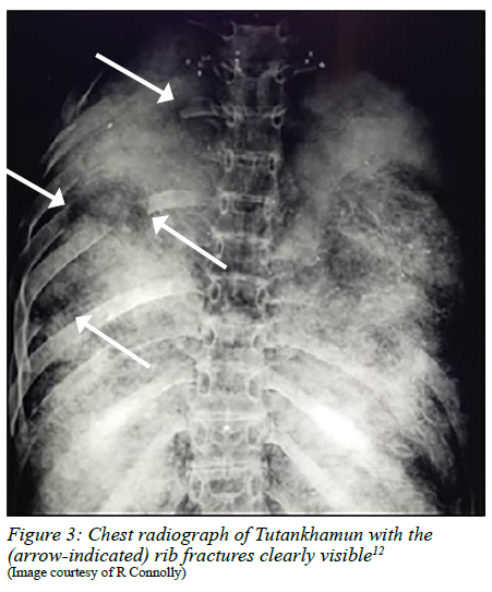
Further details of his injuries were documented in 1968 by radiographs of the mummy taken on a mobile x-ray machine by the University of Liverpool Derby Professor of Anatomy RG Harrison. The first striking findings were of multiple rib fractures (Figure 3), absence of the sternum and a number of anterior ribs. Carter's team may not have been as careful as claimed in his records. Failure to re-wrap the mummy undoubtedly led to unnecessary and preventable deterioration, due to exposure to very hot temperatures. Many of the limbs had been severed in order to remove some of the jewellery. Both hands were severed, both legs removed from the pelvis, and the head severed from the body to remove the gold mask. Several body parts went missing, in particular the right ear and penis, as earlier pictures clearly proved they were present during his initial examination. The penis was later found on a sand-tray concealed under the lower part of the mummy. Radiographs taken by Harrison detected a fractured leg, but he could not determine if this was preexisting, a result of the embalming process, or a result of Carter's examination. An abnormality of the left foot was also noted and presumed to be a congenital clubfoot. A few loose bone fragments from the vertebral column were discovered within the skull and were thought to be related to the embalming process.
1978 X-ray of skull and teeth
In 1978, the University of Michigan Professor of Orthodontics Dr James Harris conducted a closer examination of the skull and teeth with the help of newly developed radiological technology. Prior radiographs performed on the mummy in 1968 demonstrated loose bone fragments in the skull, prompting a sensational theory that the boy king had been bludgeoned to death by his political enemies during a particularly volatile time in Egyptian history. However, the scan in 1978 could not identify any skull fracture. It did, however, demonstrate that the two small bone fragments belonged to the first vertebra. Because the fragments were loose and not covered with solidified embalming resin, it is postulated that the damage occurred after the pharaoh's remains were prepared for burial and were possibly dislodged during Carter's attempts to remove the famous gold mask, densely stuck to the body by the resin. If a fracture had occurred before his death, the bone particles would likely have been fixed to the skull by resin and unable to move.
2005 CT-scan
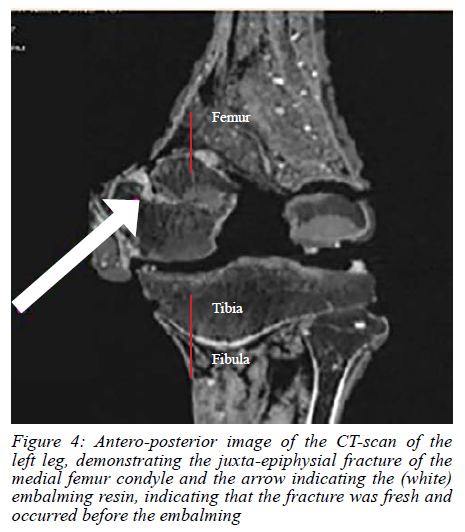
In 2005, scientists moved a mobile CT-scanner to Luxor to perform a high-resolution, full-body scan on Tutankhamun's mummy. Since then, scientists have increasingly focused on a fracture in King Tut's left thigh bone as the most likely cause of death. The CT-scan demonstrated a fracture of the left distal femur just above the knee (Figure 4). Embalming resin within the fracture indicated that this was a fresh, compound fracture and must have occurred shortly before his death as there were no signs of bone healing. Even as recently as one century ago, compound fractures had a very high mortality due to infection and other complications. Therefore 3500 years ago, with no access to antibiotics, death from a compound fracture would have been very likely. Scans of the abnormal left foot suggested a congenital clubfoot or an injury in early childhood resulting in significant bone damage. Tutankhamun has also been immortalised shooting with a bow and arrow while sitting. This fact coupled with the 134 walking sticks present in the tomb make it highly likely that the pharaoh was disabled by an abnormal foot.
Summary of medical evidence
In their comprehensive article in 2014, Ruhli and Ikram provide a plethora of medical diagnoses that have been suggested over the last 95 years. Nearly all are based on pure speculation with minimal definitive proof. Due to the extensive utilisation of resin the surface of the mummy only displays very limited information on medical conditions.
This is, however, in stark contrast with the skeleton. Although many parts of the skeleton were damaged in order to retrieve the many adornments, there remains the hard evidence of a chronic lesion of the left foot, a fresh left compound distal juxta-epiphysial femur fracture and multiple rib fractures with the sternum and part of anterior chest wall absent.
The latter point may be significant since the process of preparing the body of Tutankhamun for mummification was not performed in the usual manner. In a typical mummification, an incision in the lateral abdominal wall was used to remove the liver, stomach and intestines. The diaphragm was then breached via the abdominal cavity and the heart and lungs removed. In Tutankhamun's case, however, the abdominal incision was unusual in size and position while the diaphragm remained intact. Both heart and lungs were missing in the mummy and also missing in the canopic jars. This suggests that they were removed via the chest in a very unusual manner, possibly as a result of a severe injury.
The most teleological cause of death
The tomb size and contents provide overwhelming evidence that Tutankhamun suffered an unexpected and sudden death. The funeral proceedings and preparation of the tomb were therefore performed in a hurry. The skeletal investigations demonstrated that he had an abnormal left foot since early childhood, had suffered an acute, compound fracture of the left distal femur shortly before death and that the sternum and part of anterior chest wall were missing. The type of fracture Tutankhamun suffered, a compound distal juxta-epiphysial femur fracture, is excessively rare in children. Open fractures are typically caused by high-energy injuries, such as car crashes, falls from a height or severe sports injuries. The velocity of the injury is the main determinant of the severity of a compound fracture. The tomb contained a number of chariots with worn wheels, indicating that Tutankhamun may have enjoyed riding the chariot often and with pleasure. His clubfoot, the numerous walking sticks and the fact he was pictured using a bow and arrow while sitting provide evidence of why he would have liked the chariot as a way of locomotion. A compound femur fracture may have resulted in sudden death from acute blood loss, or an infection of the compound femur fracture after a period of days or weeks could also have been a lethal complication. Anterior chest trauma with rib and sternum fractures, and contusion of the heart and lungs may have occurred. After his demise these damaged body parts were removed during his mummification process.
Conclusion
We propose that pharaoh Tutankhamun fell from his chariot as a result of his weak left foot. During the fall he sustained multiple injuries including blunt chest trauma and a compound femur fracture. Thoracic injuries are one of the leading causes of mortality in paediatric trauma and represent the second most common lethal injury in children worldwide following a road traffic crash. The compound left femur fracture (on the side of his weak foot) provides the clue to this very conclusion. It is tragic to consider that not much has changed in nearly 40 centuries and that vehicular crashes remain very lethal and do not discriminate between the poor and the rich.
Acknowledgements
The authors would like to express their gratitude to Keith Grenville (founder) and Prof David Woods, illustrious member of The Egyptian Society of South Africa, for their expert comments on this manuscript.
Conflict of interests
The authors declare no conflict of interest.
REFERENCES
1. Clayton PA. Chronicle of the pharaohs - the reign-by-reign record of the rulers and dynasties of ancient Egypt. Thames & Hudson; 2006. p. 128. ISBN 0-500-28628-0. [ Links ]
2. Hornung E. The rediscovery of Akhenaten and his place in religion. Journal of the American Research Centre in Egypt. 1992;29:43-9. https://doi.org/10.2307/40000483. [ Links ]
3. Kemp BJ. The city of el-Amarna as a source for the study of urban society in ancient Egypt. World Archaeology. 1977;2:123-39. https://doi.org/10.1080/00438243.1977.9979691. [ Links ]
4. Zauzich K-T. Hieroglyphs without mystery. Austin: University of Texas Press; 1992. p. 30-31. ISBN 978-0-292-79804-5. [ Links ]
5. Tyldesley J. Chamber of Secrets: what lies behind the walls of Tutankhamun's tomb? Current WorldArchaeology. 2015;3:14-19. Available from: https://the-past.com/feature/chamber-of-secrets-what-lies-behind-the-walls-of-tutankhamuns-tomb/. [ Links ]
6. Harrison RG, Abdalla AB. The remains of Tutankhamun. Antiquity. 1972;46:8-14. https://doi.org/10.1017/S0003598X00053072. [ Links ]
7. Hawass Z. The death of Tutankhamun. International Journal of Humanistic Ideology. 2013;13(27):161-2. [ Links ]
8. Boyer RS, Rodin EA, Grey TC, Connolly RC. The skull and cervical spine radiographs of Tutankhamen - a critical appraisal. AJNR Am J Neuroradiol. 2003;24(6):1142-7. [ Links ]
9. Egyptian Supreme Council of Antiquities. Tutankhamun CT scan results. 2005 Mar 7. National Geographic Press Room. Available from: http://press.nationalgeographic.com/2005/03/07/tutankhamun-ct-scan-results-issued-march-7-2005-by-the-egyptian-supreme-council-of-antiquities/. Accessed 31 July 2017. [ Links ]
10. Manring MM, Hawk A, Calhoun JH, Andersen RC. Treatment of war wounds - a historical review. Clin Orthhop Relat Res. 2009;467(8):2168-91. https://doi.org/10.1007/s11999-009-0738-5. [ Links ]
11. Ruhli FJ, Ikram S. Purported medical diagnoses of pharaoh Tutankhamun, c. 1325 BC-. Homo. 2014;65(1):51-63. https://doi.org/10.1016/j.jchb.2013.08.006. [ Links ]
12. Reynolds DA. Growth changes in fractured long-bones. J Bone Joint Surg. 1981;61-B(1):83-8. https://doi.org/10.1302/0301-620X.63B1.7204480. [ Links ]
13. Van As AB, Manganyi R, Brooks A. Treatment of thoracic trauma in children - literature review. Eur J Pediatr Surg. 2013;23:434-43. https://doi.org/10.1055/s-0033-1363160. [ Links ]
 Correspondence:
email: sebastian.vanas@uct.ac.za

