


Serviços Personalizados
Artigo
 Inglês (pdf)
 Artigo em XML
 Referências do artigo
Indicadores

Links relacionados
-
 Citado por Google
 -
 Similares em Google
Compartilhar

 Permalink
South African Journal of Surgery
versão On-line ISSN 2078-5151
versão impressa ISSN 0038-2361
S. Afr. j. surg. vol.61 no.3 Cape Town 2023
http://dx.doi.org/10.36303/sajs.4039
HEAD AND NECK SURGERY
Concordance of fine needle aspiration cytology and final histology of salivary gland tumours
FR NdotoraI; BS JacksonII
IDepartment of Surgery, Steve Biko Academic Hospital, University of Pretoria, South Africa
IIDepartment of Surgery, Kalafong Provincial Tertiary Hospital, University of Pretoria, South Africa
ABSTRACT
BACKGROUND: Fine needle aspiration cytology (FNAC) is a widely used diagnostic tool to evaluate salivary gland tumours. The Milan system for reporting salivary gland cytopathology allows for standardisation and facilitates cytological-histological correlation. However, FNAC findings can still pose a diagnostic challenge. The accuracy of FNAC should be assessed at each centre. The aim of this study was to assess the concordance of FNAC and final histology of salivary gland tumours in three academic hospitals affiliated with the University of Pretoria, South Africa
METHODS: The study was a cross-sectional retrospective analytical study of 214 patients who underwent an operation for salivary gland tumours. All patients with FNAC and histology results between 2007-2017 were included. Patients were recruited from three University of Pretoria, South Africa, affiliated hospitals: Steve Biko Academic, Kalafong Provincial Tertiary and Tembisa Provincial Tertiary Hospital
RESULTS: Of the 214 patients with salivary gland tumours, the majority were located in the parotid gland (56.1%). Pleomorphic adenoma was the most common tumour (62.6%). The FNAC sensitivity, specificity and diagnostic accuracy (receiver operating characteristic) were 92.7%, 98.1% and 0.95 respectively. The concordance between salivary gland tumour FNAC and final histology was 96.95% with a Cohen's kappa coefficient of 0.91 (p = 0.0001
CONCLUSION: There is strong concordance between FNAC and histology of salivary gland tumours. FNAC is an accurate, minimally invasive diagnostic tool with high sensitivity and specificity. It provides the clinician with a reliable preoperative diagnosis determining whether the salivary gland tumour is benign or malignant
Keywords: salivary gland tumours, fine needle aspiration cytology, histology
Introduction
Salivary gland tumours are rare, accounting for 0.4-13.5 cases annually per 100 000.1 The majority of salivary gland tumours are benign, with a malignant incidence of 21.7%.2,3 The aetiology of salivary gland tumours is unknown. Distinguishing benign from malignant tumours is the main challenge which influences the management of salivary gland tumours. Clinical examination of the salivary glands is not definitive and may require complementary diagnostic investigations. Ultrasound-guided fine needle aspiration cytology (FNAC) is a widely used diagnostic tool to evaluate both neoplastic and inflammatory lesions of the salivary glands.
Although FNAC is commonly performed for salivary gland nodules, some authors support the role of cytology only in a selected group of patients, namely those with suspected malignancy, metastatic carcinoma or lymphoma.4 An accurate cytological diagnosis can avoid unwarranted surgery. FNAC is reported to have a diagnostic specificity of 73-99%, sensitivity of 81-97% and accuracy of 86-97%.59 FNAC offers valuable information for planning therapeutic management. It also assists the clinician to distinguish surgical from non-surgical treatable pathological conditions, such as lymphoma, where surgical intervention may not be the preferred primary treatment. Benign FNAC results are also beneficial in reassuring patients who are poor surgical candidates.6 Regardless of the indications for FNAC, a negative FNAC result should not supersede the clinician's judgement in management of a clinically suspected malignant or neoplastic lesion as histopathology may be required for definitive diagnosis.7
Inconsistencies in reporting of salivary gland FNAC specimens can have an effect on the correlation between cytological interpretation and surgical outcome as well as impact on patient care. This challenge was improved by standardisation of FNAC reporting using the Milan system for reporting salivary gland cytopathology.1012 Salivary gland FNAC has therefore become an accepted method of evaluating and classifying salivary gland malignancy risk preoperatively.13 The Milan system consists of six categories. The first category is a non-diagnostic sample, directing the clinician to repeat the FNAC. The categories of non-neoplastic (10%), atypia of undetermined significance (20%) and neoplasm of uncertain malignant potential (35%) have a lower malignant risk. The last two categories (5 and 6), suspicious for malignancy and malignant, have a high risk of malignancy, 60% and 90% respectively.13
By standardising the reporting of salivary gland FNAC, communication between pathologists and clinicians is improved and cytological-histological correlation is facilitated.13 However, there are still FNAC diagnostic challenges including sampling error (due to haemorrhage, necrotic tissue or fibrosis), the wide diversity of similar tumour types and the overlap of cytological features.13
It may also be limited in the identification of rare malignant tumours.14 An FNAC alone may not be sufficient for adequate preoperative assessment in all cases. Therefore, FNAC findings can still pose a diagnostic challenge and the accuracy of FNAC should be assessed at each centre. The aim of this study was to assess the concordance of FNAC and final histology of salivary gland tumours in three academic hospitals affiliated with the University of Pretoria, South Africa.
Materials and methods
The study is a retrospective analysis where historical patient records were reviewed. Patients with salivary gland tumours/masses were included if they had an FNAC and surgical excision with a histology report. The study included patients over 10 years of age between 1 January 2007 and 31 December 2017. Patients were excluded if they did not have both FNAC and histology results. Patients were recruited from three academic hospitals affiliated with the University of Pretoria: Steve Biko Academic, Kalafong Provincial Tertiary and Tembisa Provincial Tertiary Hospital. All patients who had excision of salivary gland tumours were identified from theatre records and data captured from patient files. Approval to perform the research was obtained from each hospital. Ethical approval was obtained from the Human Research Ethics Committee of the University of Pretoria, reference no: 107/2018.
Data collected included patient demographic information (age and sex) and anatomical location of the lesion, tumour FNAC and histology results. The FNAC results were reported according to the Milan system. 'Non-neoplastic', 'atypia of unknown significance', and 'neoplasm' (Milan system 2, 3 and 4) were combined and categorised as 'benign'. Milan system 5 'suspicion of malignancy', and Milan 6 'malignant' were combined as a 'malignant' category.
Statistical analyses
The concordance between FNAC and histology results of salivary gland tumours was expressed by diagnostic statistics. Data summary was described in the form of frequencies, proportion, percentages and 95% confidence intervals for FNAC and histology. Diagnostic statistics for FNAC (sensitivity, specificity, receiver operating characteristic) were determined. Cohen's kappa value was also used to compare the concordance between FNAC and histology diagnoses. Statistical analysis was done using STATA 14 statistical software.
Results
A total of 214 patients had an operation for a salivary tumour. There were 108 (50.5%) women and 106 (49.5%) men. The mean age was 45 years, with a minimum of 15 and a maximum of 83 years. The majority of tumours were located in the parotid gland (120, 56.1%), and the submandibular gland (79, 36.9%). The remainder were located in the smaller salivary glands: five (2.3%) in the buccal, eight (3.7%) in the submental and two (0.9%) in the sublingual glands.
The majority of FNAC results (156, 72.9%), were benign. Malignant FNAC results were present in 41 (19.2%) specimens. Seventeen (7.9%) FNAC specimens were inconclusive. The final histological diagnosis from 214 samples, can be seen in Table I. All salivary gland tumours had a histological diagnosis which was categorised as either 'benign' or 'malignant'. On the histology, benign salivary gland tumours were more common than malignant tumours (169 benign and 45 malignant). Pleomorphic adenoma was the most common benign diagnosis in 134 (62.6%) patients, followed by Warthihs tumour in 24 (11.1%) patients. Carcinoma ex-pleomorphic adenoma was the most common salivary gland malignancy observed in 10 (4.7%) patients.
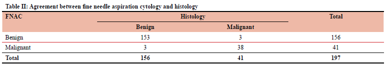
The concordance between salivary gland tumour FNAC and histology is noted in Table II. Patients with benign histology were correctly diagnosed on FNAC in 153 of 156 (98.1%) cases. Patients with malignant histology were correctly diagnosed on FNAC in 38 of 41 (92.7%) cases. Three patients with malignancy on histology were misdiagnosed as benign on the FNAC. Also, three patients with benign histology were misdiagnosed as malignant on the FNAC.
The sensitivity for FNAC was 92.7% (95% confidence interval [CI] 80.1-98.5%). The specificity for FNAC was 98.1% (95% CI 94.5-99.6%). Receiver operating characteristic (ROC) was 0.95 (95% CI 0.91-0.99). The concordance between FNAC and histology was 96.95% with a Cohen's kappa coefficient of 0.91 denoting excellent concordance between FNAC and histology (p = 0.00001).
The FNAC for parotid gland tumours demonstrated concordance of 97.3%, with Cohen's kappa coefficient of 0.88 (p = 0.0001). The FNAC for submandibular gland tumours also demonstrated concordance of 97.3%, with Cohen's kappa coefficient of 0.94 (p = 0.0001). Therefore, the concordance of FNAC and histology was not changed with the different locations of salivary glands.
Discussion
The prevalence of salivary gland tumours was similar between women (50.5%) and men (49.5%). The findings are similar in previous reports with slight predominance in women (1.1-1.8 times higher in women).15-17 The mean age of 45 years was also similar to documented reports.16,17 In this study, the parotid glands were the commonest salivary gland involved and accounted for the majority of tumours (56.1%). In the literature, the parotid tumours make up more than 80%.16
Pleomorphic adenoma (62.6%) was the most common salivary gland tumour and benign neoplasm (79.3%). These tumours originate from myoepithelial and epithelial elements.18 High exposure to radiation has been attributed to the development of pleomorphic adenomas.18 The most common malignant tumours were pleomorphic carcinoma ex-pleomorphic adenoma in 10 (22.2%) patients, however, mucoepidermoid carcinoma is usually the commonest documented salivary gland malignancy.19 Pleomorphic carcinoma ex-pleomorphic adenoma typically has a prevalence of 3-15% of all malignant salivary gland tumours, but appears to be increasing in the last decade which may explain the higher prevalence in our results.20
There was excellent concordance between FNAC and histology overall (96.95%) with Cohen's kappa coefficient of 0.91 (p = 0.0001) and the accuracy of FNAC to diagnose patients had an ROC of 0.95 (95% CI 0.91-0.99). This is in keeping with the accuracy of FNAC in other reports (91-97%).59 An FNAC diagnosed malignancy correctly in 38 (92.7%) patients. It also correctly diagnosed 153 (98.1%) benign tumours. Similar results have been reported for FNAC accuracy of malignant salivary gland tumours (78-97%).5-7 However, FNAC of benign tumours is reported as less accurate, at approximately 85%, compared to our results.5,7
The sensitivity for FNAC was high at 92.7% and specificity at 98.1% which is similar in previous reports (sensitivity of 73-93% and specificity of 94-100%).5-9 Reports have shown 73% FNAC sensitivity and 97% specificity in diagnosing malignant salivary gland tumours alone.8 Regardless of salivary gland location, i.e., parotid and submandibular glands, FNAC concordance with histology was maintained. That of the remaining salivary glands (submental, sublingual and buccal) could not be calculated because of limited numbers in those categories.
Despite the simplicity and accuracy of FNAC, non-diagnostic aspirations may occur in 6-29%.13,21 The recommended acceptable rate should be less than 10%.10 There were 17 (7.9%) cases of inconclusive/non-diagnostic FNAC cytology in this study. This may be attributed to incorrect needle positioning (especially when palpation-guided), aspiration of necrotic samples, inadequate sampling and the vascular nature of some tumours.22 Clinicians inexperienced at FNAC technique or interpreting the cytology samples may result in non-diagnostic results. It is recommended that tumours with non-diagnostic FNAC results get a repeat FNAC.12 Inadequate sampling may be improved with use of ultrasound-guided FNAC and a dedicated pathologist performing the cytological examination. Ultrasound-guided FNAC has better sensitivity, specificity and diagnostic accuracy compared to palpation-guided FNAC.23 Another option is to perform a core needle biopsy which has shown to have a sensitivity of 96% and specificity of 100%.24 Core needle biopsy also has low risk of complications, especially when performed under ultrasound guidance.24
Study limitations
Data collection was limited to three academic hospitals affiliated with the University of Pretoria. There are no previous studies in these hospitals comparing FNAC and histology of salivary gland tumours, thus the results could not be compared to assess for improved diagnostic accuracy over time.
Conclusion
There is strong concordance between FNAC and histology for salivary gland tumours. FNAC is an accurate, minimally invasive diagnostic tool with high sensitivity and specificity. It provides the clinician with a reliable preoperative diagnosis, whether the salivary gland tumour is benign or malignant.
Conflict of interest
The authors declare no conflict of interest.
Funding source
This research received no specific grant from any funding agency in the public, commercial or not-for-profit sectors.
Ethical approval
Ethical approval was obtained from the Human Research Ethics Committee of the University of Pretoria, reference no: 107/2018.
ORCID
FR Ndotora https://orcid.org/0000-0001-7337-2938
BS Jackson https://orcid.org/0000-0001-8994-8575
REFERENCES
1. World Health Organization. World Health Organization classification of tumours: pathology and genetics of head and neck tumours. 2005. Available from: https://screening.iarc.fr/doc/BB9.pdf. Accessed 27 May 2022. [ Links ]
2. Satko I, Stanko P, Longauerová I. Salivary gland tumours treated in the stomatological clinics in Bratislava. J Craniomaxillofac Surg. 2000;28(1):56-61. https://doi.org/10.1054/jcms.1999.0092. [ Links ]
3. De Oliveira FA, Duarte ECB, Taveira CT, et al. Salivary gland tumour: a review of 599 cases in a Brazilian population. Head Neck Pathol. 2009;3(4):271-5. https://doi.org/10.1007/s12105-009-0139-9. [ Links ]
4. Cohen EG, Patel SG, Lin O, et al. Fine-needle aspiration biopsy of salivary gland lesions in a selected patient population. Arch Otolaryngol Head Neck Surg. 2004;130(6):773-8. https://doi.org/10.1001/archotol.130.6.773. [ Links ]
5. Piccioni LO, Fabiano B, Gemma M, et al. Fine-needle aspiration cytology in the diagnosis of parotid lesions. Acta Otorhinolaryngol Ital. 2011;31(1):1-4. [ Links ]
6. Ali NS, Akhtar S, Junaid M, et al. Diagnostic accuracy of fine needle aspiration cytology in parotid lesions. ISRN Surg. 2011;2011:721525. https://doi.org/10.5402/2011/721525. [ Links ]
7. Jain R, Gupta R, Kudesia M, et al. Fine needle aspiration cytology in diagnosis of salivary gland lesions: a study with histologic comparison. Cytojournal. 2013;10:5. https://doi.org/10.4103/1742-6413.109547. [ Links ]
8. Gudmundsson JK, Ajan A, Abtahi J. The accuracy of fine-needle aspiration cytology for diagnosis of parotid gland masses: a clinicopathological study of 114 patients. J Appl Oral Sci. 2016;24(6):561-7. https://doi.org/10.1590/1678-775720160214. [ Links ]
9. AlGhamdi GZ, Alzahrani AK, Saati H, et al. Correlation between fine needle aspiration cytology (FNAC) and permanent histopathology results in salivary gland masses. Cureus. 2021;13(3):e13976. https://doi.org/10.7759/cureus.13976. [ Links ]
10. Rossi ED, Faquin WC, Baloch Z, et al. The Milan system for reporting salivary gland cytopathology: analysis and suggestions of initial survey. Cancer Cytopathol. 2017;125(10):757-66. https://doi.org/10.1002/cncy.21898. [ Links ]
11. Baloch Z, Field AS, Katabi N, et al. The Milan system for reporting salivary gland cytopathology. In: Faquin WC, Rossi ED, Baloch Z, et al., editors. The Milan system for reporting salivary gland cytopathology. Cham: Springer International Publishing; 2018. p. 1-9. https://doi.org/10.1007/978-3-319-71285-7_1. [ Links ]
12. Rossi ED, Faquin WC. The Milan system for reporting salivary gland cytopathology (MSRSGC): an international effort toward improved patient care-when the roots might be inspired by Leonardo da Vinci. Cancer Cytopathol. 2018;126(9):756-66. https://doi.org/10.1002/cncy.22040. [ Links ]
13. Kala C, Kala S, Khan L. Milan system for reporting salivary gland cytopathology: an experience with the implication for risk of malignancy. J Cytol. 2019;36(3):160-4. https://doi.org/10.4103/JOC.JOC_165_18. [ Links ]
14. Jackson BS, Pratt T, Van Rooyen A. Mammary analogue secretory carcinoma: a rare salivary gland tumour. S Afr Med J. 2017;107(4):304-06. https://doi.org/10.7196/SAMJ.2017.v107i4.12228. [ Links ]
15. Mahomed Y, Meer S. Primary epithelial minor salivary gland tumours in South Africa: a 20-year review. Head Neck Pathol. 2020;14(3):715-23. https://doi.org/10.1007/s12105-019-01111-4. [ Links ]
16. Bobati SS, Patil BV, Dombale VD. Histopathological study of salivary gland tumors. J Oral Maxillofac Pathol. 2017;21(1):46-50. https://doi.org/10.4103/0973-029X.203762. [ Links ]
17. Zaman S, Majid S, Chugtai O, et al. Salivary gland tumours: a review of 91 cases. J Ayub Med Coll Abbottabad. 2014;26(3):361-3. [ Links ]
18. Bokhari MR, Greene J. Pleomorphic adenoma. StatPearls. Treasure Island (FL): StatPearls Publishing LLC; 2022. [ Links ]
19. Peraza A, Gómez R, Beltran J, et al. Mucoepidermoid carcinoma. An update and review of the literature. J Stomatol, Oral Maxillofac Surg. 2020;121(6):713-20. https://doi.org/10.1016/j.jormas.2020.06.003. [ Links ]
20. Gupta A, Koochakzadeh S, Neskey DM, Nguyen SA, Lentsch EJ. Carcinoma ex-pleomorphic adenoma: a review of incidence, demographics, risk factors, and survival. Am J Otolaryngol. 2019;40(6):102279. https://doi.org/10.1016/j.amjoto.2019.102279. [ Links ]
21. Lee JJL, Tan HM, Chua DYS, Chung JGK. The Milan system for reporting salivary gland cytology: a retrospective analysis of 1384 cases in a tertiary Southeast Asian institution. Cancer Cytopathol. 2020;128(5):348-58. https://doi.org/10.1002/cncy.22245. [ Links ]
22. Wang H, Liang J, Belcher RH, et al. Nondiagnostic category of Milan system for reporting pediatric salivary gland cytopathology: outcomes and root cause analysis. Cancer Cytopathol. 2022;130(8):609-19. https://doi.org/10.1002/cncy.22571. [ Links ]
23. Khan N, Afroz N, Agarwal S, et al. Comparison of the efficacy of the palpation versus ultrasonography-guided fine-needle aspiration cytology in the diagnosis of salivary gland lesions. Clin Cancer Investig J. 2015;4(02):134-9. https://doi.org/10.4103/2278-0513.152717. [ Links ]
24. Witt BL, Schmidt RL. Ultrasound-guided core needle biopsy of salivary gland lesions: a systematic review and metaanalysis. Laryngoscope. 2014;124(3):695-700. https://doi.org/10.1002/lary.24339. [ Links ]
 Correspondence:
BS Jackson
Email: brandon.jackson@up.ac.za

