


Serviços Personalizados
Artigo
 Inglês (pdf)
 Artigo em XML
 Referências do artigo
Indicadores

Links relacionados
-
 Citado por Google
 -
 Similares em Google
Compartilhar

 Permalink
South African Journal of Surgery
versão On-line ISSN 2078-5151
versão impressa ISSN 0038-2361
S. Afr. j. surg. vol.60 no.3 Cape Town Set. 2022
http://dx.doi.org/10.17159/2078-5151/sajs3827
CASE REPORT
Genital labial phyllodes tumour recurring during pregnancy - does pregnancy impact recurrence?
L MbodiI; ZN MtshaliII; B PhakathiIII
IDepartment of Obstetrics and Gynaecology, Charlotte Maxeke Johannesburg Academic Hospital, University of the Witwatersrand, South Africa
IIDepartment of Pathology, National Health Laboratory Services, University of the Witwatersrand, South Africa
IIIDepartment of Surgery, Charlotte Maxeke Johannesburg Academic Hospital, University of Witwatersrand, South Africa
SUMMARY
Phyllodes tumours (PT) of the vulva are uncommon tumours. The gold standard treatment for these lesions is unknown but is intuitively presumed to be wide local excision. Incomplete resection is associated with recurrence although the time to recurrence is not known. Pregnancy is hypothesised to increase recurrence due to the production of steroid hormones oestrogen and progesterone. We report on a patient who had recurrence of a PT at a rare site (labia minora) and on the contralateral side from the original lesion, during pregnancy. These findings support that oestrogen and progesterone hormones could have played a role in the recurrence of the PT although margins were not free at the initial surgery.
Keywords: genital labial phyllodes tumour, pregnancy, recurrence
Case report
We previously published a case1 of a benign phyllodes tumour of the vulva excised with positive margins with a resultant risk for recurrence. In this report we present recurrence of a benign vulval phyllodes tumour in the same patient who is now 34 years old.
Following the index surgery on the right side, the patient was only seen at the 4-week follow-up clinic and was subsequently lost to follow-up for a period of 3 years, apparently with no recurrence.
She presented again during pregnancy with a slow-growing mass on the left side of the vulva involving the labium minora. The lesion was soft, not painful and was mobile. The patient confirmed that during the 3-year period from the postoperative follow-up clinic visit, there was no labial or vulva swelling, lymph node enlargement, and change in skin colour or texture
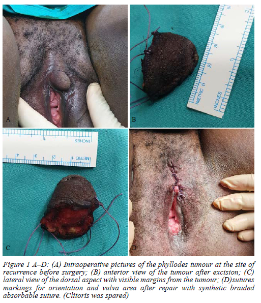
The mass was first noted during the 17th week of gestation. However, she only presented for assessment at 28 weeks gestation reporting that the mass had been slowly increasing in size. There were no pregnancy-related complaints, breast mass or pain on the vulva. On examination, a soft, mobile mass measuring 3 x 4 cm involved the left labium minora with no involvement of the labium major. After the initial assessment, pregnancy was allowed to progress and antenatal visits continued as scheduled. A term pregnancy was terminated through an uncomplicated normal vaginal delivery. At 6 weeks post-delivery, a wide local excision of the vulval lesion was performed with clear margins (Figure 1A-D). Histopathology confirmed a completely excised benign phyllodes tumour with similar histological characteristics as the primary tumour. Microscopic examination of the sections showed a well-demarcated benign neoplasm with a biphasic appearance. The tumour demonstrated extensive leaf-like papillary structures growing toward slit-like spaces under the skin (Figure 2A). There was evidence of apocrine metaplasia (Figure 2B) and lactational changes were noted (Figure 2C) in keeping with the history of recent gestation. The invaginating fronds were lined by a dual cell lining consisting of epithelial and myoepithelial cells further confirming its benign nature. The epithelial cells were cuboidal to columnar with decapitation secretions. The stromal was pauci-cellular showing bland monomorphic pale spindled cells. There was no evidence of cytologic atypia, necrosis or stromal metaplasia. The mitotic activity was very rare. The lesion was clear of the surgical margins.
The immunohistochemical (IHC) stains performed confirmed the biphasic (glandular and stromal) nature of the tumour. The IHC stains for epithelial component of the tumour were positive, i.e., pancytokeratin antibody (AE1/3) (Figure 2E). There was also immunoreactivity with gross cystic disease fluid protein 15 (GCDFP-15) (Figure 2D) in the glandular component, confirming a mammary-like nature of the anogenital sweat glands. The tumour was positive for both oestrogen and progesterone receptors. The retention of the myoepithelial cell-layer was highlighted by actin, alpha-smooth muscle type (Figure 2F).
Discussion
PTs of the vulva are rare, with about 20 cases reported in the literature.2 Most vulval PTs are located in the labium majora, with the labium minor being the least affected.3 Other locations include the interlabial sulcus, mons pubis and peri-clitoral sites. These tumours are usually solitary and rarely bilateral, with a high incidence of local recurrence.45 Genital PTs are believed to originate from the mammary-like anogenital sweat glands (with combined eccrine and apocrine features) rather than from ectopic breast tissue.6 They demonstrate capacity to branch into lobules and to form acini-like mammary glands on the anogenital area.6 These mammary-like anogenital glands are reported to be located within the interlabial sulcus, the paramedian area of the perineum and around the anus.6
Unlike in breast PT recurrence, where a 1 cm clear surgical margin is associated with the least risk of recurrence,7 the exact margin width for vulval PTs is unknown. In the index surgical procedure of this patient, the margins were involved which put her at risk for local recurrence. What was interesting was when she presented with a recurrence on the labium minor contralateral to the index lesion, a finding which is reported to be rare in the literature.8 Even with a wide local excision of the vulvar PT mass, recurrence on the ipsilateral site has been reported two years after surgery.9
The behaviour of PT of the vulva is difficult to predict based on histology alone as there is limited data or fewer reported cases.6 In the breast, reports of rapidly growing PTs during puberty and the presence of strong oestrogen and progesterone receptors positivity in 40% and 85% of epithelial cells respectively suggest that hormones could also be implicated in the recurrence.4 The recurrence of a PT in the first trimester of pregnancy in our patient supports the hypothesis that steroid hormones (oestrogen and progesterone) may play a role in recurrence. In this patient, the tumour was positive for both oestrogen and progesterone receptors.
Conclusion
PTs of the vulva are rare and most published reports are from case studies, with no conclusive factors associated with its primary occurrence or recurrence. The suggestion that pregnancy or a surge in progesterone and oestrogen hormones increases the risk of recurrence, although unproven, is not far-fetched. Our case where the PT recurred during pregnancy and displayed oestrogen receptor positivity is highly suggestive and in support of this theory.
Conflict of interest
The authors declare no conflict of interest.
Funding source
No funding was received from any companies or persons towards the publication of this case.
Ethical approval
This case was approved by the University of Witwatersrand Human Research Ethics Committee as part of the requirement for ethics approval. The case was approved by the HREC with Certificate Clearance number M210994.
ORCID
L Mbodi https://orcid.org/0000-0002-5950-791X
ZN Mtshali https://orcid.org/0000-0001-5971-6853
B Phakathi https://orcid.org/0000-0002-8991-6060
REFERENCES
1. Mbodi L, Mtshali NZ, Phakathi B. A rare case of genital labial phylloides tumour. S Afr J Surg. 2020;58(3):162. https://doi.org/10.17159/2078-5151/2020/v58n3a3275. [ Links ]
2. Venkatesh K, Jayanthy T, Patil S. Benign phyllodes tumor of the vulva. Indian J Pathol Microbiol. 2021;64(4):863-5. https://doi.org/10.4103/ijpm.ijpm_292_20. [ Links ]
3. Fujii DT, Korzen CA, Levine TC, Heitmann RJ. Phyllodes tumour of the labia minora. BMJ Case Rep. 2019;12(11):e229917. https://doi.org/10.1136/bcr-2019-229917. [ Links ]
4. Lee S, Nodit L. Phyllodes tumour of vulva: a brief diagnostic review. Arch Pathol Lab Med. 2014;138(11):1546-50. https://doi.org/10.5858/arpa.2013-0581-RS. [ Links ]
5. Way JC, Culham BA. Phyllodes tumour in pregnancy: a case report. Can J Surg. 1998;41(5):407-9. [ Links ]
6. Özbudak IH, Akkaya H, Akkaya B, et al. Phyllodes tumor of the vulva: report of two cases. Turk Patoloji Derg. 2013;29(1):73-76. https://doi.org/10.5146/tjpath.2013.01153. [ Links ]
7. Tan EY, Hoon TP, Yong WS, et al. Recurrent phyllodes tumours of the breast: pathological features and clinical implications. ANZ J Surg. 2006;76(6):476-80. https://doi.org/10.1111/j.1445-2197.2006.03754.x. [ Links ]
8. Polaquini G, Minari Bozko J, Minari C, et al. 358 contralateral recurrent phyllodes tumor of the vulva in a teenager. Int J Gynecol Cancer. 2019;29:A149. https://doi.org/10.1136/ijgc-2019-IGCS.358. [ Links ]
9. Heffernan TP, Sarode VR, Hoffman B, Lea J. Recurrent phyllodes tumour of the vulva: a case report with review of diagnostic criteria and differential diagnosis. Int J Gynecol Pathol. 2010;29(3):294-7. https://doi.org/10.1097/PGP.0b013e3181c14a8c. [ Links ]
 Correspondence:
L Mbodi
Email: mlangi2005@yahoo.co.uk

