


Services on Demand
Article
 English (pdf)
 Article in xml format
 Article references
Indicators

Related links
-
 Cited by Google
 -
 Similars in Google
Share

 Permalink
South African Journal of Surgery
On-line version ISSN 2078-5151
Print version ISSN 0038-2361
S. Afr. j. surg. vol.54 n.4 Cape Town Nov. 2016
PLASTIC SURGERY
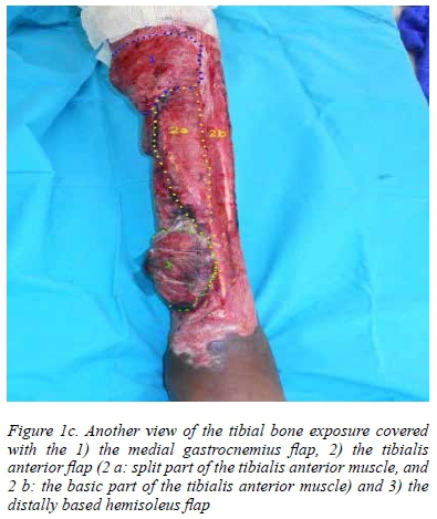
Coverage of extensive tibial bone exposure in burn patients with three local flaps
N Lahouel; P Mokwatlo; E Ndobe
Department of Plastic, Reconstructive and Aesthetic Surgery, University of the Witwatersrand, Johannesburg, South Africa
ABSTRACT
Covering tibial bone exposure from third degree burns to the lower limbs is a challenging task for the plastic surgeon. We present our experience of covering tibial exposure from burns in three different patients, where four limbs were involved and three muscular flaps were used in conjunction with one another; i.e. the tibialis anterior flap, the medial gastrocnemius flap and the hemisoleus flap. Through the use of this technique, tibial bone exposure, ranging from 15-30 cm, was successfully covered. This technique constitutes a good solution for surgically challenging wounds.
Keywords: hemisoleus flap, lower limb burns, medial gastrocnemius flap, tibial bone exposure, tibialis anterior flap
Introduction
Full-thickness lower limb third degree burns are difficult to treat, and become very challenging when the tibia is exposed. The tibial bone is subcutaneous on its anteromedial surface, extending from the tibial tuberosity to the convergence of the tibialis anterior (TA) tendon and the extensor tendons. This position makes it very vulnerable and at high risk of exposure from deep lower extremity burns.
The adult tibia ranges from in length from < 30 cm to 47 cm.1 This means that the extent of the subcutaneous tibia can easily reach up to 30 cm in a tall adult. Covering bone exposure of 30 cm is a challenging task for a plastic surgeon.
Different procedures have been carried out in an attempt to cover similar wounds. Bone drilling, to allow granulation, and thereafter performing a skin graft, is one, but this frequently leads to failure of the extensive wound as the shaft granulates very slowly.2 The application of free flaps is another option, but on numerous occasions, the extent of the bone exposure is so large that more than one flap is needed to cover all the bone. The presence of oedema, due to the nature of the injury, and the high risk of thrombosis, due to thrombocytosis which occurs in lower limb injuries, makes the risk of failure very high.3-5 Limb amputation is sometimes the only available option to this group of patients.2
We present our experience of the treatment of exposed tibia in three burn patients using three local flaps (medial gastrocnemius flap, tibialis anterior flap and hemisoleus flap) in conjunction with one another.
Anatomy and techniques
Tibialis anterior flap
TA muscle originates in the lateral condyle of the tibia, proximally two thirds of the lateral surface of the tibia, the interosseous membrane and the deep fascia of the leg.5 Its internal axial tendon courses to the medial side of the foot to insert on the medial cuneiform and the base of the first metatarsal.2 Throughout its course, the TA muscle runs superficially within the anterior compartment of the lower leg, lateral to the tibia.5
The TA is a strong inverter and dorsiflexor of the foot. It is innervated by several branches of the deep peroneal nerve.2 Hirchowitz et al.6described the arrangement of the TA muscle fibres as "circumpennate", with fibres radiating from a central tendon. Such a fibre arrangement provides considerable strength to the axial tendon. This could explain the preservation of the function of the TA after it has been used as a split turnover flap.2
According to Mathes and Nahai,7 TA muscle vascularisation is classified as a type IV pattern, with 8-12 short segmental branches from the anterior tibial artery, with connection at the anteromedial portion of the muscle.2,6 This type of vascularisation mandates the harvest of the TA flap according to the technique described by Hirchowitz et al.6and Ford et al.,8 by incising the muscle either laterally or medially, and turning it over the tibia, preserving some of the segmental pedicles.
Gastrocnemius flap
Two proximal heads converge into one distal tendon in the gastrocnemius muscle. Both heads are irrigated by sural arteries that start in the popliteal fossa. The vascularisation pattern of each head is classified as Mathes and Nahai type I,9 hence the possibility of moving it individually.10
Both the medial head and lateral head can be used, but the former is more commonly utilised because of its larger size and the absence of the fibula which can represent an obstacle with regard to stretching and reaching the distant point for coverage.11,12 The medial head provides excellent coverage of the tibial metaphysis.13
Hemisoleus flap
The soleus muscle contains two heads, one that originates on the posterior aspect of the tibia, and the other which originates on the posterior aspect of the fibula.13 The two heads are separated by an aponeurosis which converges near the Achilles tendon. The tibial head receives an artery that branches off the posterior tibial artery before or after tibiofibular bifurcation. The fibular head receives an artery that branches off the tibiofibular trunk. Accessory pedicles from the posterior tibial artery and fibular artery penetrate the two muscular heads along their course towards the Achilles tendon. The two heads receive segmentary vascularisation from the tibial posterior and fibular arteries.13 The soleus muscle is classified as type II, according to the Mathes and Nahai classification, owing to this vascular disposition. The veins are concomitant to the arteries.
The innervation is secured with one branch from the tibial nerve, which penetrates the muscle at its posterior aspect above the soleus arch, and another branch which penetrates the muscle at its anterior aspect.
The soleus flap can be harvested in full or as a hemisoleus flap. It can be proximally or distally based. However, the distally based version is less reliable owing to the vascularisation pattern, but covers the lower third of tibia better. The proximally based hemisoleus flap can cover the middle third and upper part of the lower third of the tibia.13
Cases studies
Case 1
A 26-year-old man sustained circumferential third degree burns from flames to the left leg, following alcohol intoxication. The wounds were debrided five days post burn, which left tibial bone exposure of 30 cm from the upper to the lower third of the tibia (Figure 1a).

The patient was referred to our unit one month after the burn. A decision was taken to perform three flap reconstructions (medial gastrocnemius flap, split TA flap and the distally based soleus flap) in conjunction with one another owing to the extensive bone exposure. (One large perforator was identified which allowed the distal flap to be harvested).
Two days postoperatively, all of the flaps were viable and well vascularised, except for venous congestion in the soleus flap (Figure 1 b and c).

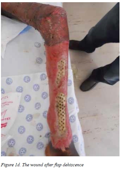
Three days later, the venous congestion resolved, the wound was clean and the bone well covered. Unfortunately, a few days later, the wound became septic due to the absence of wound care. The dressing had not been changed because of a prolonged strike, which meant that most of the hospital staff was prevented from working. Wound dehiscence occurred and the tibia became re-exposed.
Even though the patient had a peak platelet count of 1 422 x109/l, a decision was taken to perform a latissimus dorsi flap under anti-aggregant cover. Unfortunately, this, complicated by the artery and vein thrombosis which were not identified timeously by the nursing staff, led to flap loss.
We decided to drill the bone and use negative pressure wound therapy (NPWT), but granulation grew very slowly, especially at diaphysis level, > 7 weeks of NPWT therapy. It was clear that full tibia coverage would not be achieved using this option (Figure 1 d).
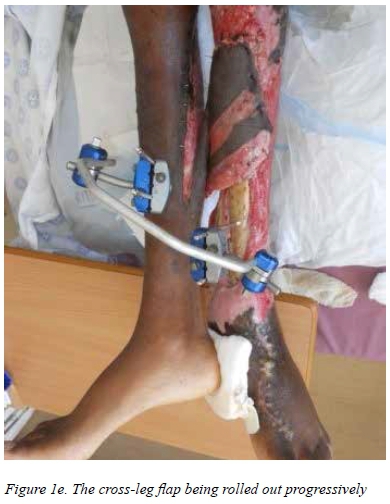
The "last weapon in our arsenal", the cross-leg flap, was then implemented. The flap was rolled out progressively. Full coverage of the tibia was achieved in five weeks' time (Figure 1 e and f). A small area of 1.5 x 2 cm of metaphysis became re-exposed later on.
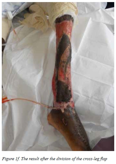
The patient was discharged after two months to another hospital in another province for personal reasons. This prevented a follow-up from being made. The patient walked with moderate limping due to pain at the time of his discharge.
Case 2
A 35-year-old man sustained circumferential third degree burns from a fire to the left leg and thigh. The patient was primarily managed at his local hospital, where he was resuscitated and underwent wound debridement. He was referred to our unit after > 2 months.
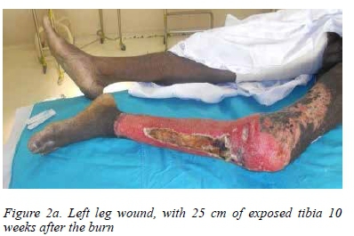
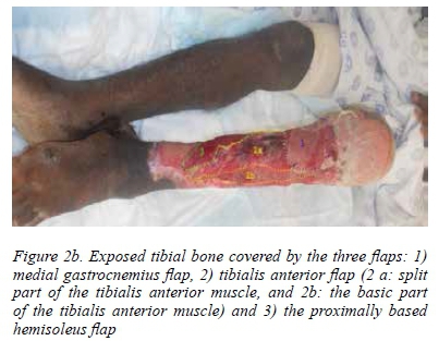
On admission, the patient had popliteal contracture and tibial bone exposure of 25 cm (Figure 2 a). Contraction release, bone drilling and the three-flap procedure (medial gastrocnemius flap, split TA flap and proximally based hemisoleus flap) were performed
The procedure allowed subtotal bone coverage, except for a small area of 1.5 x 2.0 cm of the tibial metaphysic, which was drilled and left to granulate. Postoperatively, from day 2, moderate venous congestion of the TA flap was noticed and managed with NPWT for five days. The flap was rescued with minor muscle necrosis, which led to 1.5 x 1.0 cm of diaphysis exposure (Figure 2 b).
The entire wound was grafted, except for the two small areas that were left to granulate further (the metaphysis and diaphysis areas). These areas were grafted four weeks later.
The wound healed, but continued to be oedematous, with two fistulae oozing, relating to tibial sequester. The patient was readmitted four months later for tibial sequestrectomy (Figure 2 c, d and e).
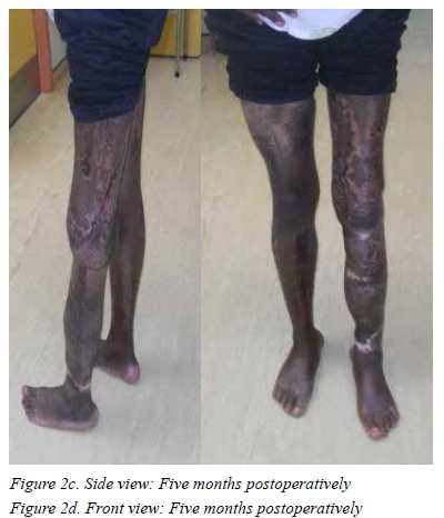
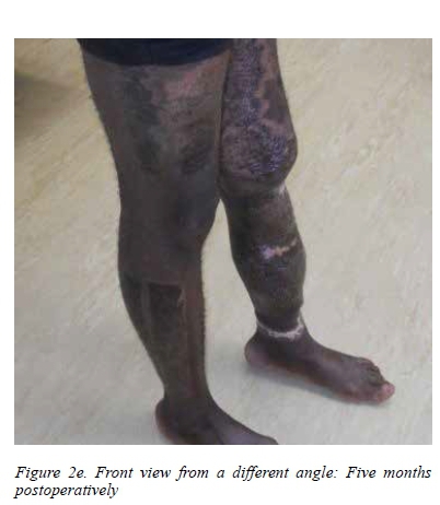
The wound healed fully, the oedema subsided and the patient was able to walk again, with slight range-of-motion limitation to the knee. He was referred for further care to a physiotherapist in this regard.
Case 3
A 43-year-old male patient sustained third degree burns from a fire to both lower limbs, from the thighs to the feet, with bilateral tibial exposure. He was managed at his local hospital and was referred to our local unit after seven months. On admission, the patient was in a wheelchair, had muscle atrophy with bilateral popliteal contraction, and a granulating wound with tibial bone exposure. Fifteen centimetres of the tibia in the left leg was exposed from the middle third to the lower third of the tibia. There was an exposed tibia of 28 cm in the right leg, from the upper third to the lower third of the tibia (Figure 3 a and b). The patient had a platelet count of 1 455 x 109/l. A decision was taken to operate, one leg at a time.

Contraction release, tibial sequestrectomy, coverage of the bone with TA and proximally based hemisoleus flaps and a split skin graft was performed on the left leg. At a later stage, tibial sequestrectomy and bony coverage using the three flaps in conjunction with one another, was performed on the right leg, which was grafted once granulation was stable. We succeeded in covering the tibial bone exposure and achieved full wound healing using this procedure (Figure 3 c and d). This resulted in the platelet count normalising at 266 x109/l.
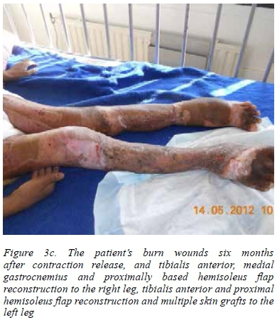
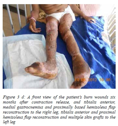
Even though we succeeded in covering all the wounds, achieving contraction release and restoring range of motion in the knee, the patient was unable to walk because of muscle atrophy in the thighs and owing to ankle joint stiffness. He couldn't sustain himself while standing. He was discharged and referred to a physiotherapist to continue rehabilitation.
Discussion
Third degree burns to the legs are challenging to treat, and become more so if the tibial bone is exposed. The difficulty is associated with limited regional soft tissue reconstructive reserves, especially for the lower third of the tibial bone. The anatomical structure of the tibia means that large areas becomes easily exposed. Limb amputation is sometimes the only available option to this group of patients.5
When the facility is available, many surgeons consider free tissue transfer as a first option,5 but this is problematic when the exposed bone extends from the upper to the lower third of the tibial bone. The extent of the subcutaneous tibia can easily reach up to 30 cm in a tall adult. Thus, more than one flap is required to cover such an extensive exposed area. In addition, microsurgical procedures are at high risk of failure in such cases owing to local oedema and the thrombocytosis that is frequently present with this kind of injury.3,4
Bone drilling and NPWT have been used with success, but usually in cases of limited tibial exposure.5 In our experience, NPWT and bone drilling do not provide optimum results. In addition, granulation is often limited owing to metaphysis and is very slow to grow on diaphysis. TA has been described as a good option for exposure of the middle third of the tibia, and is often associated with the use of gastrocnemius flap reconstruction.14
We used TA flap reconstruction alone in one case (not presented in this article), and in conjunction with the gastrocnemius and hemisoleus flap in three patients.
Failure of bone coverage was reported in one patient due to sepsis following inadequate wound care during a mass strike.
The three flaps used in conjunction with one another in the other three patients provided reliable soft tissue coverage of extensive tibial exposure.
The impact of surgery itself on the ability to walk was difficult to assess in one patient because of the delayed treatment (> 20 months) resulting in the joints becoming stiff, and in another because there was no opportunity to follow him up. However in the third case, the patient was able to walk again, without major impairment and limping.
Noticeable impairment was not noted following the use of the TA flap alone in another patient (not covered in this article).
NPWT was used as adjunction to the relief of venous congestion in the TA flap in all three cases. We drilled the bone simultaneously using the three-flap procedure to facilitate flap adhesion to the bone because our patients had experienced long periods of bone exposure which caused superficial, bone necrosis, which was difficult to demarcate. It was only possible to do so later on when the sequester was well demarcated and easy to remove with the osteotome.
The TA was an excellent solution to exposure of the middle third of the tibia bone due to burns because of its simplicity, the rapidity of harvest and its ability to preserve function. The use of the three flaps together solved the difficult problem of covering extensive tibial bone exposure.
Conclusion
A limitation to our study was that we were unable to follow-up and assess the functional impairment secondary to the harvest of the three flaps in two of the three case studies. Nevertheless, we believe that harvesting one head of the gastrocnemius muscle with the TA flap causes functional impairment.
The TA split muscle flap has minimal impact on function because of the harvest technique used. The use of the three flaps together definitely causes functional impairment, but it is still compatible with the ability to walk. However, functional impairment is still acceptable, when compared to amputation, when no other reconstructive procedure is viable. We recommend the use of this technique for extensive tibial bone exposure in deep lower limb burns as a valid solution for difficult wounds.
Operating on more cases at an early stage would enable better evaluation of the functional impairment incurred by the use of these three flaps together.
REFERENCES
1. Browner BD, Jupiter JB, Krettek C, Anderson PA. Skeletal trauma, basic science, management and reconstruction. 5th ed. Elsevier. 2003;1:2132. [ Links ]
2. Sood R, Ranieri J, Murthy V, Weber K. The tibialis anterior muscle flap for full-thickness tibial burns. J Burn Care Rehabil. 2003;24(6):386-391. [ Links ]
3. Choe EI, Kasabian AK, Kolker AR, et al. Thrombocytosis after major lower extremity trauma: mechanism and possible role in free flap failure. Ann Plast Surg. 1996;36(5):489-494. [ Links ]
4. Margiotta MS, Kasabian AK, Karp NS, et al. Humorally mediated thrombocytosis in major lower extremity trauma. Ann Plast Surg. 1998;40(5):463-468. [ Links ]
5. Nugent N, Lannon D, O'Donnell M. Vacuum-assisted closure - a management option for the burns patient with exposed bone. Burns. 2005;31(3):390-393. [ Links ]
6. Hirschowitz B, Moscona R, Kaufman T, Harshai Y. Externallongitudinal splitting of the tibialis anterior muscle for coverage of compound fractures of the middle third of the tibia. Plast Reconstr Surg. 1987;79(3):407-414. [ Links ]
7. Mathes SJ, Nahai F. Muscle flap transposition with functional preservation: technical and clinical considerations. Plast Reconstr Surg. 1980;66(2):242-249. [ Links ]
8. Ford CN, Reinhard ER, Yeh D, et al. Interim analysis of a prospective, randomized trial of vacuum-assisted closure versus the healthpoint system in the management of pressure ulcers. Ann Plast Surg. 2002;49(1):55-61. [ Links ]
9. Lineaweaver W, Hui K, Yim K, et al. The role of the plastic surgeon in the management of surgical infection. Plast Reconstr Surg. 1999;103(6):1553-1560. [ Links ]
10. Rios-Luna A, Fahandezh-Saddi H, Villanueva-Martínez M, López AG. Pearls and tips in coverage of the tibia after a high energy trauma. Indian J Orthop. 2008;42(4):387-394. [ Links ]
11. Ayyappan T, Chadha A. Supersural neurofasciocutaneous flaps in acute trauma heels reconstruction. Plast Reconstruct Surg. 2002;109(7):2307-2313. [ Links ]
12. Benito-Ruiz J, Yoon T, Guisantes-Pintos E, et al. Reconstruction of soft tissue defects of the heel with local fasciocutaneous flaps. Ann Plast Surg. 2004;52(4):380-384. [ Links ]
13. Casey R. Les lambeaux musculaires pedicules a la jambe. Techniques chirurgicales, chirurgie reparatrice. Paris: Encycl Mmed Chir, 1987, p. 28. [ Links ]
14. Chang J, Most D, Hovey LM, Yim KK. Tibialis anterior turnover flap coverage of exposed tibia in a severely burned patient. Burns. 1997;23(1):69-71. [ Links ]
 Correspondence:
N Lahouel
nebil.lahouel@gmail.com

