


Servicios Personalizados
Articulo
 Inglés (pdf)
 Articulo en XML
 Referencias del artículo
Indicadores

Links relacionados
-
 Citado por Google
 -
 Similares en Google
Compartir

 Permalink
South African Journal of Surgery
versión On-line ISSN 2078-5151
versión impresa ISSN 0038-2361
S. Afr. j. surg. vol.54 no.2 Cape Town jun. 2016
VASCULAR SURGERY
Are too many compression ultrasounds being performed for acute lower limb deep venous thrombosis in tertiary inpatients?
C R de JagerI; R J MoontII; N E PearceIII
IFaculty of Medicine, University of the Free State, Bloemfontein, South Africa
IIThe Ruth and Bruce Rappaport Faculty of Medicine, Technion, Israel Institute of Technology, Haifa, Israel
IIIFaculty of Medicine, University of the Free State, Bloemfontein, South Africa
ABSTRACT
BACKGROUND: Venous thromboembolism (VTE) is a complex and serious condition, with high morbidity and mortality, especially in hospitalised patients. Yet its diagnosis remains challenging because of its unspecific clinical presentation. The objective of this study was to apply an algorithmic combination approach to diagnosing VTE by the addition of a D-dimer assay and Wells' criteria for our hospital's internal referral forms requesting compression ultrasound (CUS), to determine the effect on the number of referrals for CUS and the incidence of deep vein thrombosis (DVT) diagnoses.
METHOD: Inpatients who had been referred to the hospital's vascular laboratory and who had undergone a CUS to exclude an acute lower limb DVT were retrospectively analysed between January 2009 and December 2013, and compared to prospectively collected data for the full year (2014) after the introduction of the new referral form. Comparisons included the mean annual number of referrals for CUS and the incidence of DVT diagnoses.
RESULTS: The hospital incidence of diagnosed DVTs for 2009-2013 was 0.17%, compared to 0.16% for 2014 (p = 0.930). In contrast, the total number of referrals for CUS as a percentage of all hospital admissions dropped from 0.84% in 2009-2013 to 0.63% in 2014 (p = 0.009, odds ratio 0.76, 95% confidence interval: 0.62-0.93.
CONCLUSION: The implementation of Wells' criteria and D-dimer to the new request form for CUS significantly decreased referrals to the hospital's vascular laboratory without impacting on the number of DVT cases diagnosed. This is a positive change which simplifies care and reduces the expense of ultrasonography investigations.
Venous thromboembolism (VTE) is a complex and serious condition encompassing deep vein thrombosis (DVT) and pulmonary embolism (PE), usually in the lower extremities.1,2 Thromboses can result from venous stasis, vascular injury or hypercoagulability, and those involving the deep veins proximal to the knee are linked to an increased risk of PE.2 It has been estimated that 26% of undiagnosed and untreated patients with PE will have a subsequent fatal embolic event, while another 26% will have a non-fatal recurrent embolic event with the potential to eventually be fatal.2
Immobilisation and dehydration are risk factors significantly associated with DVT, and explain the high incidence of DVT among hospitalised patients.3 In fact, hospitalisation for acute medical illness is known to be associated with an approximate eightfold increased risk of DVT.4 Thus, the early diagnosis of VTE is of vital importance in this most common, preventable cause of hospital mortality.2,5
The diagnosis of VTE remains a challenge in clinical practice.6 Patients with PE rarely present with the classical triad of pleuritic chest pain, breathlessness and haemoptysis, and frequently experience less specific symptoms. This diagnostic uncertainty means that PE is considered in the differential diagnosis in many acute medical admissions.6 In these patients further expensive and time-consuming radiological investigations are necessary to exclude a thrombosis, even if the clinical suspicion is low. Similarly, the clinical features of DVT are often non-specific, making it difficult to confidently exclude a thrombosis based on the physical examination alone. Consequently, rapid and accurate objective testing for VTE is crucial, especially since the consequences of a missed diagnosis are serious.
A compression ultrasound (CUS) is currently used as the noninvasive gold standard in the diagnosis of DVT, with a sensitivity of 97% and a specificity of 94%. With this modality, approximately 12-25% of patients sent for a CUS are diagnosed yearly with DVT.7 Nevertheless, alternative diagnostic strategies with the potential to reduce the need for radiological investigations in patients with a low probability of VTE are now commonly used in clinical practice. These include D-dimer concentration, a plasma marker of haemostasis. D-dimers are generated when the fibrin clot is formed, cross-linked and degraded.8 It is now widely accepted that a value less than a given threshold of D-dimer concentration rules out the presence of concurrent thrombotic pathologies. D-dimer assays are safe and helpful in excluding the diagnosis of proximal DVT and PE since they have high sensitivity (80-100% depending on the type of assay), resulting in few false negative results. However, their specificity is much lower (40-70%), and decreases further with co-morbidity, older age and a longer duration of symptoms.2,6 Therefore, clinicians should be aware of false positive results for which additional investigations are required to exclude DVT or PE.6
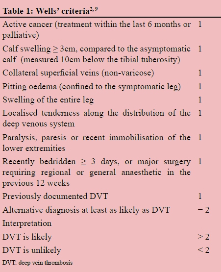
A clinical prediction rule can be used to calculate the pretest probability of VTE, based on a clinical assessment of a combined set of risk factors and physical findings. Individual clinical features do not provide good prediction. Of the various available prediction rules, Wells' criteria for DVT and PE have been most frequently evaluated in outpatients29 (Table 1). It was reported in a systematic review that outpatients with a low pretest probability and a negative D-dimer test had a three-month incidence of DVT of 0.5%, whereas those with a negative D-dimer test and moderate or high pretest probability had incidences of 3.5% and 21.4%, respectively.10 Accordingly, the latest guidelines of the American College of Chest Physicians (ACCP) (2012), recommend that risk stratification should be carried out by the clinical assessment of pretest probability of DVT and PE using Wells' criteria.2,11 A single study in elderly patients has validated the use of Wells' criteria as a risk stratification tool for DVT in hospitalised patients, where higher scores are associated with a greater probability of DVT (11.4% for low scores, 27.6% for moderate scores and 55.0% for high scores).3
The current referral form for requesting a CUS at Universitas Hospital, Bloemfontein, South Africa, includes results from a clinical examination and a lower extremity duplex evaluation. In this study, we decided to change the referral form to additionally include the patient results by applying Wells' criteria (which are currently only validated for outpatients) and a D-dimer assay. This was decided with the aim of evaluating whether such an alteration in the present study would cause a long-term change in the referral pattern with regard to requesting a CUS. Specifically, the objectives of the study were to:
• Measure if there would be a decrease in the number of referrals to the hospital's vascular laboratory for CUS procedures after the introduction of the new request form.
• Measure if the incidence of DVT diagnoses carried out by the hospital's vascular laboratory would change after the introduction of the new request form.
In this way, we aimed to increase clinical awareness of the risk factors for VTE, and the need for rapid treatment when required. It should be noted that this study was not performed to evaluate either D-dimer concentration or Wells' criteria.
Method
This was a retrospective prospective observational comparative study of referrals for a CUS prior to and following the introduction of a new CUS request form at Universitas Hospital. The new request form differed from the preceding one by the addition of Wells' criteria for DVT and the plasma concentration of D-dimer. The request form was completed by the referring doctor prior to the CUS procedure being performed by the hospital's vascular laboratory. A CUS was carried out for every request form, irrespective of the D-dimer level or Wells' criteria.
Inpatients with a suspected acute lower limb DVT who had been referred to the hospital's vascular laboratory and who had undergone a CUS to exclude acute lower limb DVT, were included in this study. Patients were excluded if they had been admitted with DVT as a primary diagnosis, had a suspected upper limb DVT or had varicose veins with superficial venous thrombosis. In cases of repeated referrals for CUS, only the first episode was recorded.
Patient records kept at the hospital's vascular laboratory were analysed for the five-year period between January 2009 and December 2013. During this period, all CUS was recorded as negative or positive. This was then compared to the period between January 2014 and December 2014 after the introduction of the new referral form.
Ethical approval was obtained from the Ethics Committee at the University of the Free State (EC UFS Number 171/2013).
Statistical analysis
Descriptive statistics were conducted of the total number of hospital admissions, the total number of CUS request forms that were sent to the hospital's vascular laboratory, and the number of positive and negative CUS results for detecting DVTs. The number of diagnosed DVTs for 2009-2013 was then calculated as a percentage of the total hospital admissions, and compared to that for 2014 using a paired t-test. Similarly, the percentage of referrals for CUS which yielded a DVT diagnosis, and the percentage of hospital admissions referred for a CUS, were compared for the two periods, i.e. before and after the introduction of the new CUS request form.
Categorical data were analysed using Fischer's exact test and the chi-square test, as appropriate. A p-value of less than 0.050 was considered to be statistically significant.
Results
For the entire study period (2009-2014), a total of 94 445 patients (18 889 patients/year) were admitted to Universitas Hospital, of whom 762 patients were referred for CUS. During the period between 2009 and 2013, 78 007 patients (15 601 patients/year) were admitted, of whom 657 patients were referred for CUS. In 2014, admissions totalled 16 438 patients, of whom 105 patients were referred for CUS (Table 2).
The number of positive DVTs for 2009-2013 as a percentage of the admissions was 0.17%, compared to 0.16% for 2014, which was not statistically significant (p = 0.930).The number of positive CUS results as a percentage of the total number of CUS request forms for 2009-2013 was found to be 20.2% (n = 133/657). This percentage of referrals for CUS which yielded a DVT increased to 25.7% (n = 27/105) in 2014 following the introduction of the new CUS request form. However, this increase did not amount to statistical significance (p = 0.320).
In contrast, when analysing the total number of referrals for CUS as a percentage of all hospital admissions, this proportion significantly dropped from 0.84% in 2009-2013 to 0.63% in 2014 (p = 0.009, odds ratio 0.76, 95% confidence interval: 0.62-0.93) (Table 2).
Discussion
It was investigated in this study whether or not a change in the hospital's standard referral request form for ultrasonography to detect suspected acute lower limb DVT would reduce the number of referrals for CUS procedures to be carried out, and alter the number of DVT diagnoses in inpatients. It was found that there was a significant drop in the number of CUS referrals, while the number of DVT diagnoses remained constant. This indicates a more efficient care procedure, where those with little risk of a DVT were not referred for further tests. It is suggested that with the addition of Wells' criteria and the D-dimer plasma level to the request form, the referring clinicians applied these new diagnostic tools effectively, and with their increased awareness of the clinical risk factors for VTE, altered their referral habits accordingly.
Algorithmic approaches using a combination of clinical assessment, D-dimer measurements and ultrasonography, such as the one introduced in this study, have been increasingly adopted in clinical practice, with the goals of standardising the diagnostic approach to DVT and reducing the number of negative ultrasound examinations.7 Clearly, this has major benefits in resources saved, while markedly reducing the time for a diagnosis and avoiding anxiety and upheaval in patients while they are acutely ill.
In this study, the number of DVTs diagnosed as a percentage of admissions was 0.16-0.17%, irrespective of the content of the CUS request form. The lack of specificity in diagnosing CUS has contributed to the lack of studies that have evaluated prevalence rates in South Africa, meaning we cannot compare our results to the national norm. When considering the few existing prevalence studies, our results showed a markedly lower incidence of DVT in hospitalised patients than that in some countries, e.g. 0.78% in an American study,12 0.93% in a Spanish report,5 and an annual 1.7-2.0% in a Chinese study.13 However, our incidence rate is in line with studies in Saudi Arabia (0.18%) and Asia (0.13%).12 This disparity may be owing to several reasons, including the different demographics of the study populations and the possibility of DVT being diagnosed without referral records. However, the retrospective part of the present study may underestimate the true incidence of DVT where many asymptomatic DVT cases may have been missed. Doppler ultrasound studies were only performed in clinically suspected DVT cases. However, if, as we suggest, the prospective part of the study increased awareness of risk factors for VTE among clinicians, we would expect more Doppler ultrasound studies to be carried out on asymptomatic patients not taking any antithrombotic prophylaxis. Yet, our results showed nearly the same incidence of DVT cases.
Conclusion
The addition of Wells' criteria and D-dimer on the new request form for CUS significantly decreased referrals to the hospital's vascular laboratory, without impacting on the number of DVT cases diagnosed with CUS. This is a positive change which simplifies care and reduces the expense of ultrasonography investigations. We hope to continue in the long term with the new referral procedure which provides practical real-life support of the recent ACCP statement: "In patients who have a low pretest probability of VTE, as defined by the Wells prediction criteria, a negative, high-sensitivity D-dimer assay for VTE has sufficiently high negative predictive value to reduce the need for further imaging studies".2
Acknowledgements
Dr Bill Venter Unit for Vascular Surgery laboratory.
Conflict of interest
The authors did not declare any conflict of interest which may have inappropriately influenced them when writing this paper.
REFERENCES
1. Brophy DF, Dougherty JA, Garrelts JC, et al. Venous thromboembolism prevention in acutely ill nonsurgical patients. Ann Pharmacother. 2005;39(7-8):1318-1324. [ Links ]
2. Qaseem A, Snow V, Barry P, et al. Current diagnosis of venous thromboembolism in primary care: a clinical practice guideline from the American Academy of Family Physicians and the American College of Physicians. Ann Intern Med. 2007;146(6):454-458. [ Links ]
3. Tiganas D, Durant R, Raschilas F, et al. Diagnostic value of the clinical probability score of deep venous thrombosis in the elderly. Rev Med Interne. 2005;26(12):931-937. [ Links ]
4. Geerts WH, Pineo GF, Heit JA, et al. Prevention of venous thromboembolism: the Seventh ACCP Conference on Antithrombotic and Thrombolytic Therapy. Chest. 2004;126(3 Suppl):338S-400S. [ Links ]
5. Barba R, Zapatero A, Losa JE, et al. Venous thromboembolism in acutely ill hospitalized medical patients. Thromb Res. 2010;126(4):276-279. [ Links ]
6. Harper PL, Theakston E, Ahmed J, Ockelford P. D-dimer concentration increases with age reducing the clinical value of the D-dimer assay in the elderly. Intern Med J. 2007;37(9):607- 613. [ Links ]
7. Zierler BK. Ultrasonography and diagnosis of venous thromboembolism. Circulation. 2004;109(12 Suppl 1):I9-I14. [ Links ]
8. Marlar RA. D-dimer: establishing a laboratory assay for ruling out venous thrombosis. MLO Med Lab Obs. 2002;34(11):28-32. [ Links ]
9. Wells PS, Anderson DR, Bormanis J, et al. Application of a diagnostic clinical model for the management of hospitalized patients with suspected deep-vein thrombosis. Thromb Haemost. 1999;81(4):493-497. [ Links ]
10. Fancher TL, White RH, Kravitz RL. Combined use of rapid D-dimer testing and estimation of clinical probability in the diagnosis of deep vein thrombosis: systematic review. BMJ. 2004;329(7470):821. [ Links ]
11. Guyatt GH, Akl EA, Crowther M, et al. Executive summary: antithrombotic therapy and prevention of thrombosis, 9th ed: American College of Chest Physicians evidence-based clinical practice guidelines. Chest. 2012;141(2 Suppl):7S-47S. [ Links ]
12. Stein PD, Patel KC, Kalra NK, et al. Deep venous thrombosis in a general hospital. Chest. 2002;122(3):960-962. [ Links ]
13. Cheng G, Chan C, Liu YT, et al. Incidence of deep vein thrombosis in hospitalized Chinese medical patients and the impact of DVT prophylaxis. Thrombosis. 2011;2011:629383. [ Links ]
 Correspondence:
CR de Jager
crdejager001@gmail.com

