


Serviços Personalizados
Artigo
 Inglês (pdf)
 Artigo em XML
 Referências do artigo
Indicadores

Links relacionados
-
 Citado por Google
 -
 Similares em Google
Compartilhar

 Permalink
South African Journal of Surgery
versão On-line ISSN 2078-5151
versão impressa ISSN 0038-2361
S. Afr. j. surg. vol.52 no.2 Cape Town Fev. 2014
GENERAL SURGERY
Is routine biopsy of sonographically benign breast lesions in black African women under 40 years of age recommended?
M L KempI; S AndronikouII; S LucasIII; G RubinIV
IMB ChB, FCRad (Diag) (SA), MMed (Rad) (D); Department of Diagnostic Radiology, Faculty of Health Sciences, University of the Witwatersrand, Johannesburg, South Africa
IIMB BCh, FCRad (Diag) (SA), FRCR (Lond), PhD; Department of Diagnostic Radiology, Faculty of Health Sciences, University of the Witwatersrand, Johannesburg, South Africa
IIIMB ChB, MMed (Rad) (D); Department of Diagnostic Radiology, Faculty of Health Sciences, University of the Witwatersrand, Johannesburg, South Africa
IVMB BCh, FCRad (Diag) (SA), MMed (Rad) (D); Department of Diagnostic Radiology, Faculty of Health Sciences, University of the Witwatersrand, Johannesburg, South Africa
ABSTRACT
INTRODUCTION: Breast lesions that appear benign on ultrasound examination continue to be biopsied, and no relevant data from Africa exist.
OBJECTIVE: To determine the histological spectrum of sonographically benign lesions measuring >3 cm in women in Johannesburg, South Africa, by age and population group, and establish associations between the histological findings and the size of the lesion and the patient's HIV status and family history.
METHODS: Biopsy results of breast masses that appeared benign on ultrasound were reviewed and the prevalence of histological subtypes was determined according to HIV status and family history. The Kruskal-Wallis test and separate logistic regression analysis were used for determining associations with size.
RESULTS: Sixty-eight of a total of 13 112 patients seen over a 3.5-year-period were included. There were 73 lesions, of which 65 (89.0%) were benign and eight (11.0%) malignant. The most common lesions were fibroadenomas (60.3%) and breast carcinomas (6.8%). Size did not predict malignancy (p=0.22). Family history and HIV status were not significant.
CONCLUSION: A high proportion (11.0%) of lesions that appeared benign on ultrasound were malignant. The size of the lesion did not correlate with histological subtype or malignancy. Further research, including training of ultrasonographers in using the Breast Imaging Reporting and Data System (BIRADS) ultrasound lexicon, standardisation of technique with assistance from established users and possibly double reading for a period, is needed to determine whether there is a true high prevalence of malignancy in sonographically benign breast lesions in our community.
The Breast Imaging Reporting and Data System (BIRADS) classification has been developed for both ultrasound and mammography.[1] 'BIRADS 2 lesions are described as benign findings including intramammary nodes and breast implants. BIRADS 3 lesions are probably benign lesions including non calcified circumscribed lesions.'[2] Lesions classified as BIRADS 3 and 4 require 6 months' follow-up and core biopsy, respectively,[2] the latter providing histological confirmation and a definitive diagnosis. For all palpable solid masses that are circumscribed and not calcified, some authors continue to recommend a tissue diagnosis even when the features indicate that the mass is probably benign.[3] This point is controversial, however, and more data are required to determine whether the alternative of short-term follow-up can replace biopsy in these patients.[3]
Patients in our breast imaging unit with benign-appearing lesions >3 cm in diameter on ultrasound are currently undergoing biopsy owing to ongoing concern that a malignancy or phyllodes tumour might be missed. Biopsies are currently being performed on lesions with benign features on ultrasound examination, without adequate data on the true prevalence of malignancy and the spectrum of disease in our specific population. To the best of our knowledge, all research on BIRADS lesions has been performed on patients in Europe and North America. We decided to investigate the spectrum of disease in sonographically benign lesions >3 cm in our local population in Johannesburg, South Africa, which comprises a majority of black patients, to determine whether biopsy is indicated owing to a true risk of cancer in sonographically benign lesions, or whether patients can be followed up over 6 months as recommended by the BIRADS system. The use of core biopsies in our unit for sonographically benign lesions measuring >3 cm in their longest axis is primarily aimed at excluding phyllodes tumours.
Objectives
To determine the prevalence of malignancy in sonographically benign lesions biopsied in South African women under 40 years of age, and to characterise the results according to histological findings, age and population group. Further objectives included determining any association between the histological features and the size of the lesion on ultrasound as well as with the HIV status and a family history of breast malignancy.
Methods
This was a retrospective descriptive study of the biopsy findings in sonographically benign breast masses seen at the Helen Joseph Mammography Unit, Johannesburg, from the beginning of 2007 to the end of 2010. Ethics clearance was obtained from the University of the Witwatersand's Ethics Department (clearance no. M110426). Patients with BIRADS 3, 4 and 5 masses on ultrasound and mammography have undergone ultrasound-guided core biopsy of the mass as routine practice in this department since 2007 (limiting the study period). Data were collected from the biopsy procedural recording sheets.
The radiologists performing the ultrasound scans were a core group of both junior and senior radiologists, most with a special interest in mammography.
Women under 40 years of age who had undergone breast ultrasound and biopsy of the breast mass were considered for inclusion. Data were included only for sonographically benign lesions, the features of which are as follows: (i) a well-circumscribed oval, round or macrolobulated mass;[4](ii) a hypoechoic mass;[4](iii) three or fewer circumscribed lobulations;[4](iv) horizontal length greater than vertical length;[4] and (v) homogeneous hyperechogenicity relative to breast fat.[5] Fig. 1 demonstrates the features of sonographically benign lesions. Patients were excluded if histological results were unavailable or equivocal, or if biopsy procedural recording sheets were illegible or incomplete.
The size of the lesion (defined as the maximum measurable diameter) was determined from ultrasound reports. The prevalence of histologically proven benign and malignant lesions was expressed as a percentage of the total number of lesions biopsied.
The STATA software package was used to determine any association between the size of the lesion and the histological group (using the Kruskal-Wallis test, as the variables were not normally distributed). A logistic regression analysis test and Pearson goodness-of-fit test were also used to assess whether there was any association between lesion size and benign or malignant status.
Results
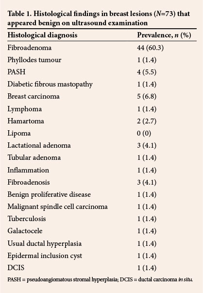
A total of 68 women (0.5%) with breast masses that appeared benign on ultrasound and who had undergone biopsy were included from a total of 13 112 patients imaged during the period from the beginning of 2007 to the end of 2010. Their ages ranged from 14 to 40 years (mean 25.9), and 62 (91.2%) were black African, four (5.9%) white and two (2.9%) coloured. There were no Asian patients. All but one of them had palpable lesions, and the non-palpable lesion was benign. The lesions ranged in size from 3 cm to over 20 cm in diameter (mean 5.2). Of the 68 patients, five had more than one lesion (73 lesions in total). Of these five patients, one had breast carcinoma and ductal carcinoma in situ (DCIS) and four had bilateral fibroadenomas. Of the 73 lesions, 65 were benign and eight were malignant. Tables 1, 2 and 3 summarise the histological spectrum of lesions according to prevalence, ethnic group and age.


The core biopsy results were not correlated with the excision results, as not all patients had had excisions.
Among the eight patients with a biopsy-proven malignancy, one patient's folder was lost so the HIV status and family history could not be obtained. Of the remaining seven, four had never had an HIV test. Of the three patients with HIV test results, two were HIV-positive. One of these had Burkitt's lymphoma of the breast, while the second had DCIS in one breast and invasive ductal carcinoma in the other. Only one patient diagnosed with breast cancer had a positive family history, and she was HIV-negative. The HIV-positive patients had no family history of breast cancer.
The prevalence of malignant lesions was 11.0%. The Kruskal-Wallis test showed that lesion size was not a significant predictor of the histological result (p=0.22). Logistic regression analysis performed independently and separately from the Kruskal-Wallis test showed that there was very little evidence that lesion size could be used to predict a histologically malignant or benign lesion. The Pearson goodness-of-fit test was used to test the validity of the logistic regression test. The p-value of 0.7111 for the goodness-of-fit test suggests that the model fits reasonably well. The sensitivity, positive predictive value and negative predictive value of ultrasound could not be calculated because there were no true negatives in the sample.
Discussion
Breast biopsy is considered the gold standard for establishing the true nature of a breast lesion identified by palpation or radiologically.
When ultrasound is used, the decision to perform a biopsy on a solid mass is often swayed by whether the mass is palpable or not,[6] regardless of recognised benign imaging features.[6] Some published reports cite benign histological findings in as many as 100% of palpable lesions with benign mammographic and ultrasound findings.[3] All but one of our patients had palpable masses, and the mass that was not palpable was benign.
Although the sample size was small, the range of biopsy results was wide, indicating that the population sample was adequate and that there was a good representation of disease. There was a high rate of malignancy, with a prevalence of 11.0%. This is in contrast to the overall cancer prevalence in the literature of 0.3% for palpable and 1.6% for non-palpable lesions with benign features on ultrasound.[7]
The high proportion of seemingly benign masses that were in fact malignant has marked significance, as many accept ultrasound features indicating that a lesion is benign as sufficient criteria for excluding malignancy. It is currently recommended[6] that short-term follow-up is a reasonable alternative to biopsy of palpable breast lesions with sonographically benign imaging features. This should be associated with a subsequent malignancy rate of less than 2%.[6]
Possible reasons for the high proportion of malignant lesions in our study include: (i) the demographics of our study population; and (ii) operator factors, i.e. errors in obtaining and interpreting the images.
Demographics
Breast carcinoma is the most common cancer in South African women,[8] accounting for 20.32% of all cancers.[9] The percentage of breast cancer is reported to be highest in Asian (35.51%) and coloured women (24.46%),[9] followed by white and black African women (18.09%).[9] Of our patient population 91.2% were black Africans, only 2.9% being coloured and 5.9% white. In the 2010 census, the total percentage of black Africans in South Africa was 79.4%.[10] The percentage of black Africans in our sample was higher than the national percentage, possibly because there was a small sample size of only eight malignancies in the total study group of 68 patients. To the best of our knowledge, there is no existing literature exploring racial influences on the likelihood of malignancy in breast lesions that appear benign on ultrasound. The prevalence of breast cancer in our subgroup of patients was just over 9% higher than that reported in the literature, and the malignant lesions were exclusively in the black African women. Although this finding may be due to the small sample size, it does suggest that malignant lesions may be more prevalent in the black African population of South Africa than was previously thought. This should be investigated further, for all breast malignancies and not only those with a benign ultrasound appearance.
Most breast cancers are sporadic, developing as a result of cumulative effects of genetic susceptibility and environmental risk factors.[11] Approximately 20% of women with breast cancer have a clear family history.[12] The association between a positive family history, presentation and outcome is uncertain, as the results of multiple studies are conflicting.[11] One of the eight patients in our study who was found to have a malignant lesion had a positive family history of breast cancer.
Several large population-based studies have demonstrated that breast cancer in younger women has unfavourable characteristics and a relatively poor prognosis, which is partially due to a lower response rate to systematic adjuvant therapy.[13] Our research included a younger population group because these patients undergo ultrasound in preference to mammography. In the process, however, we demonstrated a higher prevalence of malignancy than expected for this particular age group.
One of our HIV-positive patients was diagnosed with Burkitt's lymphoma. This is a subtype of non-Hodgkin's lymphoma with an increased prevalence in HIV-positive patients and known to be associated with extranodal lymphoma, as in our patient.[14] Only a few articles have reported an association between HIV and primary breast lymphomas,[15] which account for less than 0.7% of all non-Hodgkin's lymphomas.[15] The second HIV-positive patient had DCIS in one breast and breast cancer in the other. She was 33 years old. The mean age of HIV-positive patients presenting with breast cancer is 37.[16] Consistent with the findings in our patient, HIV-infected patients with breast cancer have an earlier age of presentation than non-HIV-infected women, a higher prevalence of bilateral disease, and a poorer outcome due to early metastatic spread.[16] There is also an accelerated and aggressive clinical course and early relapse.[16] It has been reported that the incidence of breast carcinoma in HIV-positive patients is significantly higher than that in HIV-negative women in both African[17] and developed countries.[16]
Operator factors
Characterisation of breast lesions on ultrasound, BIRADS categorisation and management recommendations have only been available for a short time.[6] The BIRADS ultrasound lexicon is only in its first edition, and guidelines for the management of ultrasound-detected lesions are less widely validated than those in mammography.[6] Raza et al.[6] found that interpretation and use of the BIRADS category 3 recommendations vary, possibly owing to the operator-dependent nature of ultrasound. We cannot determine whether the ultrasound operators in our study correctly identified benign imaging features on ultrasound, even though the BIRADS system attempts to standardise them. Raza et al.[6] reported that the BIRADS 3 category was correctly used in 86% of cases, while in the remaining 14% the recommendations for biopsy were contradictory and ultimately led to an interventional procedure. Training in BIRADS specifically has been shown to improve agreement in final mammogram assessments[18] and may also be a solution for ultrasound.
The ultrasound criteria for benign lesions are subjective, and the one objective and repeatable measure that can easily be extracted from an ultrasound scan is lesion size. However, we did not demonstrate any association between lesion size and benign or malignant histological categorisation, which is in keeping with the current literature.[19] There was also no association between lesion size and the histological subtype of the lesion. To our knowledge there is limited literature on this topic.
Limitations
Limitations of the study include the retrospective nature of the patient records, the operator-dependent nature of ultrasound, and the fact that images viewed by the attending radiologist could not be retrieved, making a retrospective review of actual images impossible.
Conclusion
We demonstrated a high prevalence (11%) of malignancies in breast lesions classified as benign by ultrasound. The only available objective measurement, the size of the lesion, did not correlate with histological subtype and benign or malignant status and cannot be used as a discriminating factor. Use of ultrasound in its current form is therefore not acceptable as a screening tool to distinguish between benign and malignant lesions in our department, and possibly in our population.
The role that HIV plays in breast cancer includes earlier presentation and a more aggressive clinical course.
Possible ways to increase the detection of malignant lesions include training of ultrasonographers in use of the BIRADS ultrasound lexicon, standardisation of technique with assistance from established users, and possibly double reading. Once this has been done, a repeat evaluation will be able to demonstrate whether black women in our patient population with breast lesions that appear benign on ultrasound truly have an increased prevalence of malignant disease. Ultrasound elastography, once approved for clinical use, may obviate dependence on structural features of benignity during ultrasound examination of breast lesions. Until then, routine biopsy of these lesions is recommended in our practice.
REFERENCES
1. Nothacker MDV Hahn M, Warm M, et al. Early detection of breast cancer: Benefits and risks of supplemental breast ultrasound in asymptomatic women with mammographically dense breast tissue: A systematic review. BMC Cancer 2009;9:335. [http://dx.doi.org/10.1186/1471-2407-9-335] [ Links ]
2. Weissleder RWJ, Harisinghani MG, Chen JW Primer of Diagnostic Imaging. 4th ed. St Louis, Mo.: Mosby Elsevier, 2007. [ Links ]
3. Graf O, Helbich TH, Fuchsjaeger MH, et al. Follow-up of palpable circumscribed noncalcified solid breast masses at mammography and US: Can biopsy be averted? Radiology 2004;233(3):850-856. [http://dx.doi.org/10.1148/radiol.2333031845] [ Links ]
4. Weinstein SP, Conant EF, Orel SG, Zuckerman JA, Bellah R. Spectrum of US findings in pediatric and adolescent patients with palpable breast masses. Radiographics 2000;20(6):1613-1621. [ Links ]
5. Stavros AT, Thickman D, Rapp CL, et al. Solid breast nodules: Use of sonography to distinguish between benign and malignant lesions. Radiology 1995;196(1):123-134. [ Links ]
6. Raza S, Chikarmane SA, Neilsen SS, Zorn LM, Birdwell RL. BI-RADS 3, 4, and 5 lesions: Value of US in management - follow-up and outcome. Radiology 2008;248(3):773-781. [http://dx.doi.org/10.1148/radiol.2483071786] [ Links ]
7. Harvey JA, Nicholson BT, Lorusso AP, Cohen MA, Bovbjerg VE. Short-term follow-up of palpable breast lesions with benign imaging features: Evaluation of 375 lesions in 320 women. AJR Am J Roentgenol 2009;193(6):1723-1730. [http://dx.doi.org/10.2214/AJR.09.2811] [ Links ]
8. Vorobiof DA, Sitas F, Vorobiof G. Breast cancer incidence in South Africa. J Clin Oncol 2001 19(18 Suppl):125S-127S. [ Links ]
9. The National Cancer Registry, Service TNHL. http://www.nioh.ac.za/?page=cancer_statistics&id=163 (accessed 16 April 2013). [ Links ]
10. Statistics South Africa. Statistical release PO302. Mid-year population estimates 2010. https://www.statssa.gov.za (accessed 10 April 2014). [ Links ]
11. Cao AY, He M, Di GH, et al. Influence of a family history of breast and/or ovarian cancer on breast cancer outcomes. Exp Ther Med 2011;2(5):917-923. [http://dx.doi.org/10.3892/etm.2011.275] [ Links ]
12. Easton DF. Familial risks of breast cancer. Breast Cancer Res 2002;4(5):179-181. [ Links ]
13. Anders CK, Hsu DS, Broadwater G, et al. Young age at diagnosis correlates with worse prognosis and defines a subset of breast cancers with shared patterns of gene expression. J Clin Oncol 2008;26(20):3324-3330. [http://dx.doi.org/10.1200/JCO.2007.14.2471] [ Links ]
14. Vaishnav K, Shah S, Pandhi S, Rathi Y. Multifocal bilateral breast masses in HIV-positive status. Indian J Cancer 2011;48(2):253-255. [http://dx.doi.org/10.4103/0019-509X.82878] [ Links ]
15. Akinwande OK, Paley R. Primary breast lymphoma: A consideration in an HIV patient when a mass is discovered by screening mammography: A case report. Cases J 2008;1(1):387. [http://dx.doi.org/10.1186/1757-1626-1-387] [ Links ]
16. Pantanowitz L, Connolly JL. Pathology of the breast associated with HIV/AIDS. Breast J 2002;8(4):234-243. [http://dx.doi.org/10.1046/j.1524-4741.2002.08409.x] [ Links ]
17. Patil P, Elem B, Zumla A. Pattern of adult malignancies in Zambia (1980-1989) in light of the human immunodeficiency virus type 1 epidemic. J Trop Med Hyg 1995;98(4):281-284. [ Links ]
18. Bowles EJ, Sickles EA, Miglioretti DL, Carney PA, Elmore JG. Recommendation for short-interval follow-up examinations after a probably benign assessment: Is clinical practice consistent with BI-RADS guidance? AJR Am J Roentgenol 2010;194(4):1152-1159. [http://dx.doi.org/10.2214/AJR.09.3064] [ Links ]
19. Sickles EA. Nonpalpable, circumscribed, noncalcified solid breast masses: Likelihood of malignancy based on lesion size and age of patient. Radiology 1994;192(2):439-442. [ Links ]
 Correspondence:
M Kemp
(marnie.kemp@gmail.com)

