


Services on Demand
Article
 English (pdf)
 Article in xml format
 Article references
Indicators

Related links
-
 Cited by Google
 -
 Similars in Google
Share

 Permalink
South African Journal of Surgery
On-line version ISSN 2078-5151
Print version ISSN 0038-2361
S. Afr. j. surg. vol.51 n.1 Cape Town Jan. 2013
http://dx.doi.org/10.7196/SAJS.1334
GENERAL SURGERY
DOI:10.7196/SAJS.1334
A comparative study assessing a new tool for occluding parenchymal blood flow during liver resection for hepatocellular carcinoma
S ZhouI; X-J XueII; R-R LiIII; D-F ChenIV; W-Y ChenV; G-X LiuVI; E-M KeVII; S-Y ZhengVIII
IMD, MS. Department of General Surgery, 175th Hospital of PLA (Southeast Affiliated Hospital of Xiamen University), Zhangzhou, Fujian Province, China
IIMD, MS. Department of General Surgery, 175th Hospital of PLA (Southeast Affiliated Hospital of Xiamen University), Zhangzhou, Fujian Province, China
IIIMD. Department of General Surgery, 175th Hospital of PLA (Southeast Affiliated Hospital of Xiamen University), Zhangzhou, Fujian Province, China
IVMD. Department of General Surgery, 175th Hospital of PLA (Southeast Affiliated Hospital of Xiamen University), Zhangzhou, Fujian Province, China
VMD, MS. Department of General Surgery, 175th Hospital of PLA (Southeast Affiliated Hospital of Xiamen University), Zhangzhou, Fujian Province, China
VIMD, MS. Department of General Surgery, 175th Hospital of PLA (Southeast Affiliated Hospital of Xiamen University), Zhangzhou, Fujian Province, China
VIIMD. Department of General Surgery, 175th Hospital of PLA (Southeast Affiliated Hospital of Xiamen University), Zhangzhou, Fujian Province, China
VIIIMD. Department of General Surgery, 175th Hospital of PLA (Southeast Affiliated Hospital of Xiamen University), Zhangzhou, Fujian Province, China
ABSTRACT
BACKGROUND: The aim of this study was to compare the efficacy of a new tool (the hepatic section vascular blocker, HSVB) with hepatic pedicle clamping and hemihepatic vascular exclusion to control bleeding during liver resection for cancer.
METHODS: Clinical data on 117 patients who underwent liver resection from 2004 to 2009 were analysed retrospectively. Forty-two patients had liver resection using the HSVB (group A), in 35 patients hemihepatic vascular exclusion was used (group B), and in 40 patients hepatic pedicle clamping with a Pringle manoeuvre was used (group C). Blood loss, operative time, postoperative hepatic function and complications were compared.
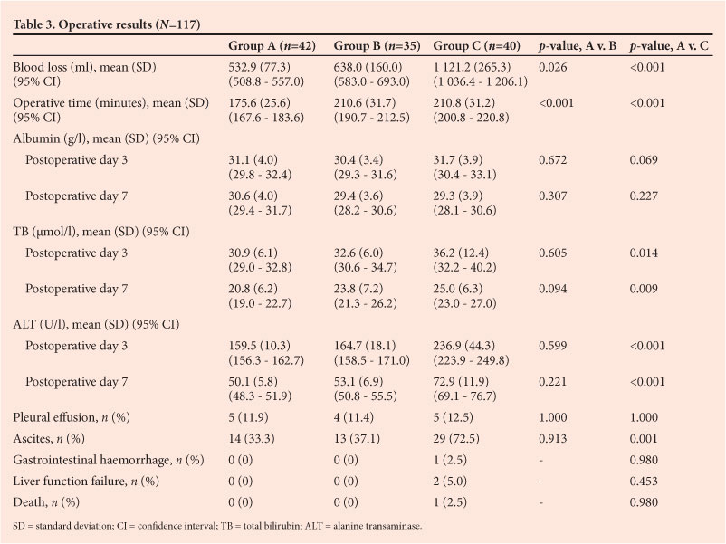
RESULTS: Mean blood loss and operative time in group A were significantly less than in groups B (p=0.026 and p<0.001, respectively) and C (p<0.001 and p<0.001). There were significant differences between groups A and C in total bilirubin (TB) and alanine transaminase (ALT) levels on postoperative days 3 and 7, and group A had better hepatic function (TB p=0.014 and p=0.009; ALT p<0.001 and p<0.001). The rate of postoperative ascites was significantly higher in group C compared with group A (p<0.001). In group C, 2 patients had liver failure, 1 had a gastrointestinal haemorrhage and 1 died.
CONCLUSIONS: Using the HSVB during liver resection effectively controlled bleeding, saved operative time and preserved hepatic function. It proved to be a safe and feasible technique.
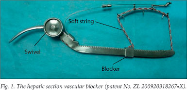
Liver surgery is associated with loss of large amounts of blood, and the amount of blood lost during liver resection has a major influence on prognosis. Vascular clamping is an efficient way of minimising bleeding during parenchymal transection. We used a new tool, the hepatic section vascular blocker (HSVB, patent No. ZL 200920318267.X, Fig. 1) to control bleeding during liver resection. In this research, comparative clinical data on 117 patients with liver cancer were analysed retrospectively.
Methods
Clinical data
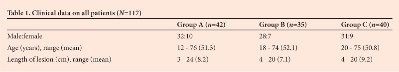
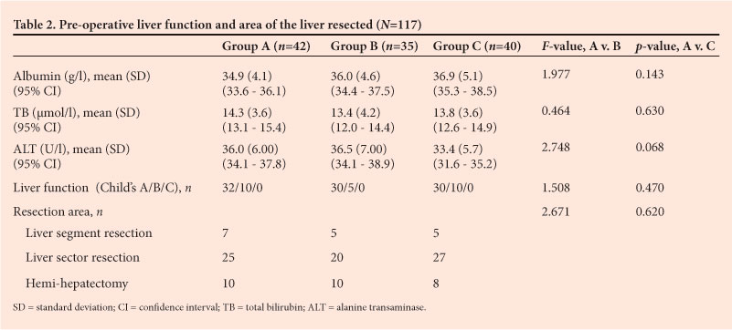
From March 2004 to June 2009, 117 patients underwent liver resection for hepatocellular carcinoma in the Department of General Surgery of the 175th Hospital of PLA, Zhangzhou, Fujian Province, China. Data on the patients are set out in Table 1. The HSVB was used to control blood loss in group A, hemihepatic vascular exclusion in group B, and hepatic pedicle clamping in group C. Serum albumin, total bilirubin (TB) and alanine transaminase (ALT) were measured in all patients pre-operatively and again on postoperative days 3 and 7. There was no significant difference in pre-operative serum albumin, TB, ALT or Child's classification of liver function between the three groups (p=0.143, p=0.630, p=0.068 and p=0.047, respectively) (Table 2).
Postoperative pathological examination confirmed that all the tumours were hepatocellular carcinomas.
Surgical methods
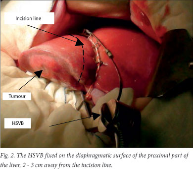
In group A, the HSVB was used to control bleeding during resection. After the abdominal cavity was opened, the ligamentum teres was ligated and divided, and the falciform ligament divided. Depending on the location of the tumour, the right or left triangular ligaments were divided to mobilise the right or left lobe, and the proposed transection line was marked 2 cm away from the edge of the tumour on the liver surface. The bed-piece of the HSVB was positioned adjacent to the inferior vena cava on the visceral surface, and the string was fixed proximally on the diaphragmatic liver surface 2-3 cm away from the proposed incision line (Fig. 2).[1] Tightening the string occluded the blood flow to the liver tissue that was to be resected. If the tumour was large, the HSVB had to be positioned close to the transection line and could slip while the parenchyma was transected. Several Kirschner wires were therefore attached to the device to fix it in position in the liver. In group B, vascular inflow was dissected depending on the location of the tumour. The left or right ipsilateral hepatic artery was controlled with a tourniquet, as well as the branches of the portal vein and biliary tract. In group C, the intermittent technique (15 - 20 minutes of clamping alternating with 5 minutes unclamped) was used to protect the hepatic parenchyma from warm ischaemic injury. The hepatic pedicle was controlled with an atraumatic flexible clamp or a tourniquet.
The study was approved by the local Ethics Committee and conducted according to the Declaration of Helsinki and Good Clinical Practice guidelines. All patients gave written informed consent prior to any study-related procedures.
Statistical analysis
Statistical analysis was done using SPSS 13.0 for Windows. Data were expressed as means (standard deviations (SDs)). Differences in the parametric data for pre-operative serum albumin, TB and ALT were examined using one-way ANOVA, and postoperative values using the Nemenyi test. Differences in non-parametric data on liver function, Child's grade and resection area were examined using the Kruskal-Wallis H-test. A p-value of <0.05 was considered statistically significant. Enumeration data on the rate of ascites were examined using the chi-square test. The significance level was á'=0.0125 after adjustment.
Results
All tumours were completely resected. There were no significant differences in liver resection area between the three groups (p=0.620) (Table 2). No patient died during the operation. Both blood loss and operative time (p<0.001) were significantly less in group A than in groups B (p=0.026) and C (p<0.001) (Table 3). There were no differences in postoperative liver function or the rates of complications, including pleural effusion, ascites, gastrointestinal haemorrhage and liver failure, between groups A and B (p>0.05) (Table 3). Levels of TB and ALT on postoperative days 3 and 7 in group C were significantly higher than those in group A (TB p=0.014 and p=0.009; ALT p<0.001 and p<0.001) (Table 3), as was the rate of ascites (72.5% v. 33.3%, p=0.001) (Table 3). In group C, 29 patients developed variable degrees of ascites, 2 patients developed liver failure, 1 had a gastrointestinal haemorrhage, and 1 died.
Discussion
The liver's dual blood supply sends approximately 1 500 ml of blood through the organ every minute. Control of bleeding is of paramount importance during liver resection. An effective technique should not only reduce bleeding and thus provide a clear operative field, but also reduce postoperative complications and mortality.[2,3] At the beginning of the 20th century, Pringle demonstrated that hepatic artery and portal vein occlusion could significantly reduce liver bleeding.[4] Intermittent hepatic pedicle clamping allows complex resections to be done using prolonged clamping, but a major problem is transection plane bleeding when the clamp is released. Hepatic pedicle clamping has no direct effect on backflow bleeding from branches of the hepatic veins, but ischaemia-reperfusion due to occlusion of the inflow to the right and left lobes has major effects on the remaining liver,[5,6] especially in the presence of hepatic dysfunction. Patients with liver cirrhosis are at risk of hepatic failure after surgery. The function of abdominal organs such as the pancreas and small intestine may also be affected.[7,8] Haemodynamic effects such as blood pressure fluctuation are also not easily avoided.[9] Pedicle clamping is well tolerated because caval flow is not interrupted, and is still extensively used in our clinic because it is simple to do in most liver resections, except for tumours located close to or in the hepatic hilum.
Hepatocellular carcinoma in China is almost invariably complicated by hepatic fibrosis because of the common association of carcinoma with viral hepatitis. These patients are at risk of hepatic failure if prolonged hepatic pedicle clamping is used. Hemihepatic vascular exclusion, i.e. control of bleeding with hepatic pedicle clamping, preserves blood flow to the remaining liver and protects the hepatic parenchyma against ischaemia and reperfusion injuries, and is suitable for patients with hepatic dysfunction.[10] The technique also preserves return blood flow to the gastrointestinal tract, protecting gut barrier function.[11] However, dissection of hepatic hilum takes time and excessive bleeding may occur, particularly when portal hypertension and large collateral veins are present in patients with hepatic cirrhosis.
The three methods of vascular control, Longmire clamping, Pringle's manoeuvre and total vascular isolation were compared by Buell et al.[12] Their results suggest that using the Longmire clamp is associated with the lowest incidence of complications. However, because of its shape the Longmire clamp cannot completely compress the liver, especially when the liver parenchyma is thick. We therefore redesigned the clamp, using a soft string and a sliding blocker to overcome its shortcomings. The string fits the shape of the liver closely and can make complete contact with the liver surface after tightening. The sliding blocker automatically slides into the optimal position, allowing the HSVB to attach completely. The string and sliding blocker control blood flow effectively, even when the hepatic parenchyma is thick.
Theoretically, using a clamp to provide local hepatic blood flow occlusion is the optimal choice. Applying the HSVB completely controlled bleeding from the hepatic artery and portal vein and backflow of the hepatic veins at the transection plane, minimising blood loss and at the same time avoiding the intrahepatic and distant spread of tumour cells that results from squeezing the tumour. There is also no time limit for occlusion, because the clamp only cuts off blood flow to the tissues to be resected. It is a simple procedure and provides a clear operative field, reducing total operative time and surgical stress. The clamp technique can selectively occlude blood flow to the lobe or segment to be resected, with little effect on liver function, and blood flow returns to the pancreas and gastrointestinal tract, resulting in fewer postoperative complications.[13,14] There is no significant effect on systemic haemodynamics, because the area occluded is relatively small.
Conclusion
In conclusion, our comparison of the HSVB with hepatic pedicle clamping and hemihepatic vascular exclusion showed that use of the HSVB is an effective and convenient way to control bleeding, with the advantages that the device can be placed and fixed in position quickly, is simple to use, completely occludes blood flow, protects liver function, and has little effect on systemic haemodynamics. The device can be used for most liver resections and in patients with liver cirrhosis or liver dysfunction, unless the tumour is close to the hepatic hilum or the case is complicated by tumour ingrowth into the portal vein or inferior vena cava.
The treatment of liver cancer includes strict attention to surgical indications, adequate pre-operative preparation, careful surgical technique, close observation of the patient after the operation, and effective supportive treatment. Control of blood loss is a secondary procedure, and in choosing a method the surgeon should take account of liver function, the results of pre-operative imaging, and intra-operative exploration.
Acknowledgements. We thank Jun-Hua Xu for making the die of the hepatic blood blocker.
References
1. Zhou S, Zheng SY, Chen DF, et al. Application of the self-made hepatic clamp for hepatic blood flow occlusion in hepatectomy. Fubu Waike Zazhi 2008;21(2):100-101. [ Links ]
2. Dixon E, Vollmer CM Jr, Bathe OF, Sutherland F. Vascular occlusion to decrease blood loss during hepatic resection. Am J Surg 2005;190(1):75-86. [http://dx.doi.org/10.1016/j.amjsurg.2004.10.007] [ Links ]
3. Batignani G, Zuckermann M. Inferior approach for the isolation of the left-middle hepatic veins in liver resections: A safe way. Arch Surg 2005;140(10):968-971. [http://dx.doi.org/10.1001/archsurg.140.10.968] [ Links ]
4. Pringle JH. Notes on the arrest of hepatic haemorrhage due to trauma. Ann Surg 1908;48(4):541-549. [http://dx.doi.org/10.1097/00000658-190810000-00005] [ Links ]
5. Brooks AJ, Hammond JS, Girling K, Beckingham IJ. The effect of hepatic vascular inflow occlusion on liver tissue pH, carbon dioxide, and oxygen partial pressures: Defining the optimal clamp/release regime for intermittent portal clamping. J Surg Res 2007;141(2):247-251. [http://dx.doi.org/10.1016/j.jss.2006.10.054] [ Links ]
6. Pietsch UC, Herrmann ML, Uhlmann D, et al. Blood lactate and pyruvate levels in the perioperative period of liver resection with hepatic pedicle clamping. Clin Hemorheol Microcirc 2010;44(4):269-281. [http://dx.doi.org/10.3233/CH-2010-1276] [ Links ]
7. Ypsilantis P, Lambropoulou M, Grapsa A, et al. Pringle maneuver deteriorates gut barrier dysfunction induced by extended-liver radiofrequency ablation. Dig Dis Sci 2011;56(5):1548-1556. [ Links ]
8. Unalp OV, Aydin U, Yazici P, et al. The effects of the Pringle maneuver on the pancreas: Can octreotide be protective? JOP 2009;10(3):284-291. [ Links ]
9. Furka A, Nemeth N, Gulyas A, et al. Hemorheological changes caused by intermittent Pringle (Baron) maneuver in beagle canine model. Clin Hemorheol Microcirc 2008;40(3):177-189. [ Links ]
10. Yang JM, Tong Y, Xie F, et al. Study of bloodless hepatectomy under occlusion of total hemihepatic vessel. Zhonghua Waike Zazhi 2007;3(20):186-188. [ Links ]
11. Qiao Z, Li R, Li ZL, Yao YM, Li JY, Lu LR. Comparative study on gut barrier dysfunction and bacterial translocation in patients following hepatolobectomy using half liver vascular occlusion with total liver vascular occlusion. Zhongguo Shiyong Waike Zazhi 2008;28(10):878-879. [ Links ]
12. Buell JF, Koffron A, Yoshida A, et al. Is any method of vascular control superior in hepatic resection of metastatic cancers? Longmire clamping, Pringle maneuver, and total vascular isolation. Arch Surg 2001;136(5):569-575. [http://dx.doi.org/10.1001/archsurg.136.5.569] [ Links ]
13. Moug SJ, Smith D, Leen E, Angerson WJ, Horgan PG. Selective continuous vascular occlusion and perioperative fluid restriction in partial hepatectomy: Outcomes in 101 consecutive patients. Eur J Surg Oncol 2007;33(8):1036-1041. [http://dx.doi.org/10.1016/j.ejso.2007.01.028] [ Links ]
14. Makino I, Chijiiwa K, Kondo K, Ohuchida J, Kai M. Prognostic benefit of selective portal vein occlusion during hepatic resection for hepatocellular carcinoma. Surgery 2005;137(6):626-631. [http://dx.doi.org/10.1016/j.surg.2005.02.008] [ Links ]
 Corresponding author:
S Zhou
(zscxy@sina.com)

