


Services on Demand
Article
 English (pdf)
 Article in xml format
 Article references
Indicators

Related links
-
 Cited by Google
 -
 Similars in Google
Share

 Permalink
South African Journal of Science
On-line version ISSN 1996-7489
Print version ISSN 0038-2353
S. Afr. j. sci. vol.119 n.11-12 Pretoria Nov./Dec. 2023
http://dx.doi.org/10.17159/sajs.2023/14509
RESEARCH ARTICLE
https://doi.org/10.17159/sajs.2023/14509
Antibacterial activity of two actinomycetes species isolated from black sand in North Egypt
Basma M. Atallah; Soliman A. Haroun; Eithar El-Mohsnawy
Botany and Microbiology Department, Faculty of Science, Kafrelsheikh University, Kafr El-Sheikh, Egypt
ABSTRACT
Increasingly high levels of multidrug-resistant (MDR) pathogens have necessitated the discovery of novel bioactive compounds. For this reason, two actinomycetes strains, Streptomyces griseorubens and Streptomyces rochei, were isolated for the first time from the black sand shores of Kafr El Sheikh in Egypt, which is home to several large fish farms. Isolates were identified via phenotypic, biochemical and 16S rRNA sequence protocols. Both strains exhibited powerful antimicrobial activity against three serious MDR pathogens: Bacillus subtilis, Salmonella enteritidis and Pseudomonas aeruginosa. The bioactive compounds of isolates' filtrates were identified using gas chromatography-mass spectroscopy (GC-MS). For S. griseorubens, the detectable antibacterial compounds were hexanoic acid, 2-ethyl-, 2-ethylhexyl ester, n-Decane, hexadecanoic acid methyl ester, benzene acetic acid, ricinolic acid, and ethylparaben, while S. rochei secretes heptadecane, 2,6-dimethyl-, benzene acetic acid, dibutyl phthalate, octacosane, hexacosane, and vitamin A aldehyde. These results strongly encourage the use of these eco-friendly isolates as a biocontrol against MDR pathogens that attack fish farms.
SIGNIFICANCE: Streptomyces spp. act as strong weapons for fighting multidrug resistance in pathogenic bacteria - one of the most important current threats to public health. They are additionally regarded as eco-friendly organisms that can be used as a biocontrol agent against infections that endanger fish farms
Keywords: antibacterial, Streptomyces griseorubens, Streptomyces rochei, black sands
Introduction
Actinomycetes are prokaryotic organisms that are widely distributed in different habitats and are characterised by a high G+C ratio (>55%) in their DNA; due to their growth pattern, they more closely resemble fungi.1 Among actinomycetes, Streptomyces is the most dominant genus in soil.2 Streptomyces species are the main sources of biologically active compounds including antibiotics, anticancer agents, anthelmintics, antioxidants, and antifungals. As a result of the resistant behaviour of major bacterial pathogens against at least one antibiotic, finding new antibacterial compounds is a main aim of researchers.3 As Streptomyces species are widely distributed and have been widely evaluated as potential sources of novel bioactive compounds, they are good weapons against multidrug resistance (MDR) in pathogenic bacteria.4
Streptomyces species produce around 7600 bioactive microbial metabolites, with rare actinomycetes producing an increasing share of novel compounds.5 Both Streptomyces griseorubens and Streptomyces rochei show high application importance, where S. rochei has a high value due to its efficient activity against various human carcinoma cell lines6, while S. griseorubens shows high biocontrol efficacy against Fusarium wilt disease of tomato.7 Here, S. griseorubens and S. rochei were isolated from black sand soils and identified using modern techniques. Also, the fractionations of their filtrates by gas chromatography prove a promising source of antimicrobials against MDR pathogens.
Materials and methods
Sampling and isolation
Soil samples were collected from several locations at Mutubas shore (31°28'04.6"N; 30°24'09.2 "E), Kafr El-Sheikh Governorate. Samples were air dried at 35 °C for 34 h, crushed, and sieved via 2 mm pores. One gram of 0.1-2 μm sieved soil particles was suspended in 9 mL of sterile distilled water8,9, followed by serial dilutions up to 10-4 dilutions. A volume of 50 μL from each dilution was spread on starch nitrate agar and incubated at 30 °C for 7 days.10
Morphological and biochemical characterisations
The morphological features including texture, aerial mycelium, substrate mycelium, growth rate, and colour of colonies on the starch nitrate medium were investigated.11 Biochemical features were performed using an API 20A kit (Biomerieux). API stripes were inoculated following the manufacturer's manual. Stripes were incubated at 30 °C for 24-48 h. After the incubation period, reagents were added to vials for 5-10 min followed by the stripes evaluation performed according to the manufacturer's instructions.
Molecular identification
Pure isolates were cultured in SN broth medium at 30 °C with shaking for 7 days. Pellets were collected by centrifugation at 3000 rpm for 20 min. Genomic DNA was extracted using EZ-10 Spin Column Bacterial Genomic DNA Miniprep Kits. Streptomyces species were partially sequenced by the 16S region. The 16S rDNA regions were amplified using the universal forward primer 27F (5'-AGAGTTTGATC (AC) TGGCTCAG--3') and the reverse primer 1492R (5'-ACGG (CT) TACCTTGTTACGACTT-3'). The polymerase chain reactions (PCR) consisted of 4 μL of dNTPs (1.0 mM each, Roche, Penzberg, Germany), 2 μL of 10X buffer (Roche), 0.2 μL of each primer (0.5 μg), 0.2 μL of Taq polymerase (5 UVL), 1 μL of 50 ng of template DNA, and sterile Milli-Q water in a final volume of 19.8 μL.
Amplification occurred through the following protocol: 94 °C for 3 min, 30 cycles of 94 °C for 30 s, 50 °C for 30 s, 72 °C for 60 s, and 72 °C for 7 min. A Qiagen PCR purification kit (Qiagen, Hilden, Germany) was used for purification of the obtained PCR fragments. The 16S rRNA genes of Streptomyces spp. were sequenced using the forward 27F primer and the Big Dye Terminator Cycle Sequencing kit v1.1. Produced DNA fragments were sequenced using the 3500xL Genetic Analyzer, Applied Biosystems, Foster City, California, USA. The resulting nucleotide sequences were aligned throughout the GenBank data, the BLAST-N program (Basic Local Alignment Search Tool - Nucleotides) from the website of the US National Center for Biotechnology Information (NCBI).12
Antimicrobial evaluation of Streptomyces species filtrates
Three multidrug - resistant (MDR) pathogens - Bacillus subtilis (ATCC6633), Salmonella enteritidis (ATCC14028), and Pseudomonas aeruginosa (ATCC 10145) - were obtained from the strains bank at the Microbial Centre of Kafrelsheikh University, Egypt. A volume of 1 mL of either S. griseorubens or S. rochei suspension was inoculated in 50 mL of autoclaved SN broth medium cultured in a 250 mL Erlenmeyer flask. Cultures were incubated at 30 °C and 150 rpm for 7 days. After incubation, the supernatant was separated by centrifugation at 3000 rpm for 20 min.
For each of the pathogens, 1 ml of old culture broth (18 h) was swabbed separately on freshly prepared nutrient agar medium. In the separately inoculated plates, wells with a diameter of 5 mm were drilled with a sterile cork drill. Each well was injected with 100 μL of each Streptomyces sp. supernatant. The plates were incubated at 37 °C for 24 h. After the incubation period, the diameter of the zone of inhibition (mm) was measured.13
Extraction of antibacterial compounds
Both S. griseorubens and S. rochei underwent fermentation. Each strain was sub-cultured separately for 5 days at 30 °C in starch nitrate broth that was used to inoculate 500 mL of fermentation broth and then incubated on a rotary shaker incubator (150 rpm) at 30 °C for 10 days. Filtration via Whatman no.1 filter paper separated the extracellular crude extract, which was then centrifuged at 8000 rpm for 15 min at 4 °C. Then the supernatant was aseptically transferred into 250 mL flasks and mixed with an equal volume 1:1 (v/v) of ethyl acetate. The mixture was agitated rapidly for 20 min before being stationary for another 15 min. By evaporation in a 40 °C oven, the aqueous and organic layers of crude extract were separated and concentrated to solvent-free content.
The residue was vacuum dried, weighed, and dissolved in 1 mg/mL methanol. The antibacterial activity of dissolved substances was tested using the well diffusion method. Methanol was employed as a control against pathogens in each test.14
GC-MS Analysis
Detection of active compounds present in the ethyl acetate extract of Streptomyces species that showed high bactericidal activity was performed using gas chromatography-mass spectroscopy (GC-MS) at the National Institute of Oceanography and Fisheries, Alexandria, Egypt. The GC-MS analysis was carried out using an Agilent 7890A GC instrument with an HP-5MS column (30 m x 250 m x 0.25 m film thickness) and an MS detector (Agilent 5975C). The oven temperature was set to 90 °C for 1 min, then increased to 300 °C at an 8 °C/min pace for 30 min. Helium was employed as the carrier gas at a flow rate of 1.5 mL/min. In the splitless mode, the sample injection volume was 1 L and the injector temperature was 290 °C. The mass spectrum was run at 70 eV with a mass range of 60-600 amu.15
Statistical analysis
Data were recorded in triplicate. Statistical analysis was carried out using the SPSS program. Obtained data are shown as means and standard errors of the means.16
Results
Identification of Streptomyces species
Phenotypic and biochemical characteristics
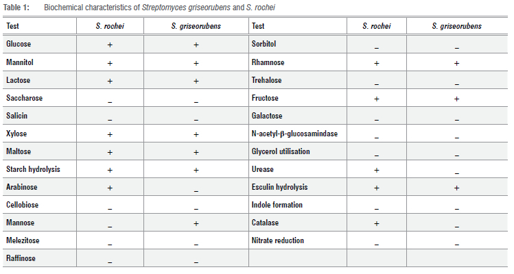
Streptomyces griseorubens showed a grey colour for substrate and aerial mycelia. S. rochei also showed a grey colour for aerial and substrate mycelia, with no production of melanin pigment and positive reactions with a Gram stain for both. Both isolates showed positive reactions against glucose, mannitol, lactose, xylose, maltose, fructose, and rhamnose. Only S. rochei was positive against arabinose and only S. griseorubens was positive against mannose. Ureases and catalases were produced by S. griseorubens only (Table 1).
Molecular identification (16S rRNA sequence analysis)
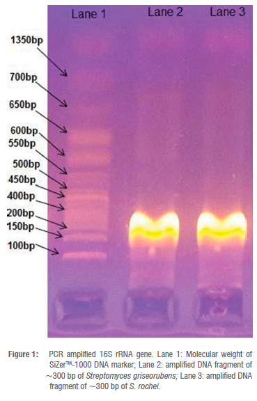
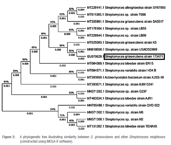
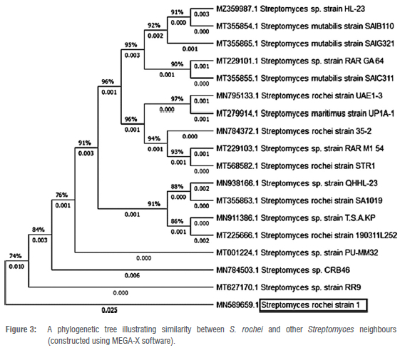
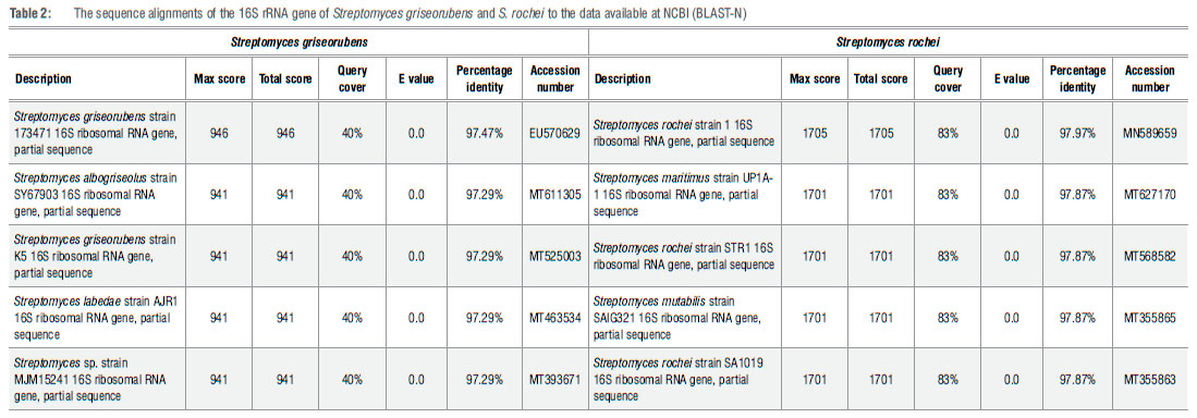
Genome amplification of the isolates' DNA using PCR with the primer 16S rRNA gene resulted in DNA fragments appearing as sharp bands at approximately 300 bp (Figure 1). The obtained sequences were analysed through an online database (NCBI) and compared with other bacterial isolates. The sequencing results showed that purified strains belong to the Phylum Actinobacteria, the Family Streptomycetaceae and the Genus Streptomyces. The data analysis confirmed isolates as Streptomyces griseorubens strain 173471 and Streptomyces rochei strain 1 (Table 2). A neighbour-joining tree was employed for both using MEGA-X software to show phylogenetic relationships between both of the Streptomyces strains and other Streptomyces neighbours (Figures 2 and 3).
Antimicrobial activity
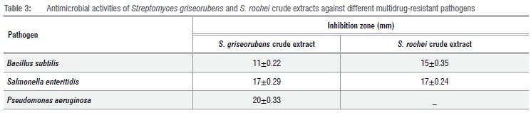
Streptomyces griseorubens filtrate exhibited significant antimicrobial activities against Bacillus subtilis, Salmonella enteritidisand Pseudomonas aeruginosa, with inhibition zone diameters of about 11 mm, 17 mm, and 20 mm, respectively, while that of S. rochei showed antimicrobial activities against B. subtilis and S. enteritidis, with inhibition zone diameters of about 15 mm and 17 mm, respectively (Table 3).
GC-MS Analysis
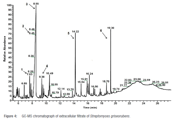
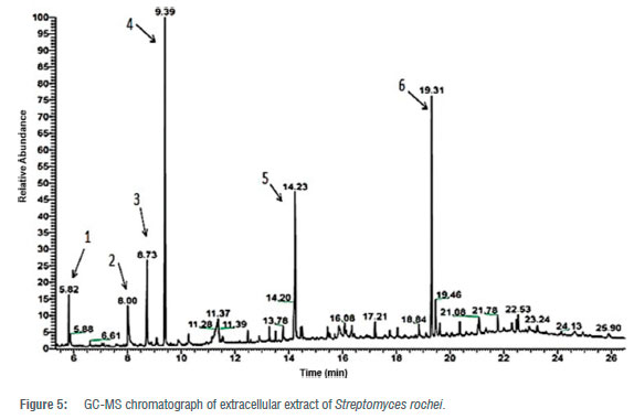
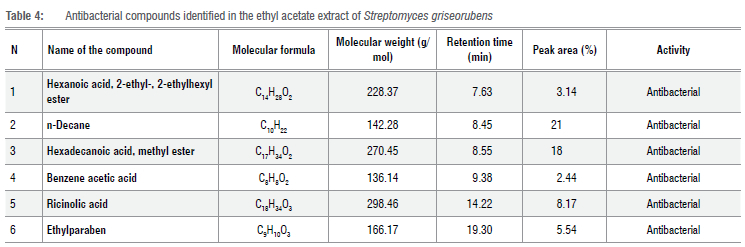
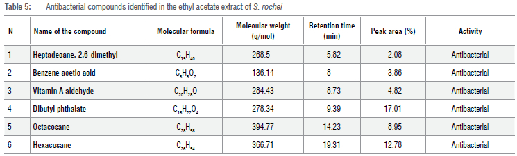
Ethyl acetate extracts of S. griseorubens and S. rochei that showed high antibacterial activities were analysed by GC-MS. The mass spectrum of GC-MS was interpreted according to the US National Institute of Standards and Technology (NIST) database, by comparing unknown spectra with the known data stored in the NIST library. For S.griseorubens, observed data confirmed the presence of six active compounds with antibacterial activity (Figure 4). The GC-MS analysis of S. rochei also confirmed the presence of six antibacterial compounds (Figure 5). The secondary metabolites from S. griseorubens that showed antibacterial activities were hexanoic acid, 2-ethyl-, 2-ethylhexyl ester (3.14%), n-Decane (21%), hexadecenoic acid methyl ester (18%), benzene acetic acid (2.44%), ricinolic acid (8.17%) and ethylparaben (5.54%) (Table 4). For S. rochei, heptadecane, 2,6-dimethyl- (2.08%), benzene acetic acid (3.86%), dibutyl phthalate (17.01%), octacosane (8.95%), hexacosane (12.78%) and vitamin A aldehyde (4.82%) were detected (Table 5).
Discussion
Two strains of Streptomyces were identified using the 16S rRNA sequence as well as traditional techniques, and their antibacterial potentiality was determined. The sequencing result of the PCR amplified 16S rRNA gene confirmed that isolate 1 belongs to Phylum Actinobacteria, Family Streptomycetaceae and Genus Streptomyces. The result revealed a 97.47% similarity with Streptomyces griseorubens strain 173471 according to NCBI GenBank and showed a similarity of about 97.29% with both Streptomyces labedae strain AJR1 and Streptomyces albogriseolus strain SY67903. According to Thirumurugan and Vijayakumar17, Streptomyces labedae substrate mycelia appeared brown on SCA medium and showed positive results with catalase production and nitrate reduction, whereas Streptomyces griseorubens could not produce catalase or reduct nitrate. In addition, Streptomyces griseorubens had a grey colour for both aerial mycelia and substrate mycelia on the SNA medium and all ISP media, with no distinctive pigments.18 According to El-Naggar et al.19, Streptomyces albogriseolus showed grey aerial mycelia and yellow/brown substrate mycelia on the SNA medium, while Streptomyces griseorubens appeared grey in both aerial mycelia and substrate mycelia on the SNA medium. In addition, Streptomyces albogriseolus can utilise sucrose and raffinose while Streptomyces griseorubens cannot.
The sequencing result of the PCR amplified 16S rRNA gene revealed that isolate 2 belongs to Phylum Actinobacteria, Family Streptomycetaceae and Genus Streptomyces. It revealed a 97.97% similarity with Streptomyces rochei strain 1 according to NCBI GenBank and showed a similarity of about 97.87% with both Streptomyces mutabilis strain SAIG321 and Streptomyces maritimus strain UP1A-1.
According to Bergey's Manual of Systemic Bacteriology18, Streptomyces mutabilis have white aerial mycelia on a starch nitrate agar medium while those of Streptomyces rochei are grey on SNA medium. In addition, Streptomyces mutabilis can utilise arabinose and sucrose while Streptomyces rochei cannot. According to Manikkam et al.20, Streptomyces maritimus have light grey aerial mycelia on a starch nitrate medium, while those of Streptomyces rochei appear dark grey on SNA medium. In addition, Streptomyces maritimus cannot utilise xylose while Streptomyces rochei can.
The antibacterial compounds - hexanoic acid, 2-ethyl-, 2-ethylhexyl ester, n-Decane, hexadecanoic acid methyl ester, benzene acetic acid, ricinolic acid, and ethylparaben - observed in Streptomyces griseorubens were found to be strong antibacterial agents. Antibacterial action has been observed for hexanoic acid, 2-ethyl-, 2-ethylhexyl ester, which contains the short chain fatty acid (hexanoic acid). Short-chain fatty acids are commonly produced by healthy gut microbiota and serve as a defender against enteric infections. Furthermore, by lowering intracellular pH and diffusing across the bacterial membrane, they show a direct antibacterial effect against bacterial pathogens.21 According to Al-Rubaye et al.22, a Streptomyces sp. isolated from Tigris River sediments in Baghdad was found to generate short-chain fatty acids. N-decane is a straight-chain alkane that was extracted by the n-butanol of Streptomyces sp. Sp1 filtrate, which has been shown to have strong antibacterial activity and could be used as a biocontrol agent against Vibrio anguillarum.23 The accumulation of such chemicals in the cell membrane can have a significant impact on its function and eventually lead to cell death.24 Disruption of the electron transport chain and oxidative phosphorylation is linked to its mechanism of action. Inhibition of enzyme activity can also cause nutrient absorption failure, production of peroxidation and auto-oxidation degradation products, and eventually leads to bacterial cell lysis.25
Hexadecanoic acid is a saturated long-chain fatty acid that was observed in a Nocardia sp. filtrate, which was found to exhibit antibacterial action against methicillin-resistant Staphylococcus aureus (MRSA).26 Benzene acetic acid is an organic compound containing a phenyl functional group and a carboxylic acid functional group. According to Al-Dhabi et al.27, Streptomyces sp. Al-Dhabi-2 isolated from a harsh environment in Saudi Arabia was found to produce benzene acetic acid. It had substantial minimum inhibitory concentration (MIC) values of less than 39 μg/mL against Bacillus cereus and Enterococcus faecalis, and 78 μg/mL against Streptococcus agalactiae. The harmful effect of benzene acetic acid is mostly mediated by acetic acid dissociation within the microbial cells, which results in lowering intracellular pH and metabolic disruption.28 Ricinoleic acid [R (Z)-12-hydroxy-9-octadecanoic acid] is a fatty acid that is considered the major component of castor oil. The antibacterial activity of ricinoleic acid against Staphylococcus aureus and Pseudomonas aeruginosa has been reported, with MICs of 2.68 μM and 2.60 μM, respectively.29 The surfactant behaviour of ricinoleic acid moiety is due to the presence of long lateral hydrophobic methylene units that could disturb and inhibit the permeability of the bacterial cell membrane which consequently inhibits the growth of bacteria.30 Ethylparaben is an ethyl ester of p-hydroxybenzoic acid. The GC analysis of a Brevibacillus brevis crude extract proved the presence of ethylparaben, which has a considerable bactericidal impact against Escherichia coli.31 Although its mode of action is still unproven, one of the suggested modes of action of parabens is to disrupt osmotic gradients in bacteria by interacting with mechanosensitive channels.32 Streptomyces rochei was detected to produce the antibacterials heptadecane, 2,6-dimethyl-, benzene acetic acid, dibutyl phthalate, octacosane, hexacosane, and vitamin A aldehyde.
Decane compounds have been shown to exhibit antibacterial action in several studies. Dodecane, n-Hexa decanoic acid, and 1-Octadecane were found in an ethyl acetate extract of Streptomyces cavouresis KUV39 by GC-MS.33 Phthalic acid esters (PAEs) are a type of lipophilic compound that is commonly found in plants and microorganisms. Di-n-butyl phthalate, diethyl phthalate, and dimethyl phthalate are the most common PAEs found in natural sources, and are reported to have antibacterial properties.34 Using ultraviolet, Fourier transform infrared, and GC-MS analyses, the bioactive chemical dibutyl phthalate was reported to be generated by the soil actinomycete isolate Streptomyces albidoflavus 321.2, showing a considerable antibacterial effect against E. coli with an MIC of 53 μg/mL and Bacillus subtilis with an MIC of 84 μg/mL.35 It was also isolated from a novel marine Streptomyces sp. that reduced Colletotrichum fragariae spore germination and mycelial growth.36 Further studies are needed to understand the related antimicrobial mechanisms of PAEs. Hexacosane is a straight-chain alkane that is a volatile oil component and a plant metabolite. Hexacosane was discovered to be among the chemicals in the medicinal plant Kielmeyera coriacea that exhibits antibacterial activity against both aerobic and non-aerobic bacteria.37 Octacosane is one of 17 chemicals found in Elsholtzia ciliate extracts that have antibacterial properties.38 Its mode of action has been linked to accumulation in the cell membrane, which affects the function of the cell and finally leads to cell death.21 Retinaldehyde is a stabilised form of vitamin A. According to Pechère et al.39, vitamin A is thought to be anti-infectious. Although retinal resistance is not unique to infection, the mechanisms underlying these post-infective actions are largely unknown and are most likely related to its pleiotropic effects on immune function. Both in vivo and in vitro, retinaldehyde has been shown to have antibacterial action against Propionibacterium acnes. P. acnes No. CIP179 and CIP53119 have MICs of 4 mg/L, while P acnes No. CIP53117 had a MIC of 8 mg/L.40
Conclusion
Two strains, Streptomyces griseorubens and Streptomyces rochei, producing novel bioactive compounds were identified via 16S rRNA sequencing and traditional techniques. Antibacterial evaluation of the crude extract showed high efficiency against very serious MDR pathogens - P aeruginosa, S. enteritidis, and B. subtilis. GC analysis of S. griseorubens revealed the presence of several strong antibacterial compounds: hexanoic acid, 2-ethyl-, 2-ethylhexyl ester, n-decane, hexadecanoic acid methyl ester, benzene acetic acid, ricinolic acid, and ethylparaben. Heptadecane, 2,6-dimethyl, benzene acetic acid, vitamin A aldehyde, dibutyl phthalate, octacosane, and hexacosane were detected in the S. rochei filtrate.
Acknowledgements
We thank Prof. Dr. Wagih A. El-Shouny for his valuable contributions to the research project, despite his unfortunate passing during its early stages. Prof. Dr El-Shouny played a pivotal role in shaping the direction of the work by suggesting the initial idea, collaborating with other group members to outline experimental plans, and providing guidance on work locations. We also thank Dr Hossam Ismail at the Faculty of Fisheries and Aquaculture Sciences, Kafrelsheikh University, for providing the MDR bacteria.
Competing interests
We have no competing interests to declare.
Authors' contributions
B.M.A.: Methodology; data collection; sample analysis; data analysis; validation; data curation; writing - the initial draft; writing - revisions. S.A.H.: Validation; data curation; writing - the initial draft; writing -revisions; student supervision. E.E-M.: Conceptualisation; data collection; data analysis; validation; data curation; writing - the initial draft; writing -revisions; student supervision; project leadership; project management.
References
1. Blunt W, Copp R, Munro G, Northcote T, Prinsep R. Marine natural products. Nat Prod Rep. 2007;24:31-86. https://doi.org/10.1039/b603047p [ Links ]
2. Sanglier J, Haag H, Huck A, Fehr T. Novel bioactive compounds from actinomycetes a short review (1988-1992). Res Microbiol. 1993;144(8):633-642. https://doi.org/10.1016/0923-2508(93)90066-B [ Links ]
3. Sharma P, Dutta J, Thakur D. Future prospects of actinobacteria in health and industry. In: Singh BP, Gupta VK, Passari AK, editors. New and future developments in microbial biotechnology and Bioengineering: Actinobacteria: Diversity and biotechnological applications. Amsterdam: Elsevier; 2018. https://doi.org/10.1016/B978-0-444-63994-3.00021-7 [ Links ]
4. Jagannathan S, Manemann E, Rowe S, Callender M, Soto W. Marine actinomycetes, new sources of biotechnological products. Mar Drugs. 2021;19:365. https://doi.org/10.3390/md19070365 [ Links ]
5. Bérdy J. Bioactive microbial metabolites: A personal view. J Antibiot. 2005;58:1-26. https://doi.org/10.1038/ja.2005.1 [ Links ]
6. El-Naggar N, El-Shweihy N. Bioprocess development for L-asparaginase production by Streptomyces rochei, purification and in-vitro efficacy against various human carcinoma cell lines. Sci Rep. 2020;10(1):7942. https://doi.org/10.1038/s41598-020-64052-x [ Links ]
7. Rashad Y Al-Askar AA, Ghoneem Kh, Saber W. Chitinolytic Streptomyces griseorubens E44G enhances the biocontrol efficacy against Fusarium wilt disease of tomato. Phytoparasitica. 2017;45:227-237. https://doi.org/10-1007/s12600-017-0580-3 [ Links ]
8. Dharmaraj S, Sumantha A. Bioactive potential of Streptomyces associated with marine sponges. World J Microbiol Biotechnol. 2009;25:1971-1979. https://doi.org/10.1007/s11274-009-0096-1 [ Links ]
9. Williams S, Goodfellow M, Alderson G, Wellington E, Sneath P Sackin M. Numerical classification of Streptomyces and related genera. Microbiol. 1983;129:1743-1813. https://doi.org/10.1099/00221287-129-6-1743 [ Links ]
10. Williams ST, Davies FL. Use of antibiotics for selective isolation and enumeration of actinomycetes in soil. J Gen Microbiol. 1965;38:251-262. https://doi.org/10.1099/00221287-38-2-251 [ Links ]
11. Shirling EB, Gottlieb D. Methods for characterization of Streptomyces species. Int J Syst Bacteriol. 1966;16:313-340. https://doi.org/10.1099/00207713-16-3-313 [ Links ]
12. Al-Dhabi N, Esmail G, Duraipandiyan V Valan Arasu M, Salem-Bekhit M. Isolation, identification and screening of antimicrobial thermophilic Streptomyces sp. Al-Dhabi-1 isolated from Tharban hot spring, Saudi Arabia. Extremophiles. 2016;20:79-90. https://doi.org/10.1007/s00792-015-0799-1 [ Links ]
13. Holmalahti J, Von Wright A, Raatikainen O. Variations in the spectra of biological activities of actinomycetes isolated from different soils. Lett Appl Microbiol. 1994;18:144-146. https://doi.org/10.1111/j.1472-765X.1994.tb00829.x [ Links ]
14. Sathiyanarayanan G, Gandhmathi R, Sabarathnan B, SeghalKiran G, Selvin J. Optimization and production of pyrrolidon antimicrobial agent from marine sponge-associated Streptomyces sp. MAPSIS Bioprocess Biosyst Eng. 2014;37:561-573. https://doi.org/10.1007/s00449-013-1023-2 [ Links ]
15. Hassan S, Shobier A. GC/MS identification and applications of bioactive seaweed extracts from Mediterranean coast of Egypt. Egypt J Aquat Biol Fish. 2018;5:1-21. https://doi.org/10.21608/ejabf.2018.17952 [ Links ]
16. Lorowitz W, Saxton E, Sondossi M, Nakaoka K. Integrating statistics with a microbiology laboratory activity. Microbiol Educ. 2005;6:14-19. https://doi.org/10.1128/me.6.1.14-19.2005 [ Links ]
17. Thirumurugan D, Vijayakumar R. Characterization and structure elucidation of antibacterial compound of Streptomyces sp. ECR77 isolated from East Coast of India. Curr Microbiol. 2015;70:745-755. https://doi.org/10.1007/s00284-015-0780-3 [ Links ]
18. Goodfellow M, Kâmpfer P Busse H-J, Trujillo ME, Suzuki K-I, Ludwig W, et al. editors. Bergey's manual of systematic bacteriology. Volume 5: The Actinobacteria. New York: Springer; 2012. https://doi.org/10.1007/978-0-387-68233-4 [ Links ]
19. El-Naggar NEA, Abdelwahed NA, Saber WI, Mohamed AA. Bioprocessing of some agro-industrial residues for endoglucanase production by the new subsp.; Streptomyces albogriseolus subsp. cellulolyticus strain NEAE-J. Braz J Microbiol. 2014;45:743-756. https://doi.org/10.1590/S1517-83822014005000049 [ Links ]
20. Manikkam R, Ganesan V Kaari M, Venugopal G, Arumugam S, Joseph J. Antibacterial efficacy of Streptomyces maritimus SACC-E6 against carbapenem-resistant Klebsiella pneumoniae ATCC BAA-1705. J Appl Pharm Sci. 2021;11:1. https://doi.org/10.7324/JAPS.2021.110110 [ Links ]
21. Lamas A, Regal P Vázquez B, Cepeda A, Franco M. Short chain fatty acids commonly produced by gut microbiota influence Salmonella enterica motility, biofilm formation, and gene expression. Antibiotics. 2019;8(4):265. https://doi.org/10.3390/antibiotics8040265 [ Links ]
22. Al-Rubaye S, Risan M, Al-Rubaye D. Gas chromatography-mass spectroscopy analysis of bioactive compounds from Streptomyces spp. isolated from Tigris river sediments in Baghdad city. J Biotechnol Research Center. 2020;14:1. https://doi.org/10.24126/jobrc.2020.14.1.590 [ Links ]
23. Kawuri R, Darmayasa IBG. Bioactive compound from extract filtrate Streptomyces sp.Sp1. as biocontrol of vibriosis on larvae of Macrobrachium rosenbergii shrimps. HAYATI J Biosci. 2019;26:1. https://doi.org/10.4308/hjb.26.1.15 [ Links ]
24. Sikkema J, de Bont JA, Poolman B. Mechanisms of membrane toxicity of hydrocarbons. Microbiol Rev. 1995;59:201-220. https://doi.org/10.1128/rnr.59.2.201-222.1995 [ Links ]
25. Dhakal D, Rayamajhi V, Mishra R, Sohng JK. Bioactive molecules from Nocardia: Diversity, bioactivities and biosynthesis. J Ind Microbiol Biotechnol. 2019;46(3-l):385-407. https://doi.org/10.1007/s10295-018-02120-y [ Links ]
26. Andrew P Smith V. Antibacterial free fatty acids: Activities, mechanisms of action and biotechnological potential. Appl Microbiol Biotechnol. 2010;85(6):1629-1642. https://doi.org/10.1007/s00253-009-2355-3 [ Links ]
27. Al-Dhabi N, Esmail G, Duraipandiyan V Arasu VM. Chemical profiling of Streptomyces sp. Al-Dhabi-2 recovered from an extreme environment in Saudi Arabia as a novel drug source for medical and industrial applications. Saudi J Biol Sci. 2019;26(4):758-766. https://doi.org/10.1016/j.sjbs.2019.03.009 [ Links ]
28. Treek J, Mira N, Jarboe L. Adaptation and tolerance of bacteria against acetic acid. Appl Microbiol Biotechnol. 2015;99(15):6215-6229. https://doi.org/10.1007/s00253-015-6762-3 [ Links ]
29. Narasimhan B, Mourya VK, Dhake AS. OSAR studies of antibacterial ricinoleic acid derivatives. Pharm Chem J. 2005;41(3):16-21. https://doi.org/10.1007/s11094-007-0030-5 [ Links ]
30. Khan AA, Husain A, Jabeen M, Mustafa J, Owais M. Synthesis and characterization of novel n-9 fatty acid conjugates possessing antineoplastic properties. Lipids. 2012;47(10):973-986. https://doi.org/10.1007/s11745-012-3707-9 [ Links ]
31. Jianmei C, Bo L, Zheng C, Huai S, Guohong L, Cibin G. Identification of ethylparaben as the antimicrobial substance produced by Brevibacillus brevis FJAT-0809-GLX. Microbiol Res. 2015;172:48-56. https://doi.org/10.1016/j.micres.2014.11.007 [ Links ]
32. Nguyen T, Clare B, Guo W, Martinac B. The effects of parabens on the mechanosensitive channels of E. coli. Eur Biophys J. 2005;34:389-395. https://doi.org/10.1007/s00249-005-0468-x [ Links ]
33. Al-Dhabi NA, Ghilan AKM, Esmail GA, Arasu MV, Duraipandiyan V, Ponmurugan K. Bioactivity assessment of the Saudi Arabian marine Streptomyces sp. Al-Dhabi-90, metabolic profiling and its in vitro inhibitory property against multidrug resistant and extended-spectrum beta-lactamase clinical bacterial pathogens. J Infect Public Health. 2019;12(4):549-556. https://doi.org/10.1016/j.jiph.2019.01.065 [ Links ]
34. Huang L, Zhu X, Zhou S, Cheng Z, Shi K, Zhang C, et al. Phthalic acid esters: Natural sources and biological activities. Toxins (Basel). 2021;13(7):495. https://doi.org/10.3390/toxins13070495 [ Links ]
35. Roya R, Laskarb S, Sena S. Dibutyl phthalate, the bioactive compound produced by Streptomyces albidoflavus 321.2. Microbiol Res. 2006;161:121-126. https://doi.org/10.1016/j.micres.2005.06.007 [ Links ]
36. Li X, Jing T, Zhou D, Zhang M, Qi D, Zang X, et al. Biocontrol efficacy and possible mechanism of Streptomyces sp. H4 against postharvest anthracnose caused by Colletotrichum fragariae on strawberry fruit. Postharvest Biol Technol. 2021;75:111401. https://doi.org/10.1016/j.postharvbio.2020.111401 [ Links ]
37. Martins CM, Nascimento E, de Morais S, de Oliveira A, Chang R, Cunha L, et al. Chemical constituents and evaluation of antimicrobial and cytotoxic activities of Kielmeyera coriacea Mart. & Zucc. essential oils. eCAM. 2015;2015:842047. https://doi.org/10.1155/2015/842047 [ Links ]
38. Ma J, Xu R, Lu Y, Ren D, Lu J. Composition, antimicrobial and antioxidant activity of supercritical fluid extract of Elsholtzia ciliate. J Essent Oil-Bear Plants. 2018;21(2):556-562. https://doi.org/10.1080/0972060X.2017.1409657 [ Links ]
39. Pechère M, Pechère C, Siegenthaler G, Germanier L, Saurata, J. Antibacterial activity of retinaldehyde against Propionibacterium acnes. Dermatology. 1999;199(1):29-31. https://doi.org/10.1159/000051375 [ Links ]
40. Rusdy RSN, Legiawati L, Sitohang IBS, Sirait SMHAP. Adjuvant therapies of acne: Review of literatures. Bali Dermatol Venereol J. 2022;5(1):17-22. https://doi.org/10.15562/bdv.v5i1.75 [ Links ]
 Correspondence:
Basma Atallah
Email: basma.2015@yahoo.com
Received: 04 Aug. 2022
Revised: 20 July 2023
Accepted: 25 Aug. 2023
Published: 29 Nov. 2023
Editors: Priscilla Baker, Amanda-Lee Manicum
Funding: None

