


Servicios Personalizados
Articulo
 Inglés (pdf)
 Articulo en XML
 Referencias del artículo
Indicadores

Links relacionados
-
 Citado por Google
 -
 Similares en Google
Compartir

 Permalink
South African Journal of Science
versión On-line ISSN 1996-7489
versión impresa ISSN 0038-2353
S. Afr. j. sci. vol.117 no.5-6 Pretoria may./jun. 2021
http://dx.doi.org/10.17159/sajs.2021/8747
RESEARCH ARTICLE
One hominin taxon or two at Malapa Cave? Implications for the origins of Homo
Yoel RakI, II; Eli GeffenIII; William HylanderIV; Avishag GinzburgI; Ella BeenI,V
IDepartment of Anatomy and Anthropology, Faculty of Medicine, Tel Aviv University, Tel Aviv, Israel
IIInstitute of Human Origins and School of Human Evolution and Social Change, Arizona State University, Tempe, Arizona, USA
IIIDepartment of Zoology, Tel Aviv University, Tel Aviv, Israel
IVDepartment of Evolutionary Anthropology, Duke University, Durham, North Carolina, USA
VSports Therapy Department, Ono Academic College, Kiryat Ono, Israel
ABSTRACT
A report on the skeletons of two individuals from the Malapa cave site in South Africa attributes them both to a new hominin species, Australopithecus sediba. However, our analysis of the specimens' mandibles indicates that Australopithecus sediba is not a Homo-like australopith', a transitional species between Australopithecus africanus and Homo. According to our results, the specimens represent two separate genera: Australopithecus and Homo. These genera are known to have jointly occupied sites, as seen in several early South African caves, so one cannot rule out the possibility that Malapa also contains remains of the two taxa. Our results lead us to additionally conclude that all the Australopithecus species on which the relevant mandibular anatomy is preserved (not only the 'robust' australopiths but also the 'gracile' - more generalised - ones) are too specialised to constitute an evolutionary ancestor of Homo sapiens. Furthermore, given that the Malapa site contains representatives of two hominin branches, one of which appears to be Homo, we must seek evidence of our origins much earlier than the date assigned to Malapa, approximately 2 million years before present. Support for this claim can be found in Ethiopian fossils attributed to the genus Homo and dated at 2.4 and 2.8 million years before present.
SIGNIFICANCE:
• The proposed hominin species Australopithecus sediba, from the Malapa Cave in South Africa, seems to actually consist of two species, each of which represents a different hominin genus: Homo and Australopithecus. If, indeed, this is the case, Homo must have originated prior to the Malapa remains, contrary to the scenario suggested in the original report on Au. sediba.
Keywords: Australopithecus sediba, Homo spp., Malapa hominins, hominin phylogeny, South Africa
Introduction
The proposal of a new hominin species, Australopithecus sediba, announced and described by Berger1, Berger et al.2, and de Ruiter et al.3, is based primarily on the analysis of two partial skeletons, MH1 and MH2. The taxon is claimed to exhibit many features that suggest that it represents an intermediate species between Australopithecus africanus and Homo. This assertion was recently reiterated in a special issue of PaleoAnthropology dedicated to Australopithecus sediba.4,5However, a careful assessment of the mandibular remains leads us to conclude that the proposed Au. sediba species actually encompasses two species representing separate genera - Australopithecus and Homo - and as such cannot play a role in the origin of the latter. The discovery of two hominin species at one site is not unheard of in South Africa.
The two mandibles from Malapa plainly exhibit different patterns of ramal morphology: MH1 resembles australopith morphology, and MH2 displays the generalised morphology exhibited by Homo sapiens and other Homo species.
The morphology of the ascending ramus of the mandible in hominins has been found to be a diagnostic character6 (note that Wolpoff and Frayer7 claim that the upper part of the ramus is not diagnostic enough to distinguish between H. sapiens and H. neanderthalensis, but they cannot refute our argument because they have not applied our method to their sample); as such, the ramal morphology clearly distinguishes between Australopithecus and H. sapiens8. In the latter, the condylar and coronoid processes are relatively slender in a lateral view, they are similar in size, and they are separated by a broad, scooped out mandibular (sigmoid) notch, whose deepest point lies about halfway between the tips of the two processes (Figure 1). This configuration lends the notch a somewhat symmetrical appearance. In Australopithecus, on the other hand, the coronoid process is tall and broad, occupying about three-fourths of the ramal breadth. The process's superior end is rather flat, with a hook-like profile, and overhangs the relatively small mandibular notch, which is shallow in relation to the mandibular condyle. As a result, the outline of the notch is confined and asymmetric.
Similarly, H. sapiens and australopith rami seem to differ vis-ä-vis the preangular notch. In H. sapiens, as in many other primates (i.e. the generalised configuration), the concave anterior margin of the ramus forms this notch, which is also present in MH2 (Figure 1). In the australopiths, as in MH1, the anterior margin of the ramus usually slopes diagonally in a straight line until it meets the mandibular body. Some exceptions to this dichotomy can be noted - for example, the presence of the preangular notch on MLD 40, Sts 52, Sts 7 and SKW 5, despite their assignment as Au. africanus. These exceptions somewhat diminish the diagnostic power of the preangular notch.
Because H. sapiens shares its ramal morphology with many other primates (for example, chimpanzees, orangutans, vervets and colobines), that morphology is clearly the primitive one, whereas the Australopithecus ramal configuration is derived - a synapomorphic character that combines Au. robustus, Au. africanus and Au. afarensis (and possibly other australopiths, such as Au. anamensis and Au. boisei, neither of which has a ramus that is sufficiently preserved to permit study) into what seems to be a monophyletic group. To suggest that the derived configuration, that of Australopithecus, evolved into the modern human configuration violates the principle of parsimony.
Although we are convinced that the discrepancies that we have observed in ramal morphology stem from profound biomechanical differences, elucidation of the functionality at play (of the derived configuration) is a major project and beyond the scope of this study. Because the morphological differences are manifested in very young individuals9,10, as described later, one can be certain that these morphologies are embedded in the genome and not generated by some activity during an individual's lifetime. In any case, the functional issue has no bearing on the taxonomic question treated here.
In this paper, our aim is not to determine which species of Australopithecus or Homo the Malapa mandibles belong to, but to determine how the two mandibles differ and what those differences mean. To accomplish these goals, we show that the differences are beyond what is expected in a trait's normal range of distribution in a given population. Our null hypothesis is that the two mandibles of Au. sediba represent a single taxon (as claimed, for example, by Berger1; Berger et al.2; de Ruiter et al.3; de Ruiter et al.5; Ritzman et al.9; and Williams et al.4). The alternative hypothesis is that the mandibles of Au. sediba represent a mix of taxa; in this scenario, a statistical analysis would classify one mandible with the Australopithecus cluster (but not provide any species assignment), and the other mandible with the generalised cluster (bearing a shared morphology). Indeed, our evidence supports this alternative hypothesis.
Materials and methods
Our sample includes 115 mandibles from mature extant primates, both male and female (Supplementary table 1): 41 modern humans, 58 chimpanzees (29 each of Pan paniscus and Pan troglodytes, grouped into one class following the results of previous analyses8), and 16 orangutans (Pongo pygmaeus). The H. sapiens specimens emanate from geographically varied regions: Australia (Aboriginal peoples), India, the Levant, and northern Canada (Inuit). Regarding the size of the modern H. sapiens sample, see the Results section. Fossil hominins in the sample consist of four rami from mature Australopithecus individuals (A.L. 288-1, SK 23, MAK-VP 1/83 and SK 34) and two rami from Australopithecus juveniles (SK 63 and A.L. 333-43). The juvenile specimens help increase the sample and were added after it became apparent that no ontogenetic change occurs in ramal morphology9,10 (Figures 2-4). Another young individual, DIK11, from the Ethiopian Dikika site, exhibits the same ramal morphology, as seen on a photograph of the specimen (no cast has been available to us as yet). In addition, one Ardipithecus ramidus ramus, specimen GWM5sw/P5611 (Figure 3), was included as an unknown. Five Homo fossils (three H. erectus specimens from Choukoutien, restored by Franz Weidenreich12; KNM-WT 15000 -H. ergaster; and ATD696, a mandible from Gran Dolina, Spain) were also analysed, although they proved to be of limited value (see Discussion).
Gorillas were excluded from our analysis. It was demonstrated in a 2007 study that the ramal morphology of gorillas is similar - although not identical - to that of Australopithecus.8As noted in that study:
given a phylogeny in which chimpanzees and modern humans are sister groups, parsimony dictates that we view the similarity in ramal morphology between Australopithecus afarensis [in fact, all the australopiths that provide ramal evidence] and gorillas as a homoplastic character, a character that appears independently and as such has no phylogenetic value.8(p.6570)
The similarity between the gorilla ramus and that of Au. robustus may well stem from the very tall ramus in both groups.
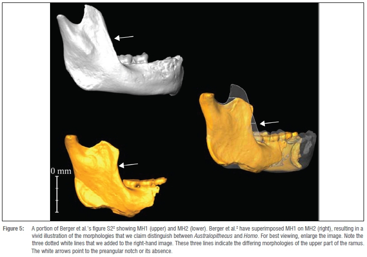
Regarding reconstruction, MH1 requires none. In MH2, the tip of the coronoid process is damaged; nevertheless, its reconstruction is straightforward, as seen in Figure 5. The three dotted white lines on the superimposed images were added by us. The lines demonstrate that there is no way to reconstruct the coronoid process in MH2 to resemble the robust configuration.
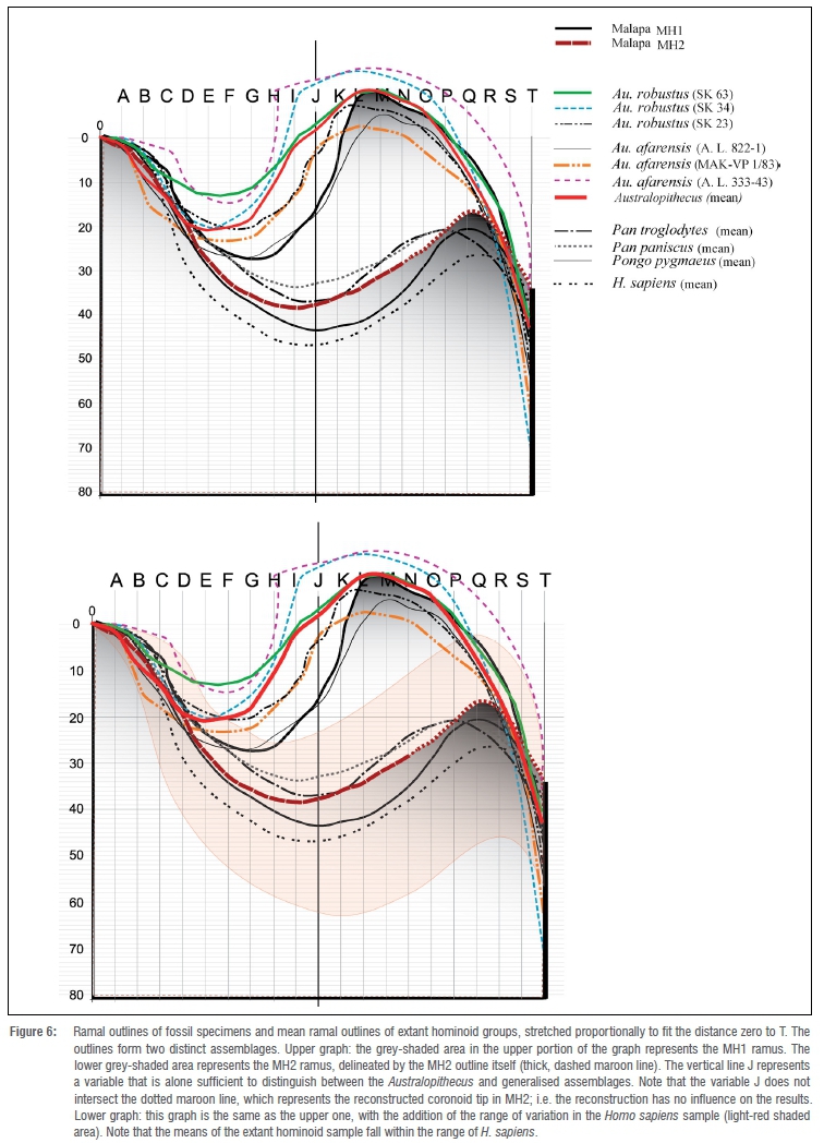
We quantified the upper ramal contours of the specimens through a simple method described by Rak et al.8(p.6571):
To convey the anatomical differences in the upper ramal contour, we adopted a method... which consisted of capturing a digital image of the mandibular ramus with the camera centred at the vertical level of the mandibular notch and held perpendicular to the lateral surface of the ramus. ... We traced the digital image of each ramus from the tip of the condylar process to the anterior margin of the ramus. .
.We stretched the contour proportionally on the vertical and horizontal axes by dragging the contour's lower right corner until it occupied the entire width of the area of the fixed coordinates in the background template. This part of the procedure eliminated differences in size in the analysis [leaving shape only]. The posterior margin [of the ramus] was aligned with the vertical line at 0, and the anterior margin was aligned at T. The posterior ramal margin in the entire sample exhibits a slight concavity between the posterior end of the condyle and the insertion site of the posterior fibres of the masseter and medial pterygoid muscles; using these two posteriorly protruding structures, we were able to orient the posterior margin on a vertical line throughout the sample. The intersection of the ramal contour with each of the vertical lines, A through T, yielded 20 numeric variables for each ramus.8(p.6571)
We define variable T as the maximum horizontal distance between the condyle and two-thirds of the anterior ramal margin's height. In this way, we accentuate the most diagnostic part of the ramal outline (A-T). Note that the use of the point defining T (or any other point on the ramus) does not affect the height measurement of the coronoid process in the mandibles under study.
The intersection of each contour with a vertical line and a horizontal line (i.e. coordinates) is assigned a value representing the distance of the intersection point from the zero horizontal line (for example, 10, 20 or 30) (Figure 6). These are the numerical values used for the statistics. Note that as long as all the contours are on the same grid, units of measure are irrelevant, as is the distance between the lines (provided that it is constant).
We chose the same orientation for the posterior margin of all the rami in our sample because that orientation seems to be fixed in relation to the base of the skull, the Frankfurt horizontal, and the zygomatic arch (indicating functional significance), as demonstrated in Figure 7. Alternatively, positioning all the mandibles with a horizontal orientation of the occlusal plane or of the base of the mandibular body would introduce variation in the shape of the mandibular notch.
The 20 (AT) variables served as independent variables in a general discriminant analysis to classify two unknown fossil specimens (MH1 and MH2). We used Jump version 15 software for all analyses. General discriminant analysis applies the general linear model approach to discriminant analysis and can use both continuous and categorical independent variables. Our reference classes consisted of Australopithecus, Pan, Pongo pygmaeus and H. sapiens mandibles. The prior probability of classification was set as equal. The key assumption in discriminant analysis is that the variables used are not completely redundant.
To reduce dimensionality and eliminate the dependence between the variables, we used two independent approaches. First was the best-subset approach. Of 1 048 556 possible models, we inspected the 100 models that accounted for most of the variation (i.e. that exhibited the lowest misclassification rate) (Supplementary figure 1). Out of those models, we selected the one with the least number of parameters and used its functions to predict the state of unknowns. This approach considerably reduces the number of variables in the analysis and keeps the power of classification nearly the same. Thus, the retained variables are those that are most multidimensionally informative for the classification.
Our second approach was a principal component analysis to accommodate the effects of collinearity among the variables. All 20 variables were collapsed into principal components. Five components - those with an eigenvalue greater than or equal to 1 (after varimax rotation) - were retained. We used the components' scores as independent variables in the general discriminant analysis, and the resulting discriminant functions served to classify the unknown fossil specimens. For cross-validation, we applied the leave-one-out procedure. Through the two approaches just described, we classified the unknown fossil specimens. We also reran the analysis under a two-class model: Australopithecus and taxa with a generalised ramus (Pan, Pongo and Homo).
Results
Even in the absence of the coronoid process on MH2, the differences between it and MH1 are readily visible, as was shown in 2010 by Berger et al.2 themselves (reproduced and modified here in Figure 5). The unreconstructed outline of the mandibular (sigmoid) notch in MH2 diverges quite clearly from the comparable area in MH1. When the two specimens are adjusted to the same scale (Figure 6), the deepest part of the notch in MH2 is situated much more anteriorly than in MH1 and descends much farther relative to the zero point, i.e. the mandibular condyle, as in the generalised configuration. The statistical analysis tells us that the difference between the height of the two coronoid processes, reconstructed or not, is of less importance than the outline of the mandibular notch itself and has little effect on the results.
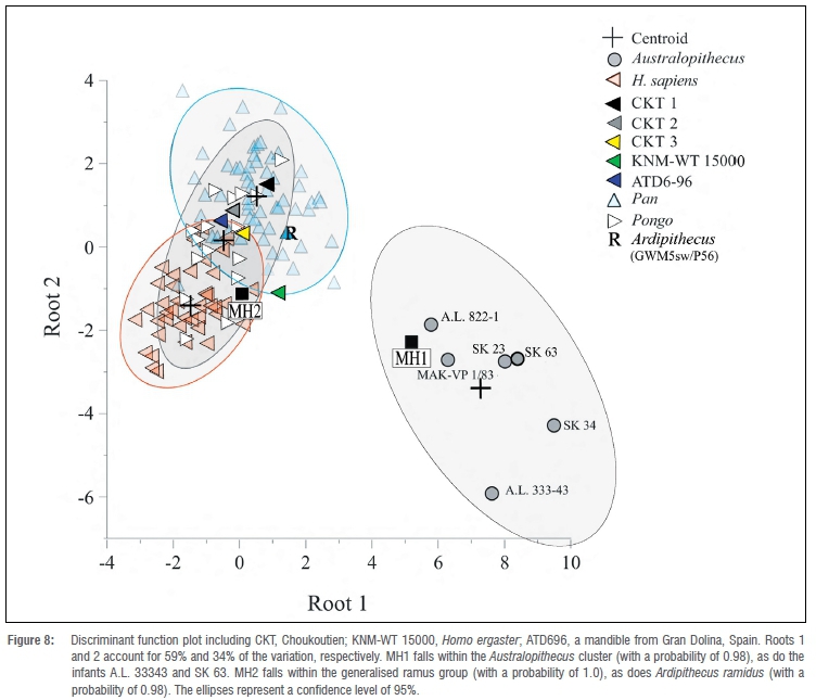
The best-subset approach yielded a classification success ranging from 94.6% to 92.5%. Most of these models share variables A, G, H, I, R R and T (Figure 8). The smallest subset model consists of eight effects (A, F, G, I, O, R R and T; Figure 8), which correctly classify 93.3% of the cases (the leave-one-out cross-validation classification success is 87.4%). According to the posterior probabilities (p(ki)) from the smallest subset model, MH2 falls in the generalised group (assigned as most likely an orangutan, with p(ki) = 0.76), whereas MH1 is assigned as most likely Australopithecus (p(ki) = 1.00). Finally, when only Australopithecus and the generalised ramus group are considered, the two best models consist of a single variable (I or J) that correctly classifies all cases (100%) (Figure 6). This variable corresponds to the deepest point of the notch in the generalised group; compare with the position of the homologous point in the specialised group (Figure 6). According to this model, MH1 is assigned to the Australopithecus cluster (p(ki) = 0.98) and MH2 to the generalised ramus group (p(ki) = 1.00). The ramus of the Ar. ramidus mandible11 (Figure 3) falls in the generalised cluster, with a probability of 0.98.
Note that the size of the H. sapiens sample (41 individuals) is not what counts; rather, the statistical analysis regards the entire generalised sample, consisting of 115 individuals, as one group, because the real issue is whether the Sediba mandibles fall in the generalised cluster, the specialised one, or both.
In the principal component approach, the four factors with an eigenvalue greater than or equal to 1 together accounted for 91.7% of the variation in the data. (The first four principal components accounted for 50%, 17%, 17% and 8% of the variance, respectively, totalling 92%.) The eigenvalues of the factors were 9.9 (49.7%), 3.4 (17.2%), 3.4 (17.2%) and 1.5 (7.6%). All four factors were significantly different from each other (Bartlett test, p < 0.0001 for each of the factors).
The general discriminant analysis correctly classifies 74.2% of cases (with the leave-one-out crossvalidation classification success at 69.7%). Rosterior probabilities of this model assign MH1 as Australopithecus (p(ki) = 1.0) and MH2 as most likely an orangutan (p(ki) = 0.755). The latter is in contrast to a probability of 0 as Australopithecus. Ar. ramidus is classified as most likely a chimpanzee (p(ki) = 0.49) or orangutan (p(ki) = 0.49). Finally, when only Australopithecus and the generalised ramus group are considered, MH2 and Ar. ramidus are assigned to the generalised ramus group (p(ki) = 1.00 in both cases) and MH1 is classified as Australopithecus (p(ki) = 1.00).
The morphological overlap between the comparative taxa is high only in the groups displaying the generalised morphology (not surprisingly, given that 'generalised', by definition, is shared). On the other hand, there is no overlap whatsoever between the generalised group and the derived one (Figure 6 and Figure 8). Note that no attempt has been made to assign MH1 and MH2 to particular species (because one of these configurations is synapomorphic and the other is generalised). The fact that MH2 is classified as most likely an orangutan is of little relevance, nor does it come as a surprise. What counts is that MH2's generalised configuration puts it in the generalised cluster.
Discussion
The data, as seen in both the distribution of the actual contours (Figure 6) and the plot of the discriminant analysis (Figure 8), clearly demonstrate that the MH2 mandible falls in the group that exhibits the generalised configuration, a group that includes H. sapiens. The MH1 mandible, on the other hand, is clearly clustered with the australopiths.
Although we were limited to specimens that are complete enough to be included in our analysis, other, more fragmentary, specimens of Australopithecus (A.L. 333100, A.L. 333w15, A.L. 333n1, A.L. 333108, A.L. 4381g, A.L. 288-1i and DNH 8) exhibit what is undoubtedly the derived configuration of the ramus. Although not a single ramus of Au. africanus is complete enough to be included in the analyses, one can clearly see the derived morphology on a forgotten fragment, Sts 7, that is still embedded in matrix (Supplementary figure 2). (Note that Kimbel and Rak's study13 of the face of MH1 led them to conclude that the specimen is Au. africanus.)
Not surprisingly, the fossil Homo specimens that were included in the sample fall in the same cluster as the generalised hominoids (Figure 8). Nevertheless, the Homo fossils are of little value to the analysis because in order to serve as an outgroup, they must be assigned to a branch that predates the emergence of the so-called Au. sediba species (i.e. fossils that are nested between the H. sapiens branch and the Australopithecus clade are of no use in this context) - a scenario that we find hard to accept. Ar. ramidus is the only hominin that is helpful in this respect; indeed, like chimpanzees and orangutans, Ar. ramidus displays a generalised configuration of the ramus (Figure 8).
A recent study9 examines a claim that has been presented in several forums14-16 and that we offer here in detail: that two taxa are present in the Au. sediba hypodigm. In their analysis, Ritzman et al. state that9(p.54):
while the difference between MH1 and MH2 is large relative to within-species comparisons, it does not generally fall outside of the confidence intervals for extant intraspecific variation. However, the MH1-MH2 distance also does not plot outside and below the between-species confidence intervals. Based on these results, as well as the contextual and depositional evidence, we conclude that MH1 and MH2 represent a single species and that the relatively large degree of variation in this species is due to neither ontogeny nor sexual dimorphism.
Ritzman et al. do, however, acknowledge that 'the possibility that it [Au. sediba] samples two taxa cannot be completely refuted'9(p.62).
The reason that Ritzman et al.9 cannot clearly distinguish between the gorilla cluster and that of modern humans, for example, nor between MH1 and MH2, is rather simple: in their study's method, a large percentage of the variables (semi-landmarks) are identical in humans, gorillas, and all the other groups in their sample because of the straight anterior outline of the rami in all. In other words, only a small percentage of the semi-landmarks are of diagnostic value and worth comparing. The Ritzman et al.9 analysis is thus incompatible with our analysis, which takes into consideration only the relevant, more diagnostic, part of the anatomy.
Furthermore, the absence of an australopith sample in Ritzman et al.'s9 study seriously detracts from their conclusions. Indeed, the inclusion of australopiths as a distinct known group in our statistical analysis demonstrates clearly that MH1, when treated as unknown, falls in the australopith cluster, whereas MH2, when treated as unknown, falls in the generalised group, as noted earlier (Figure 8). (An additional factor affecting the analysis by Ritzman et al.9 is their inclusion of gorillas, due to the different goals of their study.)
The discovery of a hypodigm represented by only two individuals whose morphologies fall at the extreme opposite ends of the range of a population with a normal distribution is highly unlikely. Is it possible that, in keeping with the common primate pattern, the specialised mandible (MH1) represents a male, exhibiting more specialised cranial anatomy, and MH2, with its generalised mandible, represents a female? This scenario is also unlikely, given that primate mandibles usually do not exhibit sexual dimorphism to such magnitude in characters other than size (see also Ritzman et al.9). Even species with pronounced sexual dimorphism, such as the gorilla, demonstrate no sexual dimorphism in ramal shape. The specialised cluster in our study includes specimens that are clearly female (for example, A.L. 822), as well as young individuals, which, although expected to display the generalised anatomy, are nevertheless characterised by a specialised morphology. Most important - even if MH1 and MH2 do represent one highly dimorphic, transitional species - the presence of a specialised mandible (as commonly defined) in that hypodigm would be sufficient to invalidate Berger et al.'s3 original claim that the proposed species is part of our ancestry. Hence, the credibility of the MH2 reconstruction is of no relevance to the phylogenetic issue once we recognise that the complete MH1 mandible is specialised. (See also Du and Alemseged17.)
Regarding the question of the Homo species at play, note that the main concern is the distinction between Australopithecus at large and Homo. Hence, we do not deal with nomenclature on a species level; it is sufficient to demonstrate that one of the Malapa mandibles belongs to the genus Homo and the other to the genus Australopithecus - that is, that representatives of both genera existed at Malapa.
The presence at one site of two hominin species of two genera ought not to be surprising. The coexistence of Homo and Australopithecus at South African cave sites has already been documented. In the mid-20th century, a Homo mandible with small teeth was discovered in Swartkrans Cave, which has yielded specimens that are mostly Au. robustus.18Later, Clarke and Howell19 recognised that the Swartkrans specimens SK 80 and SK 47 actually constitute one Homo specimen, SK 847. In the nearby Sterkfontein site, the presence of both Homo and Au. africanus was acknowledged with the discovery of the Homo specimen StW 5320, although admittedly, the latter specimen may be several hundred thousand years younger than the Au. africanus specimens from Sterkfontein. The StW 53 skull includes a neglected fragment of a badly preserved ramus, whose morphology supports the claim by Hughes and Tobias20 that the specimen represents Homo. However, Sts 19, a controversial specimen that emanates from the Sterkfontein type site itself along with numerous Au. africanus specimens, has been identified by some researchers as Homo rather than Australopithecus.21-24The Sterkfontein group of Homo specimens probably includes the immature individual StW 151 as well.25 Another South African site, Drimolen, provides additional evidence of the coexistence of these two taxa.26,27 For a meticulous discussion of the chronology of the hominin-bearing layers in South Africa, see Herries et al.28 and Herries and Shaw29.
Conclusions
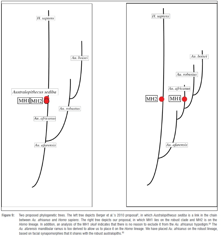
The phylogenetic scenario that Berger1 and Berger et al.2 propose, in which Au. sediba is a link in our evolutionary chain, ought to be ruled out because one of the Malapa mandibles is too derived to be positioned in the human lineage. Furthermore, one need not dismiss in limine the presence of Homo at sites contemporary with Malapa or even earlier, or question their geological age, as does Berger in his discussion of a 2.4-million-year-old Homo specimen par excellence, A.L. 666, from the Afar in Ethiopia.1 Malapa itself clearly contains a generalised specimen, and Homo, given the size and shape of the fossil, is the only candidate. The split between Homo and the robust clade must have occurred earlier than the occupation of Malapa.
Hence, Au. sediba is not a species that 'shares more derived features with early Homo than any other australopith species', as claimed by Berger et al.2 In fact, their Au. sediba seems to represent a mixture of two hominin taxa, leading Berger et al. to refer to the new species as a transitional one (Figure 5). Moreover, by viewing Au. sediba as an ideal link between Au. africanus and H. habilis, they ignore all the synapomorphic features that the former already shares with the robust clade and to which attention was drawn many years ago (for example, by Aguirre30, Johanson and White31 and Rak32) (Figure 9).
All the australopiths on which the relevant ramal morphology is preserved (Au. afarensis; Au. africanus, including the Australopithecus specimen at Malapa; and certainly Au. robustus) are actually too derived to play the role of a H. sapiens ancestor. Given that Malapa already contains representatives of two hominin branches, one of which appears to be Homo, we must seek the latter's origin in geological layers that are earlier than those at Malapa, which are dated at approximately 2 million years before present.33 Support for such a scenario can be found in earlier Ethiopian fossils attributed to the genus Homo: A.L. 666, dated at 2.4 million years34, and LD 3501, dated at 2.8 million years35.
Acknowledgements
We thank J. Moggi-Cecchi, WH Kimbel and Erella Hovers for their comments on the manuscript. We also thank Lee Berger for his hospitality and for graciously providing YR. with access to the original fossils. This research did not receive any specific grant from funding agencies in the public, commercial, or not-for-profit sector.
Competing interests
We declare that there are no competing interests.
Authors' contribution
Y.R.: Study conception and design, acquisition of data, analysis and interpretation of data, drafting of manuscript. W.H.: Study conception and design, critical revision. E.B.: Study conception and design, critical revision. A.G.: Acquisition of data, analysis and interpretation of data. E.G.: Analysis and interpretation of data, drafting of manuscript.
References
1. Berger LR. Australopithecus sediba and the earliest origins of the genus Homo. J Anthropol Sci. 2012;90:117-131. https://doi.org/10.4436/jass.90009 [ Links ]
2. Berger LR, De Ruiter DJ, Churchill SE, Schmid P Carlson KJ, Dirks PHGM, et al. Australopithecus sediba: A new species of Homo-like australopith from South Africa. Science. 2010;328:195-204. https://doi.org/10.1126/science.1184944 [ Links ]
3. De Ruiter DJ, DeWitt TJ, Carlson KB, Brophy JK, Schroeder L, Ackermann RR, et al. Mandibular remains support taxonomic validity of Australopithecus sediba. Science. 2013;340:12329971-12329974. https://doi.org/10.1126/science.1232997 [ Links ]
4. Williams SA, DeSilva JM, De Ruiter DJ. Malapa at 10: Introduction to the special issue on Australopithecus sediba. PaleoAnthropology. 2018:49-55. https://doi.org/10.4207/PA.2018.ART111 [ Links ]
5. De Ruiter DJ, Carlson KB, Brophy JK, Churchill SE, Carlson KJ, Berger LR. The skull of Australopithecus sediba. PaleoAnthropology. 2018:56-155. https://doi.org/10.4207/PA.2018.ART112 [ Links ]
6. Rak Y, Ginzburg A, Geffen E. Does Homo neanderthalensis play a role in modern human ancestry? Am J Phys Anthropol. 2002;119:199-204. https://doi.org/10.1002/ajpa.10131 [ Links ]
7. Wolpoff MH, Frayer DW. Unique ramus anatomy for Neandertals? Am J Phys Anthropol. 2005;128:245-251. https://doi.org/10.1002/ajpa.10432 [ Links ]
8. Rak Y Ginzburg A, Geffen E. Gorilla-like anatomy on Australopithecus afarensis mandibles suggests Au. afarensis link to robust australopiths. Proc Natl Acad Sci USA. 2007;104:6568-6572. https://doi.org/10.1073/pnas.0606454104 [ Links ]
9. Ritzman TB, Terhune CE, Gunz P Robinson CA. Mandibular ramus shape of Australopithecus sediba suggests a single variable species. J Hum Evol. 2016;100:54-64. https://doi.org/10.1016/j.jhevol.2016.09.002 [ Links ]
10. Terhune CE, Robinson CA, Ritzman TB. Ontogenetic variation in the mandibular ramus of great apes and humans. J Morphol. 2014;275:661-677. https://doi.org/10.1002/jmor.20246 [ Links ]
11. Semaw S, Simpson SW, Quade J, Renne PR, Butler RF, McIntosh WC, et al. Early Pliocene hominids from Gona, Ethiopia. Nature. 2005;433:301-305. https://doi.org/10.1038/nature03177 [ Links ]
12. Weidenreich F. The mandibles of Sinanthropus pekinensis: A comparative study. Palaeontology. 1936;Sinica D 7, 1e132. [ Links ]
13. Kimbel WH, Rak Y. Australopithecus sediba and the emergence of Homo: Questionable evidence from the cranium of the juvenile holotype MH 1. J Hum Evol. 2017;107:94-106. https://doi.org/10.1016/j.jhevol.2017.03.011 [ Links ]
14. Been E, Rak Y. The lumbar spine of Australopithecus sediba indicates two hominid taxa. In: Paleoanthropology Society Meeting Abstracts; 2014 April 8-9; Calgary, Canada. PaleoAnthropology. 2014:A2. https://doi.org/10.4207/PA.2014.ABS12 [ Links ]
15. Rak Y, Been E. Two hominid taxa at Malapa: The mandibular evidence. In: Paleoanthropology Society Meeting Abstracts; 2014 April 8-9; Calgary, Canada. PaleoAnthropology. 2014:A20. https://doi.org/10.4207/PA.2014.ABS12 [ Links ]
16. Rak Y Been E. What do we really know about the origin of humans? (Abstract). In: Proceedings of the 6th Annual Meeting of the European Society for the Study of Human Evolution (PESHE); 2016 September 14-17; Madrid, Spain. p. 199. [ Links ]
17. Du A, Alemseged Z. Temporal evidence shows Australopithecus sediba is unlikely to be the ancestor of Homo. Sci Adv. 2019;5(5), eaav9038. https://doi.org/10.1126/sciadv.aav9038 [ Links ]
18. Robinson JT. Telanthropus and its phylogenetic significance. Am J Phys Anthropol. 1953;11:445-502. https://doi.org/10.1002/ajpa.1330110402 [ Links ]
19. Clarke RJ, Howell FC. Affinities of the Swartkrans 847 hominid cranium. Am J Phys Anthropol. 1972;37:319-335. https://doi.org/10.1002/ajpa.1330370302 [ Links ]
20. Hughes AR, Tobias PV. A fossil skull probably of the genus Homo from Sterkfontein, Transvaal. Nature. 1977;265:310-312. https://doi.org/10.1038/265310a0 [ Links ]
21. Clarke RJ. The cranium of the Swartkrans hominid, SK 847, and its relevance to human origins [PhD dissertation]. Johannesburg: University of the Witwatersrand; 1977. [ Links ]
22. Dean MC, Wood BA. Basicranial anatomy of Plio-Pleistocene hominids from East and South Africa. Am J Phys Anthropol .1982;59:157-174. https://doi.org/10.1002/ajpa.1330590206 [ Links ]
23. Kimbel WH, Rak Y The importance of species taxa in paleoanthropology and an argument for the phylogenetic concept of the species category. In: Kimbel WH, Martin LB, editors. Species, species concepts, and primate evolution. New York: Plenum; 1993. p. 461-484. https://doi.org/10.1007/978-1-4899-3745-2_18 [ Links ]
24. Kimbel, WH, Rak Y Johanson DC. The skull of Australopithecus afarensis. Oxford: Oxford University Press; 2004. https://doi.org/10.1093/oso/9780195157062.001.0001 [ Links ]
25. Moggi-Cecchi J, Tobias PV Beynon AD. The mixed dentition and associated skull fragments of a juvenile fossil hominid from Sterkfontein, South Africa. Am J Phys Anthropol. 1998;106:425-465. https://doi.org/10.1002/(SICI)1096-8644(199808)106:4<425::AID-AJPA2>3.0.CO;2-I [ Links ]
26. Keyser AW, Menter CG, Moggi-Cecchi J, Pickering TR, Berger LR. Drimolen: A new hominid-bearing site in Gauteng, South Africa. S Afr J Sci. 2000;96:193-197. [ Links ]
27. Moggi-Cecchi J, Menter CG, Boccone S, Keyser A. Early hominin dental remains from the Plio-Pleistocene site of Drimolen, South Africa. J Hum Evol. 2010;58:374-105. https://doi.org/10.1016/j.jhevol.2010.01.006 [ Links ]
28. Herries IR, Curnoe D, Adams JW. A multi-disciplinary seriation of early Homo and Paranthropus bearing palaeocaves in southern Africa. Quat Int. 2009;202:14-28. https://doi.org/10.1016/j.quaint.2008.05.017 [ Links ]
29. Herries IR, Shaw J. Palaeomagnetic analysis of the Sterkfontein palaeocave deposits: Implications for the age of the hominin fossils and stone tool industries. J Hum Evol. 2011;523-539. https://doi.org/10.1016/j.jhevol.2010.09.001 [ Links ]
30. Aguirre E. Identificación de 'Paranthropus' en Makapansgat [Identification of 'Paranthropus' in Makapansgat]. Crónica del XI Congreso Nacional de Arqueología (Mérida). 1969;98-124. Spanish. [ Links ]
31. Johanson DC, White TD. A systematic assessment of early South African hominids. Science. 1979;201:321-330. https://doi.org/10.1126/science.104384 [ Links ]
32. Rak Y The australopithecine face. New York: Academic Press; 1983. https://doi.org/10.1016/B978-0-12-576280-9.50006-7 [ Links ]
33. Pickering R, Dirks PHGM, Jinnah Z, De Ruiter DJ, Churchill SE, Herries AIR, et al. Australopithecus sediba at 1.977 Ma and implications for the origins of the genus Homo. Science. 2011;333:1421-1423. https://doi.org/10.1126/science.1203697 [ Links ]
34. Kimbel WH, Johanson DC, Rak Y Systematic assessment of a maxilla of Homo from Hadar, Ethiopia. Am J Phys Anthropol. 1997;103:235-262. https://doi.org/10.1002/(SICI)1096-8644(199706)103:2<235::AID-AJPA8>3.0.CO;2-S [ Links ]
35. Villmoare B, Kimbel WH, Seyoum C, Campisano CJ, DiMaggio EN, Rowan J, et al. Early Homo at 2.8 Ma from Ledi Geraru, Afar, Ethiopia. Science. 2015;347:1352-1355. https://doi.org/10.1126/science.aaa1343 [ Links ]
36. Walker A, Leakey R, editors. The Nariokotome Homo erectus skeleton. Berlin: Springer; 1993. https://doi.org/10.1007/978-3-662-10382-1 [ Links ]
 Correspondence:
Ella Been
Email: beenella1@gmail.com
Received: 08 Aug. 2020
Revised: 04 Nov. 2020
Accepted: 09 Nov. 2020
Published: 28 May 2021
Supplementary Data
The supplementary data is available in pdf: [Supplementary Data]
Editor: Margaret Avery
Funding: None

