


Services on Demand
Article
 English (pdf)
 Article in xml format
 Article references
Indicators

Related links
-
 Cited by Google
 -
 Similars in Google
Share

 Permalink
South African Journal of Science
On-line version ISSN 1996-7489
Print version ISSN 0038-2353
S. Afr. j. sci. vol.113 n.11-12 Pretoria Nov./Dec. 2017
http://dx.doi.org/10.17159/sajs.2017/20170057
RESEARCH ARTICLE
Finding fossils in Malapa breccia - medical CT scanning or micro-CT scanning?
Jacqueline S. SmilgI, II, III
IDepartment of Radiation Sciences, University of the Witwatersrand, Johannesburg, South Africa
IIEvolutionary Studies Institute, School of Geosciences, University of the Witwatersrand, Johannesburg, South Africa
IIIDepartment of Radiology, Charlotte Maxeke Johannesburg Academic Hospital, Johannesburg, South Africa
ABSTRACT
Computed tomography (CT) imaging of fossils has revolutionised the field of palaeontology, allowing researchers to gain a better understanding of fossil anatomy, preservation and conservation. Micro focus X-ray computed tomography (µXCT) has been far more extensively used for these purposes than medical CT (XCT) - mostly because of the exquisite detail that the µXCT scanning modality, using slices of micron thicknesses, can produce. High energy X-rays can potentially penetrate breccia more effectively than lower energy beams. This study demonstrates that lower energy beams produce superior images for prioritising breccia for preparation. Additionally, XCT scanners are numerous, accessible, fast and relatively cost-effective when compared to µXCT scanners - the latter are not freely available, scanning times are much longer and there are significant limitations on the size and weight of scannable objects. Breccia blocks from Malapa were scanned at high and lower energy and images were analysed for image quality, artifact and certainty of diagnosis. Results show that lower energy images are deemed superior to higher energy images for this particular application. This finding, taken together with the limitations associated with the use of µXCT for the imaging of the large breccia from Malapa, shows that XCT is the better modality for this specific application. The ability to choose fossil-bearing breccia, ahead of manual mechanical preparation by laboratory technicians, would allow for the optimal use of limited resources, manual preparatory skills as well as the curtailment of costs.
SIGNIFICANCE:
•'Blind' manual preparation of fossil-bearing breccia is a costly and time-consuming exercise - and often results in a low yield.
•The ability to triage fossil-bearing breccia ahead of manual preparation would allow for the optimal use of limited resources.
•Medical CT is better than micro-CT to triage breccia to allow for prioritisation of rocks for manual preparation.
Keywords: Australopithecus sediba; manual preparation; kVp; beam energy; image quality
Background
The use of computed tomography (CT) in the analysis of fossils has become common place1-4, although most of the CT work to date has been performed on prepared or partially prepared specimens3,5,6. The application of CT for matrix that potentially contains fossils has lagged behind these many advances in the visualisation and study of prepared fossils.
Micro CT (µXCT) scanners have become increasingly popular in the imaging of prepared fossils as a result of the combination of their ability to produce high-resolution images because of much smaller slice thicknesses (in the micron range), increased spatial resolution and more variable energy capabilities (especially in the higher energy ranges) than medical CT (XCT) scanners; in fact, µXCT dominates the current fossil literature as the X-ray modality of choice for virtual analysis of prepared fossils.7-10
The site of Malapa has yielded hundreds of breccia blocks, which have the potential to contain fossils of the hominin Australopithecus sediba. Traditionally, breccia has undergone manual preparation - with a single block taking months to adequately prepare, with no guarantee of obtaining any fossils. This process is costly - both in time and money - and is not a prudent use of scarce preparatory skills.
New ways have been sought to better deal with the many Malapa breccia requiring assessment. The idea of triage (prioritising for preparation) of breccia by imaging prior to manual preparation has been explored. Mass use of XCT to image Malapa breccia has been undertaken and the predicted findings from imaging have been compared to the actual post-preparation findings.11 Smilg and Berger11 demonstrated good correlation and suggested that XCT is a valuable imaging modality in the triage process. There is considerable advantage to being able to know the contents of a rock ahead of costly, time-consuming 'blind' manual preparation. Knowledge of breccia contents before manual preparation allows decisions to be made as to the most efficient use of resources, personnel and funds, in addition to allowing planning of the course of action desired for the preparation of fossil material.
The effectiveness of XCT with regard to image quality and object penetration was explored. The quality of an X-ray beam is the measurement of the penetrating power of the photons, which depends on the energy of the photons, the atomic number (Z), the density and the thickness of the object being scanned.12 Peak kilovoltage (kVp) governs the penetrating power of photons - the higher the kVp, the more the beam penetrates the object. Hence the question was posed as to whether higher kilovoltage beams would produce images from the breccia that would be better quality than those produced from lower kilovoltage beams. XCT is limited in its kilovoltage, with the maximum being 140 kV, whereas µXCT is capable of higher kilovoltage. As a consequence of the better penetrative ability of high energy X-rays,13 it may be expected that high energy scanning would be superior to lower energy scanning when it is applied to large, dense breccia blocks.
High and lower energy images of breccia were obtained to address this question of image quality from the Malapa breccia for triage purposes. µXCT was used to obtain the higher kilovoltage values that XCT was unable to generate. The spectrum of potential imaging characteristics available from µXCT was not researched for this particular application.
The image quality of lower energy and high energy images when applied to objects within breccia blocks was compared. Material for this experiment was selected from the fossil hominin bearing site of Malapa in the Cradle of Humankind World Heritage site because of the importance of fossils from this site, the many blocks collected from which there are no fossils visible on the surface, and the sheer number of blocks retrieved that require assessment.
The possibility of the use of neutron microtomography (NCT or n-µCT) or magnetic resonance imaging (MRI) was considered. NCT differs from X-rays in that neutrons can penetrate materials that are opaque to X-rays and organic material strongly attenuates these neutron beams. NCT may thus be appropriate for imaging organically preserved fossils as a complement to XCT or µXCT.14 NCT has shown promising results in being able to differentiate otherwise similar dense materials - a recent study has documented the use of NCT to view a fossil encased in breccia.15
However, NCT may induce hazardous levels of radioactivity in some geological materials which leaves these imaged samples radioactive and necessitates that the samples be isolated for a long time after the imaging. Additionally, there are currently no functioning NCT machines in South Africa, where this breccia triage is needed. NCT necessitates much longer scanning times when compared to XCT and is also limited to smaller fields of view.
MRI maps properties related to the chemical environment of certain elements, rather than mapping radiation attenuation. MRI has been considered to be poorly suited to geological material16 and at present does not compete with µXCT or NCT. MRI machines are not easily accessible to palaeontological researchers, scans are very expensive and scan times are long compared with those of XCT. For these reasons, both NCT and MRI were not considered suitable for the mass screening of breccia from Malapa.
Fossils may have the potential to contain ancient DNA and whilst the effect of radiation on living tissue has been well investigated, little has been done to research the impact that radiation may have on ancient DNA.17 Recent work has shown that radiation of fossils may have a detrimental effect on ancient DNA when the total surface dose exceeds 200 Gray, so these researchers recommended using as low a dose as possible when scanning fossils as well as using resolution no higher than necessary to achieve the desired outcome.18 The value of 200 Gray is far higher than any dose from a XCT or µXCT scan (8000 times higher than the highest dose for a medical CT scan).18
The Malapa site
The site of Malapa (site UW88)19 represents an unusually rich early hominin locality in Africa20-24, dating to 1.977±0.002 million years ago (Ma)25. The site contains remains of several individuals, all ascribed to A. sediba.20 These remains are found alongside an abundant, well-preserved fauna.24 It is postulated that this well-preserved material was accumulated during a seemingly rapid depositional event that occurred over a few days, weeks or months.26 The site of Malapa is located in the region of the Cradle of Humankind World Heritage Site, northwest of Johannesburg, South Africa. The locality is recognised as a de-roofed cave of at least 25 x 20 m, in an area where limited limestone mining had taken place, probably during the late 19th or early 20th century, almost certainly before Robert Broom began exploring the area in the mid-1930s.26
Materials and methods
A total of 15 breccia blocks from the site of Malapa were chosen to be scanned at differing kVp using both XCT and µXCT. Blocks were selected taking cognisance of the limitations on weight and volume presented by the University of the Witwatersrand's micro-CT scanner (a maximum permissible weight of 50 kg).
Lower energy scanning was performed on a Philips Brilliance 128 slice CT at Charlotte Maxeke Johannesburg Academic Hospital (South Africa), whilst high energy scanning was performed on a Nikon Metrology XTH 225/320 LC dual source industrial CT system at the University of the Witwatersrand. Both data sets were analysed on AMIRA 5.4.5 Ink.
This experiment was intended to simulate a real-world situation. After discussions with the principal scientists in charge of the Malapa project, it was decided that if mass screening of breccia was to be implemented, it would be impractical to adjust energy levels and scanning parameters for every block in order to optimise image quality, because of the desire to scan large numbers of rocks in a single session and the possibility that the scanning would be overseen by technicians not familiar with CT images. Hence the imaging parameters would have to be constant and pre-decided. The parameters selected are given in Table 1. A single energy parameter was selected for each system based upon initial tests that resulted in a good quality image from both machines. Reference was also made to prior work done with low energy scanning.11 Micro-CT images were reconstructed with micro-CT Pro v2.2 associated with the Nikon Metrology XTH 225/320 LC dual source industrial CT system.

Three objects, thought to represent potential fossils, were chosen from within each block for evaluation. The same object was identified on images from each modality and displayed on a two-dimensional image with the same orientation and alignment specific to the object under assessment. The viewing parameters (including greyscale setting and magnification) were fixed for all readers, with no manipulation allowed. Readers were supplied with identical static two-dimensional images. Qualitative visual assessment was done by each reader independently.
Each object was evaluated for overall image quality, certainty of diagnostic accuracy and imaging artifact. There is no objective definition of image quality - it is a matter of the observer's subjective judgement. CT artifacts can affect the quality of the images, sometimes to the point of making them diagnostically unusable. Artifact is any distortion or error in the image that is unrelated to the subject being studied.12 Artifact production can degrade the CT image and hinder interpretation, but modern CT machines are developed with built-in artifact reduction features, including filters, calibration correction, automatic tube current modulation and scanner software.27
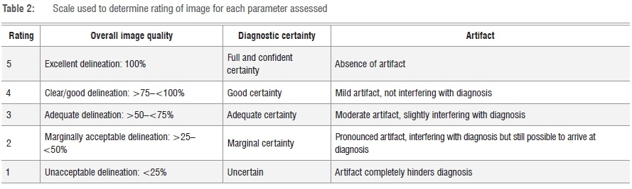
The images were rated according to a qualitative, visual five-point scale for each parameter given in Table 2.
The objective was to compare these parameters between lower kilovoltage and higher kilovoltage images. The scores from an individual reader for each object, per criterion, for each energy were compared, rather than analysing inter-reader variability. Results were evaluated by two diagnostic radiologists and three palaeontological scientists. These results are summarised in Appendices 1 and 2 in the supplementary material.
Results
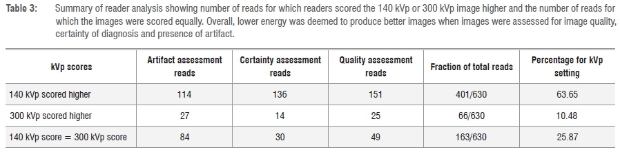
Results were assessed to determine which kVp choice scored higher (indicating better overall image) per reader, per object and per criterion, and whether the reader assigned the same score per criterion per object - the latter indicating an indeterminate result of neither kilovoltage setting producing a superior image (Table 3).
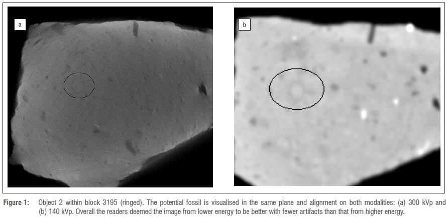
When considering the presence or absence of artifacts, the readers found that the lower energy scans produced better interpretive images in 114 of 225 reads (Figure 1). In 27 of 225 reads, high energy scanning was found to produce better interpretive images and 84 of 225 reads were indeterminate as to one scanning energy being better than the other.
Overall quality was deemed to be better in 151 of 225 reads for lower energy scans, whilst higher energy scans were deemed better in 25 of 225 reads (Figure 2). An indeterminate result, with both energy levels scoring equally, was obtained in 49 of 225 reads.

One reader did not rate for certainty of diagnosis for either modality because of a lack of confidence in cross-sectional identification. Results here identified lower energy scans as being superior in 136 of 180 reads (Figure 3). Higher energy scans were deemed better in 14 of 180 reads and results were indeterminate in 30 of 180 reads.

Overall percentages indicate that for all three criteria, the images obtained from lower energy scanning produced better, more diagnostic and more useful images with fewer artifacts in 63.65% of reads. Higher energy was deemed to deliver better images in 10.48% of reads and both modalities yielded equitable images in 25.87% of reads.
Discussion
In producing a CT image, both milliamp seconds (mAs) and kVp must be chosen to ensure sufficient delivery of X-rays to achieve acceptable image quality.28 The selection of kVp sets the energy of the X-rays reaching the object. The energy of an X-ray defines its penetrative ability13, as well as the expected attenuation of the ray as it passes through materials of different densities. High energy X-rays, when compared to lower energy rays, may penetrate certain objects more effectively but are less sensitive to changes in material density and composition.
Each object varies in density and atomic number - both of which impact the X-ray beams' attenuation. Lower energy settings decrease the overall signal and result in increased noise, which can degrade the image. Increasing technical factors, such as mAs or kVp, decrease image noise but also increase dose. Because of the inanimate and inorganic nature of the material being studied, dose was not as important as it would be with living material. As X-rays traverse an object they are attenuated by scattering and absorption. This attenuation is a result of three processes13: photoelectric absorption, Compton scattering and pair production. Medical scanners (which use lower X-ray energy up to 140 kV) have more photoelectric effect and lower Compton scatter, whereas in higher energy µXCTs, attenuation is greater from Compton scatter.13 This difference is important as it explains why low energy X-rays are more sensitive to differences in composition than higher energy ones. The photoelectric absorption is proportional to Z4-5 (atomic number of atom in the material to the power 4-5) whereas Compton absorption is proportional only to Z (atomic number).29 Thus two materials could be differentiated in lower energy CT, but at higher energies may be indistinguishable. Despite a better penetration ability, high energy scanning may not differentiate objects as well as lower energy scanning.
The quality of a CT image is affected by several factors; enhancing or suppressing any of these factors depends upon the imaging interests.12 CT image quality is dependent upon balancing these parameters to produce the best possible image for the object being scanned. CT parameters can be manipulated to either decrease or eliminate the adverse effects of these characteristics. Generally, there is a trade-off when CT parameters are manipulated.
Although CT scanners using high kVp (µXCT) could potentially be more effective at penetrating large breccia and can produce images with increased resolution as a result of much thinner slices, their disadvantages are their limitations in the size/weight of scannable objects, the long scan times needed as well as their limited availability to many scientists. In addition, the huge databases generated from the scan data necessitate specialised computer hardware and software for analysis, which are costly and not readily available to many researchers.
In contrast, medical CT machines are accessible as a consequence of their widespread use in clinical medical situations and are found in most hospitals and radiology practices, have a weight restriction of up to 200 kg and offer fast, reliable imaging and greater throughput of scanned objects. The databases are manageable on basic modern computers and data can be assessed with easily available (often free) software packages. Finally, at present, medical CT scanning, on a case by case basis, is typically significantly cheaper than micro-CT scanning, largely because of differences in initial machine cost as well as the presence of a greater number of medical CT machines in the community.
To be effective for the screening of Malapa breccia, the imaging modality used should:
• be cost effective, both in time and money
• be quick
• be repeatable
• use predetermined parameters that would negate the need for specialist attendance at scanning sessions
• be regularly readily accessible
• be able to accommodate large pieces of breccia.
The µXCT machines in the Gauteng area that are available to the Malapa project are limited in that:
• they have prolonged application and waiting times
•t hey are costly
• scanning is time consuming (±6 h/block)
• there are significant limitations in the weight and dimensions of objects to be scanned
• they generate large databases which make assessment time consuming and post processing limited to specialised programmes.
Conclusions
Fossils, particularly of hominins, are highly sought-after objects in the search for human origins, but 'blind' manual mechanical preparation of fossil-bearing breccia is a costly and time-consuming exercise - often with a low yield. The ability to triage breccia ahead of manual preparation would allow for the optimal use of limited resources, manual preparatory skills as well as the curtailment of costs.
Microcomputed tomography is well established as an imaging modality for the imaging of prepared fossils. Because of its potential to penetrate breccia more effectively by using high energy X-rays, it might be considered the modality of choice for breccia triage.
This study shows that in the application of breccia triage, the penetrating ability of a lower energy beam is not detrimental to the outcome.
Given the many other limitations of µXCT faced by the Malapa team, it is of significant advantage to researchers interested in a high throughput of potentially low value material, in search of high value material (as is the case with using CT scanners to triage potentially fossil-bearing blocks), that the differential penetration of lower and higher energy beams does not have a significant impact on image quality and that XCT is overall the better choice over µXCT for this application. The study does not seek to generalise the contribution of XCT and acknowledges that for other applications, µXCT may be the modality of choice. But for the purpose of fossil identification within large rocks and for breccia triage for the breccia originating from the Malapa site, it has been shown that XCT is superior to µXCT for this particular palaeontological application. Application of these findings can now be expanded to breccia from other fossil sites.
Additionally, as the effects of radiation on ancient DNA are still not clear, consideration should be given to the accumulative dose of radiation to which an individual fossil is exposed. It is recommended that the lowest possible dose necessary to achieve the desired outcome is used, as well as the lowest resolution possible to achieve the desired result. The dose increases at about the square power of the increase of resolution.18 Thus, another reason to advocate the application of lower energy scanning over higher energy scanning for breccia triage is that the potential and ability to access ancient DNA from fossil specimens still needs research and elucidation.
Use of medical CT scanning of fossil-bearing breccia is thus an alternative to random block preparation. In order to maximise the use of limited resources and manual preparatory skills, as well as to curtail costs, this research shows that, prior to manual preparation, blocks should undergo scanning with medical CT scanners and virtual assessment of contents should be undertaken to allow for prioritisation of rocks for manual preparation.
Triage of fossil-bearing breccia using medical CT scanners will shorten the time from breccia removal from the field to the retrieval of relevant fossil specimens for interrogation, publication and dissemination of their information.
Acknowledgements
L. Berger, B. de Klerk, K. Jakata, J. Mukanku, E. Odes, P. Randolph-Quinney, F. Thackeray and R. van der Merwe of the Evolutionary Studies Institute at the University of the Witwatersrand (Johannesburg, South Africa) and National Palaeosciences Centre of Excellence are acknowledged for their assistance. J. Haberfeld and Q. Letsoalo of the Department of Diagnostic Radiology at Charlotte Maxeke Johannesburg Academic Hospital (Johannesburg, South Africa) are acknowledged for their assistance. The Virtual Imaging in Palaeontology Laboratory, the Microfocus X-ray Computed Tomography Facility at the Evolutionary Studies Institute and Charlotte Maxeke Johannesburg Academic Hospital are acknowledged for providing access to their facilities.
References
1. Sutton MD, Rahman IA, Garwood RJ. Techniques for virtual palaeontology. London: John Wiley & Sons; 2013. https://doi.org/10.1002/9781118591192 [ Links ]
2. Lautenschlager S. Reconstructing the past. Methods and techniques for the digital restoration of fossils. R Soc Open Sci. 2016;3(10), Art. #160342, 18 pages. https://doi.org/10.1098/rsos.160342 [ Links ]
3. Balzeau A, Crevecoeur I, Rougier H, Froment A, Gilissen E, Grimaud-Herv D, et al. Applications of imaging methodologies to paleoanthropology: Beneficial results relating to the preservation, management and development of collections. CRPalevol. 2010;9(6):265-275. https://doi.org/10.1016/j.crpv.2010.07.006 [ Links ]
4. Wu X, Schepartz LA. Application of computed tomography in palaeoanthropological research. Prog Nat Sci. 2009;19:913-921.https://doi.org/10.1016/j.pnsc.2008.10.009 [ Links ]
5. Odes EJ, Randolph-Quinney PS, Steyn M, Throckmorton Z, Smilg JS, Zipfel B, et al. Earliest hominin cancer: 1.7-million-year-old osteosarcoma from Swartkrans Cave, South Africa. S Afr J Sci. 2016;112(7/8), Art. #2015-0471, 5 pages. https://doi.org/10.17159/sajs.2016/20150471 [ Links ]
6. Randolph-Quinney PS, Williams SA, Steyn M, Meyer MR, Smilg JS, Churchill SE, et al. Osteogenic tumour in Australopithecus sediba: Earliest hominin evidence for neoplastic disease. S Afr J Sci. 2016;112(7/8), Art. #2015-0470, 7 pages. https://doi.org/10.17159/sajs.2016/20150470 [ Links ]
7. Biolaurus. 8 Spectacular examples of micro-CT being used to analyse fossils [homepage on the Internet]. No date [updated 2017; [ Links ] cited 2017 Jan 25]. Available from: http://biolaurus.com/8-spectacular-examples-micro-ct-used-analyze-fossils/
8. Liu Y, Scholtz G, Hou X. When a 520 million-year-old Chengjiang fossil meets a modern micro-CT - a case study. Sci Rep. 2015;5, Art. #12802, 8 pages. https://doi.org/10.1038/srep12802 [ Links ]
9. Cunningham JA, Rahman IA, Lautenschlager S, Rayfield EJ, Donoghue PJC. A virtual world of paleontology. Trends Ecol Evol. 2014;29(6):347-357. https://doi.org/10.1016/j.tree.2014.04.004 [ Links ]
10. Abel RL, Laurini CR, Richter MA. A palaeobiologist's guide to 'virtual' micro-CT preparation. Palaeontol Electron. 2012;15(2):496-500. [ Links ]
11. Smilg JS, Berger LR. Discovering hominins - Application of medical computed tomography (CT) to fossil-bearing rocks from the site of Malapa, South Africa. PLoS ONE. 2015;10(12), e0145340, 19 pages. https://doi.org/10.1371/journal.pone.0145340. [ Links ]
12. Reddinger W. CT image quality [document on the Internet]. c1998 [cited 2017 Feb 22]. [ Links ] Available from: https://www.coursehero.com/file/9938632/CT-Image-Quality/
13. Ketcham RA, Carlson WD. Acquisition, optimization and interpretation of X-ray computed tomographic imaging: Application to the geosciences. Comput GeoSci. 2001;27(4):381-400. https://doi.org/10.1016/S0098-3004(00)00116-3 [ Links ]
14. Winkler B. Applications of neutron radiography and neutron tomography. Rev Mineral Geochem. 2006;63:459-471. https://doi.org/10.2138/rmg.2006.63.17 [ Links ]
15. Beaudet A, Braga J, De Beer F, Schillinger B, Steininger C, Vodopivec V, et al. Neutron microtomography-based virtual extraction and analysis of a cercopithecoid partial cranium (STS 1039) embedded in a breccia fragment from Sterkfontein member 4 (South Africa). Am J Phys Anthropol. 2016;159(4):737-745. https://doi.org/10.2/ajpa.22916 [ Links ]
16. Sutton MD. Tomographic techniques for the study of exceptionally preserved fossils. Proc R Soc B Biol Sci. 2008;275(1643):1587-1593. https://doi.org/10.1098/rspb.2008.0263 [ Links ]
17. Grieshaber BM, Osborne DL, Doubleday AF, Kaestle FA. A pilot study into the effects of X-ray and computed tomography exposure on the amplification of DNA from bone. J Archaeol Sci. 2008;35(3):681-687. https://doi.org/10.1016/j.jas.2007.06.001 [ Links ]
18. Immel A, Le Cabec A, Bonazzi M, Herbig A, Temming H, Schuenemann VJ, et al. Effect of X-ray irradiation on ancient DNA in sub-fossil bones - Guidelines for safe X-ray imaging. Sci Rep. 2016;6, Art. #32969, 14 pages. https://doi.org/10.1038/srep32969 [ Links ]
19. Zipfel B, Berger LR. New cenozoic fossil-bearing site abbreviations for collections in the University of the Witwatersrand. Palaeont Afr. 2009;44:77-81. [ Links ]
20. Berger LR, De Ruiter DJ, Churchill SE, Schmid P, Carlson KJ, Dirks PHGM, et al. Australopithecus sediba: A new species of Homo-like australopith from South Africa. Science. 2010;328(5975):195-204. https://doi.org/10.1126/science.1184944 [ Links ]
21. Kibii JM, Churchill SE, Schmid P, Carlson KJ, Reed ND, De Ruiter DJ, et al. A new partial pelvis of Australopithecus sediba. Science. 2011;333(6048):1407-1411. https://doi.org/10.1126/science.1202521 [ Links ]
22. Zipfel B, De Silva JM, Kidd RS, Carlson KJ, Churchill SE, Berger LR. The foot and ankle of Australopithecus sediba. Science. 2011;333(6048):1417-1420 https://doi.org/10.1126/science.1202703 [ Links ]
23. Kivell TL, Kibii JM, Churchill SE, Schmid P, Berger LR. Australopithecus sediba hand demonstrates mosaic evolution of locomotor and manipulative abilities. Science. 2011;333(6048):1411-1417. https://doi.org/10.1126/science.1202625 [ Links ]
24. Kuhn BF, Werdelin L, Hartstone-Rose A, Lacruz RS, Berger LR. Carnivoran remains from the Malapa hominin site, South Africa. PLoS ONE. 2011;6(11), e26940, 11 pages. https://doi.org/10.1371/journal.pone.0026940 [ Links ]
25. Pickering R, Dirks PHGM, Jinnah Z, De Ruiter DJ, Churchill SE, Herries AIR, et al. Australopithecus sediba at 1.977 Ma and implications for the origins of the genus Homo. Science. 2011;333(6048):1421-1423. https://doi.org/10.1126/science.1203697 [ Links ]
26. Dirks PHGM, Kibii JM, Kuhn BF, Steininger C, Churchill SE, Kramers JD, et al. Geological setting and age of Australopithecus sediba from southern Africa. Science. 2010;328(5975):205-208. https://doi.org/10.1126/science.1184950 [ Links ]
27. Barret JF, Keat N. Artifact in CT: Recognition and avoidance. Radiographics. 2004;24(6):1679-1691. https://doi.org/10.1148/rg.246045065 [ Links ]
28. Courtney A, Coursey MD, Donald P, Frusch MD. CT and radiation: What the radiologist should know. Appl Radiol. 2008;37(3):22-29. [ Links ]
29. Van Grieken RE, Markowicz AA. Handbook of X-ray spectrometry: Methods and techniques. New York: Marcel Dekker; 1993. [ Links ]
 Correspondence:
Jacqueline Smilg
Email: jsmilg@yahoo.com
Received: 22 Feb. 2017
Revised: 19 Apr. 2017
Accepted: 10 July 2017
FUNDING: Malapa Project, Evolutionary Studies Institute, University of the Witwatersrand (grant no. 98839)
Supplementary Data
The supplementary data is available in pdf: [Supplementary data]

