


Servicios Personalizados
Articulo
 Inglés (pdf)
 Articulo en XML
 Referencias del artículo
Indicadores

Links relacionados
-
 Citado por Google
 -
 Similares en Google
Compartir

 Permalink
South African Journal of Science
versión On-line ISSN 1996-7489
versión impresa ISSN 0038-2353
S. Afr. j. sci. vol.112 no.7-8 Pretoria 2016
http://dx.doi.org/10.17159/sajs.2016/a0167
SCIENTIFIC CORRESPONDENCE
The possibility of lichen growth on bones of Homo naledi: Were they exposed to light?
J. Francis Thackeray
Evolutionary Studies Institute, School of Geosciences, University of the Witwatersrand, Johannesburg, South Africa
Keywords: Homo naledi; manganese; lichen; taphonomy
Dirks et al1 report on the taphonomy of bones from the Dinaledi Chamber in the Rising Star cave complex, situated in the Cradle of Humankind in South Africa. Fossils of Homo naledi have been recovered from this chamber.2 An age estimate for the Homo naledi fossils, based on morphometric analyses of crania, is in the order of 2 million years,3 although it is recognised that a younger date may apply if morphological patterns persisted for an unknown period of time.
Dirks et al.1 state that 'some bones and teeth are dotted with black iron-manganese oxy-hydroxide deposits and coatings'. The question arises as to what factors contributed to the 'dotted' or spotty distribution of manganese oxy-hydroxide on bone surfaces. Abiotic geological factors may account for continuous (matted) deposition of manganese oxy-hydroxide coatings in some cases, but why should these be dotted or spotted in the instance of many bones from the Dinaledi Chamber - or from other nearby sites, such as Sterkfontein? To try to answer this question, it is appropriate to refer to observations by Thackeray et al.4 in which we noted, firstly, that in the Cradle of Humankind, lichen can grow on certain substrates (including bone or rock) with a dotted or spotted distribution. Secondly, the spotted distribution of lichen is sometimes associated with dotted distributions of manganese oxy-hydroxide on the same surfaces.4 The source of manganese would include dolomite and chert, 2 billion years old, relating to the shallow saline sea that existed at that time.
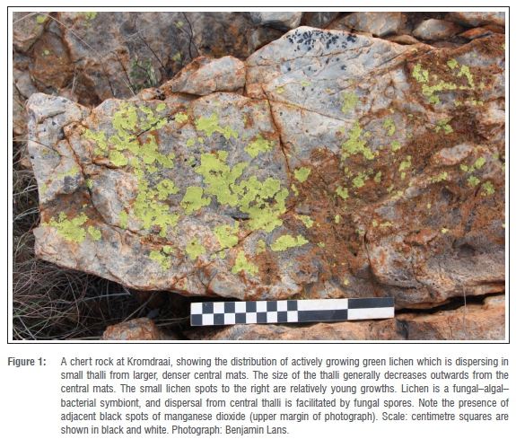
Lichen are symbionts5 that include a fungus (a mycobiont) and a photosynthetic partner (a photobiont). This partner could be a cyanobacterium or a green alga. The growth of the symbiont results in a lichen body called a thallus. On substrates such as chert or bone, under suitable micro-environments the spottiness of lichen may relate to the dispersal of fungal spores. An example of lichen growth is shown on chert at Kromdraai in the Cradle of Humankind (Figure 1). In this case, small thalli of lichen - possibly Rhizocarpon - grow adjacent to older central thalli in which the lichens form continuous mats. The size of lichen thalli generally decreases outwards from the central thalli. Very small spots at the edge of lichen growth are relatively young growths of lichen.
In the Cradle of Humankind, lichen can grow not only on chert but also on dolomite. However, weathering of the latter substrate may inhibit extensive lichen growth, which certainly does occur on more durable chert as well as on bone.
As mentioned above, lichens are algal-fungal-bacterial symbionts. Most bacteria include a manganese-containing superoxide dismutase or Mn-SOD6, and the association with manganese is of special interest in the study of lichen in the Cradle of Humankind4. Elsewhere, Pentecost et al.7 reported lichen on a substrate rich in manganese. Geomicrobial transformation of manganese compounds by fungi can also occur. Hansen et al.8 demonstrate that ascomycetes are capable of oxidising manganese. Beckett et al.9 indicate the existence of redox cycling in lichens, and refer to a process which 'will increase the photosynthetic capacity of thalli during winter when light intensities are low'. This may apply also to micro-environments in dolomitic caves under limited light conditions.
In Figure 1, lichen is shown in close association with spots of manganese oxy-hydroxide on chert in the Cradle of Humankind. Figure 2 shows a chert rock at the same site (Kromdraai), which is more extensively coated by spots of manganese oxy-hydroxide, apparently dispersing (with diminishing size) from a central area. The central area is potentially analogous to a continuous mat of a central thallus of lichen, as shown in Figure 1.
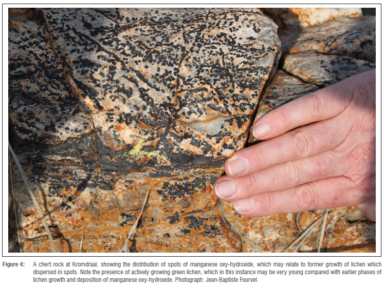
In the case of durable rocks such as chert at Kromdraai, lichen may have absorbed manganese from substrates.4 Because magnesium is potentially toxic, the lichen may then have subsequently deposited and fixed the spots or dots of manganese dioxide on surfaces such as rock or bone.4 Episodes of lichen growth could have occurred over long periods of time, such that the same rock might show relatively old spots of manganese oxy-hydroxide as well as relatively younger lichen thalli. Good examples of the association between modern lichen and manganese oxy-hydroxide at Kromdraai are shown in Figure 3 and Figure 4. These images record the spotty distribution of modern lichen, and the spotty or dotted distribution of black manganese oxy-hydroxide that may relate to former (spotty) growth of lichen.
Hominid cranial specimens from Sterkfontein, such as Stw 505 (Australopithecus africanus) and Sts 71 (attributed to A. prometheus), have small dots or spots of manganese oxy-hydroxide on bone surfaces, even within the inner cranial wall of these skulls. These spots of manganese oxy-hydroxide may represent areas where lichens were able to grow in a partially sunlit micro-environment and for a relatively short time, before the deposition of sediments that covered the crania, thereby halting lichen growth through the absence of light.4 Figure 5 shows such spots on specimen Stw 505. Because this relates partly to a biological process, and manganese has a radioactive isotope that decays with a half-life in the order of 1 million years, there is exciting potential to use this theory or process to date fossils in the Cradle of Humankind.4
Modern dolomitic cave systems in the Cradle of Humankind include the Wonder Cave, which is situated within 10 km of the Rising Star cave complex. One encounters the following set of circumstances as one walks from the entrance of the cavern into the darkness of the dolomitic solution cavity:
1. Where there is intense light and heat on dry exposed surfaces outside the cave, there is little or no lichen growth.
2. As one walks into moister and slightly darker regions of the cave, there is an area where light and moisture appear to be optimal for the present lichen colonists, and lichen growth is extensive on cave walls if not also on bone.
3. As one enters the darkness of the cave where there is little light, there is a decrease in the mean size of lichen thalli until there is no lichen growth at all; this is the case even where there may be some moisture as a result of water dripping through the phreatic maze of the dolomitic cave system.
As mentioned above, the growth of lichen includes a photobiont (the photosynthetic partner to the fungal mycobiont), and this photobiont is dependent on at least some degree of exposure to light for growth to occur. It has been proposed that the Dinaledi Chamber in prehistory could be accessed only by means of very narrow and circuitous passages, in complete darkness.1 The question arises whether the Dinaledi Chamber had an additional entrance. In response to this question, I hypothesise that in the case of the Dinaledi Chamber, bones such as tibia shaft specimen UW 101-1070 (Figure 6) may show spotty or dotted coatings of manganese oxy-hydroxide, of the kind reported by Dirks et al.1, at least in part because of the growth of lichen.
As mentioned, lichen are bacterial-fungal-algal symbionts, including a photobiont. The growth of lichen on bone surfaces, even for a limited period of time, may have occurred in subdued but essential lighting. If correct, this theory may mean there was at some time a second entrance to the Dinaledi Chamber in the phreatic maze cave system. Such an entrance might have allowed penetration of at least some light into the cave, facilitating the growth of lichen and subsequent deposition of manganese oxy-hydroxide on bones. It is further hypothesised that such an entrance, if it existed, was temporary. A rock fall in the phreatic maze cave system may subsequently have sealed the entrance at some stage in the dolomitic solution cavity, whereupon lichen growth would have terminated, in darkness.
If there was more than one entrance into the Dinaledi Chamber, as suggested here and by Val11, the 'intentional depositional model' of Dirks et al.1 would need to be re-assessed. Such re-assessment would need to account for the occurrence of 15 individuals of Homo naledi in the Dinaledi Chamber.1 More recently, Dirks et al.12 have responded to comments by Val11, and those comments are relevant to this discussion. However, it should be noted that the age distribution of the 15 individuals seems to correspond to a family group. Perhaps this group died as a result of a crisis near an entrance to the Dinaledi Chamber, and/or a roof collapse in a phreatic maze, such that the group was trapped.
It is certainly accepted that abiotic factors may account for deposition of manganese oxy-hydroxide on bone surfaces. However, the question is why manganese oxy-hydroxide should be dotted or spotted, as in the case of bones from Dinaledi Chamber1 or other nearby sites such as Sterkfontein4. The answer may lie in a biological process associated with lichen requiring light, rather than an entirely abiotic (geological) process whereby manganese oxy-hydroxide may have been distributed as a continuous mat.
Dominguez-Rodrigo and Barba12 conducted experimental work on modern bone to investigate taphonomic processes associated with fungi and bacteria. They reported that 'bone surfaces are affected by dark circular marks caused by mycelial fungi, with bacteria playing a decidedly smaller role'.12 They also noted that the excretions deposited on bone included 'organic and inorganic acids, enzymes, pigments, and toxins'.12 However, their investigation did not relate to lichen and manganese in the context of a bacterial-fungal-algal symbiotic relationship as discussed here. Long-term experimental work on lichen on bone substrates in the Cradle of Humankind is planned for a 10-year period, in and around cave environments.
The concepts advocated here can be assessed in the context of empirical data already available. The mean size of manganese oxy-hydroxide spots on a selection of bones from the Dinaledi Chamber is 1.11 ± 0.31 mm, for n = 72 measurements (on mandibular and cranial bone as well as tibiae, a femur and a humerus). This mean size does not differ significantly from that of manganese oxy-hydroxide spots on the inner cranial bone of Stw 505 (1.32 ± 0.42 mm; n = 25 measurements). The smaller spot size for H. naledi specimens may perhaps relate to a lower degree of light exposure that allowed at least temporary growth of lichen on bone surfaces.
Apart from the possibility of lichen growth, it is pertinent to mention that Dirks et al.1 reported taphonomic alteration to bones from the Dinaledi Chamber in the form of damage by snails and beetles. Snails and beetles may be dependent to some extent on the presence of leaf litter, and leaves in turn depend on light. Therefore, such damage may also indicate there was at least a temporary opening into the Dinaledi Chamber, in addition to the opening accessed by modern explorers. The Rising Star cave complex is a phreatic maze where episodic openings and rock falls can be common within long periods of time.
Acknowledgements
This research was supported by the National Research Foundation and the Andrew W. Mellon Foundation and the Centre of Excellence for the Palaeosciences. I am grateful to Benjamin Lans and Jean-Baptiste Fourvel for the photographs; to Bernhard Zipfel and the Access Committee for access to H. naledi specimens to take measurements of manganese oxy-hydroxide spots, based on photographs with a scale; to Don Cowan, Richard Beckett, Mike Wingfield, Sarah Elton and anonymous commentators for helpful recommendations on an earlier version of this manuscript; and to Laurent Bruxelles for useful discussions on phreatic maze cave systems.
References
1. Dirks PHGM, Berger LR, Roberts EM, Kramers JD, Hawks J, Randolph-Quinney PS, et al. Geological and taphonomic context for the new hominin species Homo naledi from the Dinaledi Chamber, South Africa. eLife 2015;4:e09561 http://dx.doi.org/10.7554/eLife.09561 [ Links ]
2. Berger LR, Hawks J, de Ruiter DJ, Churchill SE, Schmid P Delezene LK, et al. Homo naledi, a new species of the genus Homo from the Dinaledi Chamber, South Africa. eLife 2015;4:e09560 http://dx.doi.org/10.7554/eLife.09560 [ Links ]
3. Thackeray JF. Estimating the age and affinities of Homo naledi. SA J Sci. 2015;111:3-4. http://dx.doi.org/10.17159/sajs.2015/a0124 [ Links ]
4. Thackeray JF, Sénégas F, Laden G. Manganese dioxide staining on hominid crania from Sterkfontein and Swartkrans: Possible associations with lichen. SA J Sci. 2005;101:28. http://reference.sabinet.co.za/webx/access/electronic_journals/sajsci/sajsci_ v101_n1_a4.pdf [ Links ]
5. Purvis W. Lichens. The Natural History Museum, London; 2000. [ Links ]
6. Law N, Caudle M, Pecoraro V. Manganese redox enzymes and model systems: Properties, structures, and reactivity. Adv Inorg Chem 1998;46:305-440. http://dx.doi.org/10.1016/S0898-8838(08)60152-X [ Links ]
7. Pentecost A, Spiro B, Williamson B. A note on the relationship between some saxicolous lichens and manganese ore in North Wales UK. Geomicrobiol J. 2010;27:349-352. http://dx.doi.org/10.1080/01490451003707668 [ Links ]
8. Hansel CM, Zeiner CA, Santelli CM, Webb SM. Mn(II) oxidation by an ascomycete fungus is linked to superoxide production during asexual reproduction. Proc Natl Acad Sci USA. 2012;109(31):12621-12625. http://dx.doi.org/10.1073/pnas.1203885109 [ Links ]
9. Beckett RP Ntombela N, Scott E, Gurjanov OP Minibeyeva FV Liers C. Role of laccases and peroxidases in saprotrophic activities in the lichen Usnea undulata. Fungal Ecol. 2015;14,71-78. http://dx.doi.org/10.1016/j.funeco.2014.12.004 [ Links ]
10. Val A. Deliberate body disposal by hominins in the Dinaledi Chamber, Cradle of Humankind, South Africa? J Hum Evol. 2016;96:145-148. http://dx.doi.org/10.1016/j.jhevol.2016.02.004 [ Links ]
11. Dirks PHGM, Berger LR, Hawks J, Randolph-Quinney PS, Backwell LR, Roberts EM. Comment on 'Deliberate body disposal by hominins in the Dinaledi Chamber, Cradle of Humankind, South Africa?' J Hum Evol. 2016;96:149-153. http://dx.doi.org/10.1016/j.jhevol.2016.04.007 [ Links ]
12. Dominguez-Rodrigo M, Barba R. Five more arguments to invalidate the passive scavenging version of the carnivore-hominid-carnivore model: A reply to Blumenschine et al. (2007a). J Hum Evol. 2007;53:427-433. http://dx.doi.org/10.1016/j.jhevol.2007.05.010 [ Links ]
 Correspondence:
Francis Thackeray
Evolutionary Studies Institute, University of the Witwatersrand
Private Bag 3, Wits 2050
South Africa
Francis.thackeray@wits.ac.za

