


Servicios Personalizados
Articulo
 Inglés (pdf)
 Articulo en XML
 Referencias del artículo
Indicadores

Links relacionados
-
 Citado por Google
 -
 Similares en Google
Compartir

 Permalink
South African Journal of Science
versión On-line ISSN 1996-7489
versión impresa ISSN 0038-2353
S. Afr. j. sci. vol.112 no.3-4 Pretoria mar./abr. 2016
http://dx.doi.org/10.17159/sajs.2016/20150152
RESEARCH ARTICLE
Antimutagenic and antioxidant effects of a South African traditional formulation used as an immune booster
Mlungisi NgcoboI; Nceba GqaleniII; Victor NdlovuIII
ITraditional Medicine Laboratory, School of Nursing and Public Health, College of Health Sciences, University of KwaZulu Natal, Durban, South Africa
IIDepartment of Public Management and Economics, Durban University of Technology, Durban, South Africa
IIIMakhonya Natural Health Products (Pty) Ltd, Durban, South Africa
ABSTRACT
The traditional medicines sector in South Africa is still largely unregulated despite legislation aimed at regulating the practice being in place. The HIV and AIDS epidemic has fuelled demand for traditional medicines, with many patients consulting traditional health practitioners who offer different treatments, including herbal immune boosters. This study investigated the mutagenic and antioxidant effects of the widely sold herbal immune booster, uMakhonya®. The Ames test was used for analysis of the genototoxic effects while the adenosine triphosphate (ATP) assay was used to evaluate cell cytotoxicity in peripheral blood mononuclear cells (PBMCs) and THP-1 monocytes. To evaluate the antioxidant effects the malondialdehyde (MDA) quantification, the nitric oxide and 1,1-diphenyl-2-picryl-hydrazyl (DPPH) assays were used. UMakhonya® doses of up to 5000 μg/mL were not genotoxic in the Ames test. UMakhonya® was shown to induce dose-dependent cytotoxicity in both PBMCs and THP-1 cells with doses ranging from 500 μg/ mL to 1000 μg/mL, showing significant (p<0.05) toxicity. UMakhonya® was able to significantly (p<0.05) reduce nitrite radicals at 100 μg/mL while lower doses were not effective when compared to samples stimulated by lipopolysaccharide only. Non-cytotoxic doses of uMakhonya® showed significant (p<0.05) lipid peroxide scavenging ability in supernatants while this scavenging ability was considerably reduced intracellularly. In the DPPH assay, when both uMakhonya®and ascorbic acid were reconstituted in buffered saline, the traditional herbal remedy showed better radical scavenging abilities. Therefore further studies on the genotoxicity of uMakhonya®, when metabolically activated, and its antioxidant effects in in-vivo models are warranted.
Keywords: traditional medicines; regulations; genotoxicity; antioxidants; immune cells
Introduction
Traditional healers have for centuries advocated the value of using a combination of herbal remedies, single extracts and combined extracts to switch on the body's defence mechanisms, self-healing and protective processes.1 Herbal plants are mostly used for this purpose and are often referred to as bitter tonics, adaptogens, stimulants, immune stimulants, immune boosters and strengthening mixtures. These African tonic plants are used for specific outcomes such as reducing fatigue, improving general health (during or after illness), reducing stress and cleansing the blood. Some aphrodisiacs for men are also made from tonic plants.2 Stress related bio-chemicals are known to play a significant role in immune suppression and mental interventions using traditional methods of healing and can play an important role in relieving the effects of such suppression.3 Therefore the study of the relationship between the effects of immune stimulants on the inflammatory response and the possible induction of oxidative stress and mutagenicity is important.
As complementary and alternative medicine has become more popular and attractive in the developed world, there has been an increasing interest by the scientific community to study the safety, efficacy and the mechanism of action of multiple herbal medicine.4 Most of the studied African herbal medicinal extracts are made from a single plant, while most of the traditional medicines are made up of a combination of two or more plants. Scientific information regarding plants used in African traditional medicine in the form of mixtures and their effect on human health or on genetic material is poorly understood. Of those traditional medicines that have been researched and studied, the medicinal plants have been shown to be safe and effective in improving the health status of patients and hence have warranted further research.5 Therefore, the study of the relationship between the effects of immune stimulants on the inflammatory response and the possible induction of oxidative stress and mutagenicity is important. Cellular mechanisms and external factors involved in the production of oxidative stress include the inflammatory response, peroxidation of cell membrane lipids and pro-oxidant activities of toxins.6 Consumption of herbal products has been linked to reduced risk of conditions such as cancer and cardiovascular disease.7 These potential benefits were demonstrated in a recent study of traditional herbal preparations sold in South Africa which showed that these preparations possess high antioxidant potential, reverse transcriptase inhibition or acetylcholinesterase enzyme inhibitory activity greater than 50%.7 These observed activities not only demonstrate the potential benefits of these herbal preparations but necessitate further research studies.
Plants are known to be a rich source of secondary metabolites. Of all current pharmaceutical products, 25-50% are derived from plants.8 Compounds from plants could act as protective agents with respect to human carcinogenesis, acting against the initiation, promotion or progression stages of this process or, perhaps by destroying or blocking the DNA-damaging mutagens outside the cells, thus avoiding cell mutations.9 Many naturally occurring compounds with antioxidant activity are known to protect cellular components from oxidative damage and prevent diseases.10 A number of such compounds can activate the phase II detoxification enzymes, which can remove the toxic elements from the system.11 This study investigated the mutagenic and antioxidant effects of a South African commercial traditional medicinal product, uMakhonya®, which is used as an immune tonic. This product is formulated by combining five different medicinal plants, including Artemisia afra, Menthol, Psidium guajava liquid extract, Chondrus crispus, and Uncaria tomentosa. Artemisia atra is used extensively to treat respiratory ailments and fever, suggesting an ability to increase resistance.2 Menthol is used in foods, topical therapeutic preparations, oral hygiene and dentifrice formulations, and tobacco products by virtue of its pleasant minty flavour and the cooling sensation it imparts when in contact with the skin or oral membranes.12Psidium guajava leaf, root, and bark extracts are used traditionally for the treatment of diarrhoea, leukorrhea, cholera, external ulcers and skin diseases.13 Seaweed like Chondrus crispus contains bioactive substances like polysaccharides, proteins, lipids and polyphenols, with antibacterial, antiviral and antifungal properties.14,15 Uncaria tomentosa (cat's claw) is a medicinal plant from the Amazon forest in South America used for treatment of a wide range of diseases, including arthritis, gastritis, osteoarthritis, diabetes, and cancer.16 According to statistics provided by the owner of uMakhonya®, over 16 500 1 L containers of this immune tonic were sold in 2014.
Material and Methods
Ethical clearances
This study received ethical approval from the Biomedical Research Ethics Committee of the University of KwaZulu-Natal (Reference number: BE168/11). Human whole blood samples were kindly donated by the South African National Blood Service (SANBS) (Human Research Ethics Committee Certificate Number: 2012/07).
Materials
The THP-1 monocyte cells were a gift from Mr Saiyur Ramsugit of the discipline of Medical Microbiology, University of KwaZulu Natal. Roswell Park Memorial Institute (RPMI)-1640 medium with L-glutamine, foetal calf serum (FCS), penicillin-streptomycin-fungizone (PSF), L-glutamine, Opti-MEM, Histopaque 1077 and Hepes buffer were purchased from Lonza (Johannesburg, South Africa). The lipopolysaccharide (LPS) from Salmonella typhosa, Peptidogylcan from Staphylococcus aureus, Cyclosporine, 1,1-Diphenyl-2-picryl-hydrazyl (DPPH), Ascorbic acid, 4-nitroquinoline-1-oxide (4-NQO), phorbol 12-myristate 13-acetate (PMA) and Polymyxin B sulphate were purchased from Sigma Aldrich (St Louis, MO, USA). The Promega CellTiter-Glo™ Luminescent Cell Viability assay kit and the Griess Reagent System were purchased from Promega (Fitchburg, WI, USA). The OxiSelect™ TBARS Assay Kit (MDA Quantitation) kit was purchased from Cell Biolabs Inc (San Diego, CA, USA). The automated cell counter was from BioRad (Hercules, CA, USA). Glo-Max Modulus™ Microplate Luminometer was from Turner BioSystems (Sunnyvale, CA, USA). The colometric plate reader was from Zenyth200 (Cambridge, UK). All other reagents and equipment were purchased from standard commercial sources and were of the highest available purity analytical grade.
Methods
Preparation of the uMakhonya® formula
UMakhonya® formulation samples (Batch number: R0101, National Pharmaceutical Product Index (NAPPI) Code: 710345-001) were donated by the owner of the traditional medicine formulation. He was actively involved in the research process and gave the researchers a tour of the industrial plant where the product is formulated and manufactured. Samples of these plants were given to our research group as reference. The herbal plants are listed on the packaging of the formula when sold in supermarkets and each 5 mL of uMakhonya® contains Artemisia afra (90 mg), Menthol (8.5 mg), Psidium guajava liquid extract (0.3 mL), Chondrus crispus (0.08 mL), and Uncaria tomentosa (Cats-claw tincture, 0.06 mL) and the rest is water. The formulation is listed with the Medicines Control Council (MCC) of South Africa and exclusive rights are reserved. It is manufactured by UMakhonya Natural Health Products (Pty) Ltd (Pinetown, South Africa). At the manufacturing phase, the plants are mixed proportionally in a 25 L tank and then extracted using tap water by boiling the contents of the tank at 100 °C overnight. The extract is then cooled, filtered once with a steel sift followed by removal of finer particles using a sifting net and then packaged into 1 L or 5 L containers.
To prepare the extract for in-vitro studies, the liquid extract was further sterile filtered and then freeze-dried to powder. The powdered plant material was then reconstituted at 10 mg/mL in phosphate buffered saline (PBS) (pH 7.2) and this was further sterile filtered with 0.22 μm filters. Working concentrations ranging between 1000 μg/mL and 10 μg/mL were then made using complete culture media. Endotoxin contamination was measured using the Limulus Amebocyte Lysate QVL-1000™ (Lonza, USA) with a sensitivity of 0.1 endotoxin units per mL. Polymyxin B sulphate (10 μg/mL) was added to reduce the immunostimulatory effects resulting from endotoxin contamination.
Ames test
The bacterial strains used for the mutagenicity testing were the histidine-requiring Salmonella typhimurium tester strains TA98 (detects frameshift mutagens) and TA100 (detects mutagens that cause base-pair substitution) without metabolic activation. Freeze-dried samples of uMakhonya®were reconstituted in deionised water to known concentrations (5000 μg/mL, 500 μg/mL and 50 μg/mL) prior to biological activity testing. The test was carried out using the plate incorporation procedure described by Maron and Ames17. Briefly, 100 μL of bacterial stock was incubated in 20 mL of Oxoid Nutrient Broth for 16 h at 37 °C on an orbital shaker. The overnight culture (100 μL) was added to 2 mL of top agar (containing traces of biotin and histidine) together with 100 μL of test solution (uMakhonya® doses, solvent control or positive control) and 500 μL of PBS (for exposure without metabolic activation). The top agar mixture was poured over the surface of the agar plate and incubated for 48 h at 37 °C. After incubation, the number of revertant colonies (mutants) was counted. All cultures were made in triplicate (except the solvent control where up to five replicates were made) for each assay. The positive control used was 4-nitroquinoline-1-oxide (4-NQO) at a concentration of 2 μg/mL. Substances are considered mutagenic if the number of induced revertant colonies is twice the revertant colonies of the negative control (blank).17
Cell culture
Normal human whole blood was carefully layered onto equal amounts of Histopaque 1077 then centrifuged at 600 g for 30 min at 25 °C. After centrifugation, the buffy coat layer containing PBMCs was isolated and washed twice in PBS (5 mL) and centrifuged again (300 g for 20 min at 25 °C). The final pellets were re-suspended in complete culture media (CCM) at 1x10⁶ cells/mL and then left untreated or incubated for 2 h with 20 μg/mL of cyclosporine A. Without removing the immunosuppressive effect of cyclosporine, the cells were aliquoted to 6 well plates and treated with doses of uMakhonya® ranging from 1000 μg/mL to 10 μg/ mL at a ratio of 1:1. The treated PBMCs were then incubated for 24 h at 37 °C, 5% CO2 and 95% humidity. At the end of the incubation period the cells and their supernatants were used for further experiments.
THP-1 monocytes were cultured in RPMI-1640 with L-glutamine, 10% FCS and 1% PSF in an incubator set at 37 °C with 5% CO2 and 95% humidity. The cells were passaged every 2-3 days and new media was added. To generate adherent macrophage-like cells, THP-1 cells at a density of 1x10⁶ cells/mL were treated with 0.5 μg/m1 phorbol 12-myristate 13-acetate (PMA) for 24 h and rested for a further 24 h before stimulation. Confluent wells of cells (1x10⁵ cells/mL) were left untreated or activated with 10 μg/mL of LPS from S. typhosa for 2 h. Without removing the LPS stimulation, the THP-1 cells were aliquoted to 6 well plates and treated with doses of uMakhonya®ranging from 1000 μg/mL to 10 μg/mL at a ratio of 1:1. Cell viability was evaluated after 24 h using the luminescent cell viability ATP assay kit from Promega (USA) while the supernatants of treated cells were used for the nitric oxide free radical scavenging assay. Cyclosporine A was used a positive control for cytotoxicity and nitric oxide secretion suppression.
Cell viability assay
The luminescent cell viability ATP assay kit from Promega uses recombinant luciferase to catalyse the following reaction:
ATP + d-Luciferan + O2 - Oxyluciferan + AMP + PPi + CO2 + Light (560 nm).
When ATP is the limiting component in the reaction, the intensity of the emitted light is proportional to the concentration of ATP. Based on these principles, the levels of ATP in cyclosporine immunosuppressed PBMCs, unstimulated, and LPS-stimulated THP-1 cells treated with doses of uMakhonya®were analysed according to the manufacturer's instructions. Briefly, a sample (100 μL) of 24 h treated/control cell suspension was pipetted into three different wells of a white opaque 96-well plate. The working CellTiter-Glo™ Reagent (cat number: G7570) was prepared immediately before use and was added to the wells with treated cells at 100 per well. The plate was shaken on a plate shaker for 2 min at 150 g. This plate was then incubated in darkness for 10 min at room temperature. Background signals of cell culture media and uMakhonya®doses (negative control) were subtracted from each average read. A dose response curve was also generated for the ATP levels using RLU versus different concentrations of samples. The cell viability assay was performed in triplicate and repeated three times before the follow up assays were undertaken.
Nitric oxide free radical scavenging activity
The Griess reagent system from Promega (USA) was used to measure nitrite (NO2-), which is one of two primary, stable and nonvolatile breakdown products of NO. To perform the assay, 50 of separate supernatants from peptidoglycan stimulated, cyclosporine treated (20 μg/mL) and control PBMCs were plated in triplicate 96 well plates. The samples were left to equilibrate to room temperature after which 50 of Sulfanilamide solution was dispensed to all experimental samples and incubated for 10 min at room temperature away from light. The NED solution (50 μL) was then dispensed to all sample wells and the plate incubated for another 10 min at room temperature protected from light again. Absorbance was then measured within 30 min in a Zenyth200 plate reader at 540 nm. Nitrite standards at doses ranging from 100 to 1.56 were included as part of the samples and were used to draw a reference curve. All samples and standards were prepared in triplicate and the experiments were repeated twice.
Malondialdehyde quantification
To measure lipid peroxide levels in treated PBMCs and supernatants, the thiobarbituric acid reactive substances (TBARS) assay was used. Firstly, the treated and control PBMCs in PBS were homogenised on ice and supernatants were centrifuged at 10 000 g for 5 min to remove insoluble particles. Cyclosporine A, at 20 μg/mL, was used as a positive control for lipid peroxidation. MDA standards (100 of each sample) at doses ranging from 125 to 7.8 were added into separate microcentrifuge tubes followed by 100 of sodium dodecyl sulphate lysis solution. The tubes were mixed thoroughly and incubated for 5 min at room temperature. The TBA reagent (250 μL) was added to each sample and standard and the tubes were incubated at 95 °C for 1 h. After this, the tubes were cooled in an ice bath and then centrifuged at 700 g for 15 min. The samples and standards supernatants (200 per well of a 96 well plate) were analysed using a Zenyth200 spectrophotometer at 532nm. All samples and standards were read in triplicate and each experiment was repeated twice. A blank control was included to substrate background noise.
1,1-Diphenyl-2-picryl-hydrazyl free radical scavenging activity
The DPPH assay was performed according to Sharma and Bhat18 with a few deviations. Non-cytotoxic doses of uMakhonya® ranging from 10 pg/mL to 100 pg/mL as determined by the ATP cell viability assay on both PBMCs and THP-1 cells were used for this assay. Briefly, samples (3000 of different non-cytotoxic doses of uMakhonya®, PBS or positive control) and methanolic DPPH solution (1000 μL, 200 μM) were combined and kept in the dark at 37 °C for 30 min. The absorbance of samples was measured at 517 nm on a Zenyth200 plate reader. All tests were performed in triplicate. Ascorbic acid was used as a positive control and was reconstituted in PBS at a concentration of 1 mM (175 μg/mL).
Statistical analysis
Data analysis was done in Microsoft Excel to obtain descriptive statistics. The different levels of significance within the separate treated groups were analysed using one-way analysis of variance (ANOVA) and the differences between the treated cells, the untreated cells and the negative control samples were analysed using GraphPad Prism (version 5) software with the Tukey-Kramer multiple comparison test. Differences of p< 0.05 were considered statistically significant.
Results
Ames test
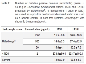
Results obtained from the mutagenicity test of uMakhonya®using S. typhimurium TA98 and TA100 strains were expressed as a mean ± s.e.m. (Table 1) and are based on a number of induced revertant colonies. Based on Table 1, uMakhonya® doses of up to 5000 μg/mL were not mutagenic in the Salmonella/microsome tester strains TA98 and TA100 when compared to the negative control and 4-NQO. The positive control, 4-NQO, induced significantly (p < 0.05) higher numbers of colonies and therefore demonstrated its mutagenic activities.
Cell viability assay
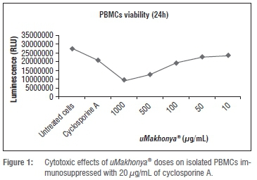
The cytotoxicity of uMakhonya® was evaluated in normal human PBMCs and malignant THP-1 monocytes to establish noncytotoxic doses for the antioxidant assays. In PBMCs immunosuppressed with 20 μg/mL of cyclosporine, uMakhonya®induced a dose dependent cytotoxic effect with high doses (1000 μg/mL and 500 μg/mL) significantly (p<0.05) increasing immunosuppression. Lower doses were less cytotoxic and these were used for the evaluation of MDA levels (Figure 1).
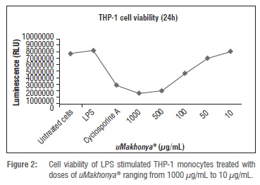
THP-1 monocytes stimulated with LPS from S. typhi and then treated with doses of uMakhonya® showed a similar trend to PBMCs. Higher doses were significantly (p< 0.05) cytotoxic to stimulated and unstimulated monocytes.
At these higher doses uMakhonya®was more cytotoxic than the immuno-suppressive drug cyclosporine (Figure 2). Supernatants from stimulated monocytes treated with lower doses were used for the evaluation of nitrite radicals as a measure of the radical scavenging potential of uMakhonya®.
Nitric oxide free radical scavenging activity
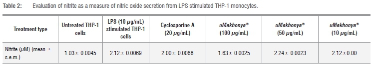
Nitrite (NO2-) is one of two primary, stable and non-volatile breakdown products of NO. Stimulation of THP-1 monocytes with LPS significantly (p<0.05) increased the secretion of NO as represented by levels of NO2-. Treatment of LPS stimulated THP-1 monocytes with non-cytotoxic doses of uMakhonya® did not significantly (p> 0.05) change the levels of nitrite radicals in treated supernatants with the highest dose tested (100 μg/mL) showing potential radical scavenging potential but this was not significantly (p>0.05) different from LPS stimulated THP-1 monocytes (Table 2).
Malondialdehyde quantification
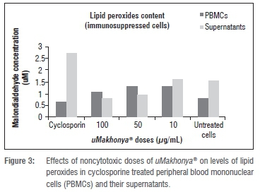
Immunosuppression of PBMCs with cyclosporine was meant to increase lipid peroxidation and changes in levels of peroxides after treatment with uMakhonya®were a measure of the possible antioxidant effects of this traditional medicinal product. In PBMCs, treatment with uMakhonya® doses increased lipid peroxides and this was significant (p<0.05) at the lowest dose (10 μg/mL) when compared to PBMCs treated with cyclosporine only. In supernatants, uMakhonya®doses significantly (p<0.05) decreased lipid peroxides at all doses tested when compared to supernatants from PBMCs treated with cyclosporine A only. Lipid peroxide levels from supernatants of immunosuppressed PBMCs were similar to those of untreated supernatants (Figure 3). Therefore uMakhonya®increased intracellular levels of lipid peroxides while displaying potent abilities to scavenge for these radicals in the surrounding media of the treated PBMCs.
1,1-Diphenyl-2-picryl-hydrazyl free radical scavenging activity
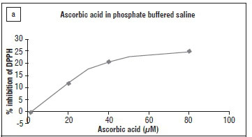
The DPPH assay is one of the quick methods used to evaluate antioxidant activity on DPPH, a stable free radical and widely used index. DPPH salt at 200 μM in methanol was mixed with non-cytotoxic doses of uMakhonya® in PBS ranging from 100 to 10 μg/mL and the changes in absorbance were measured as an indicator of free radical scavenging activity. Ascorbic acid was reconstituted in the same medium as uMakhonya®(PBS) to ensure that the results of the free radical scavenging activities were comparable.
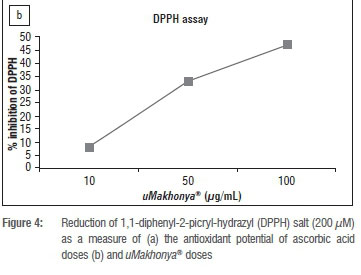
Ascorbic acid had a reduced antioxidant effect when dissolved in PBS with a plateau being reached at 80 μM (14 μg/mL) at 25% reduction of DPPH. Therefore, the IC50 for free radical scavenging activity for ascorbic acid was not reached when PBS was used as reconstitution medium (Figure 4a). There was a dose dependent increase in DPPH reduction by uMakhonya® with the highest dose tested (100 μg/mL) showing significantly (p<0.05) better antioxidant potential than ascorbic acid at 14 μg/mL. Similarly to ascorbic acid, uMakhonya®also did not reach an IC50 dose when doses shown to be noncytotoxic to PBMCs and THP-1 monocytes used were tested (Figure 4b).
Discussion
We tested the possible mutagenic and antioxidant effects of uMakhonya®, one of the popular commercial immune boosters based on traditional medicine knowledge. Even though it is a difficult task to convince manufacturers or herbalists to reveal the ingredients as well as the recipes of their products as they are well guarded secrets,7 this study collaborated with the owner of the traditional medicinal product. Novel potential drugs are usually screened for their possible mutagenic activities in many systems, including the Salmonella typhimurium microsome assay.19 Using the Ames test, uMakhonya® doses of up to 5000 μg/mL were not mutagenic in the Salmonella/microsome tester strains TA98 and TA100 when compared to the solvent negative control and 4-NQO (Table 1). These results showed that uMakhonya® can be assumed to have no direct in-vitro mutagenic effects when not metabolically activated. Similar results were shown in a study of commercial traditional herbal products sold in KwaZulu Natal where the Ames test results revealed that all 14 herbal preparations were non-mutagenic towards the Salmonella typhimurium strains TA98 and TA100 without metabolic activation.20 Mutagenic effects of any extract can be manifested in two other ways: the extract can be promutagenic by requiring metabolic activation or comutagenic by enhancing the mutagenic response of a known mutagen, irrespective of being mutagenic or not.21 Therefore, the risk assessment of uMakhonya® is not complete until this traditional medicinal product is tested for promutagenic and comutagenic effects.
Cell defences against oxidative stress are also known to decrease through changes in gene expression. The greatest protection against mutagenic activity observed in the promutagenic assay may be related to the activation of cytochrome P450 which mediates the oxidation of promutagens. Cytochromes function as antioxidants which scavenge and neutralise compounds that generate oxygen radicals, free radicals and reactive oxygen species.9 Most medicinal plants are known to possess innate antioxidant activities without the need for metabolic activation. Antioxidants from dietary and medicinal plant sources, particularly those containing phenolic compounds, have significant antioxidant activity.6 Mutagenic and antimutagenic activities have been linked to the presence of certain phytochemical substances such as flavonoids and tannins.9 The lack of direct cytotoxicity of uMakhonya® at doses of up to 5000 μg/mL observed in the Ames test was contrasted with direct cytotoxicity on isolated immune cells to establish non-cytotoxic doses for radical scavenging activities. In immunosuppressed PBMCs, uMakhonya®induced a dose-dependent cytotoxic effect at high doses above 100 μg/mL thus increasing immunosuppression significantly (Figure 1). A similar trend was observed in LPS stimulated THP-1 macrophages with similarly high doses as cytotoxic as the immunosuppressive drug cyclosporine A (Figure 2). UMakhonya® doses ranging from 10 μg/mL to 100 μg/mL were chosen to assess the antioxidant activities of this traditional medicine product.
Engagement of cellular receptors by LPS in stimulated immune cells leads to synthesis of new proteins through alteration in the pattern of gene expression.22 NO synthesis and secretion increases considerably after exposure to immunological stimuli such as bacterial LPS.23 Secretion of NO was induced by stimulation of THP-1 monocytes with LPS from S. typhi followed by treatment with doses of uMakhonya® for 24 hours. The Griess reagent system measures nitrite radicals as a reflection of the amount of NO secreted by cells. Treatment with non-cytotoxic doses of uMakhonya® showed a dose dependent effect on the levels of nitrite radicals in treated supernatants with the highest dose tested (100 μg/mL) showing possible radical scavenging potential by significantly (p <0.05) decreasing nitrite radicals when compared to LPS stimulated THP-1 monocytes. NO is a physiological mediator produced by many cells involved in immunity and inflammation. When generated in high concentrations, NO is rapidly oxidised to reactive nitrogen oxide species (RNOS) that mediate most of the immunological effects of NO. RNOS can reduce thiols to modify key signalling molecules such as kinases and transcription factors.24 Cyclosporine A, a known inhibitor of induced nitric oxide synthase, did not cause a significant (p>0.05) decrease in nitrite radicals when compared to LPS stimulated THP-1 cells. This may be related to the dose of cyclosporine A used which in this case was not effective in suppressing NO secretion. From the observed results, uMakhonya®did not show potent antioxidant activity, with only the highest non-cytotoxic dose showing nitrite radical scavenging potential.
The antioxidant activity of a given extract depends not only on its chemical constituents but also on the type of generated radical it can neutralise.6 For this reason we also tested the radical scavenging activity of uMakhonya®using the TBARS and DPPH assays. The occurrence of lipid peroxidation in biological membranes causes impairment of membrane functioning, changes in fluidity, inactivation of membrane-bound receptors and enzymes, and increased non-specific permeability to ions such as calcium (Ca2+).25 Lipid peroxidation was induced by treating PBMCs with cyclosporine A followed by incubation with various non-cytotoxic doses of uMakhonya®. This traditional medicinal product showed significant antioxidant potential by reducing lipid peroxides in supernatants of immunosuppressed cells to levels of the untreated control supernatants. Such antioxidant activity was not seen inside the immunosuppressed PBMCs treated with several doses of uMakhonya®. In the DPPH assay uMakhonya®showed a dose dependent increase in radical scavenging activity with the highest dose (100 μg/mL) showing the greatest potential (Figure 4b). The radical scavenging profile of ascorbic acid reached a plateau at 80 (14 μg/mL) without reaching an IC50 concentration when this known antioxidant was reconstituted in PBS (Figure 4a), the same medium used to reconstitute freeze dried samples of uMakhonya®. Other studies have shown that ascorbic acid has IC50 values of 11.8 μM18 and 56 μM26 when dissolved in methanol. Therefore, the use of an aqueous solution like PBS to dissolve both ascorbic acid and uMakhonya®might limit their radical scavenging activities.
Lipid peroxides are one of the aldehyde products of reactive oxygen species degradation of membrane lipids and these aldehydes can cause cross links in nucleic acids leading to DNA damage.27 The results of this study showed that uMakhonya®did not induce significant intracellular lipid peroxidation while showing significant radical scavenging activities in the supernatants of treated immunosuppressed PBMCs and in the DPPH assay. A recent study of a South African traditional herbal product with similar claims to uMakhonya®but of unknown composition, showed promising antioxidant potential in the DPPH assay, ferric reducing power and β-carotene-linoleic acid model system.7 Different studies have shown that the individual medicinal plants constituting uMakhonya®possess both antioxidant and antimutagenic activities. Dichloromethane and 90% methanol extracts of A. afra did not show mutagenicity in strain TA98 with and without metabolic activation.28 Volatile oils from A. afra have also been shown to possess considerable antioxidant effects in preventing the discoloration of β-carotene and linoleic acid and also showed significant radical scavenging potential in the lipid peroxidation assay.29,30C. crispus displayed antioxidant activities in the DPPH and ferric-reducing antioxidant power (FRAP) assays and contained phenolics, condensed tannins and flavonoids.30,31 C. crispus extracts have not been shown to possess any genotoxic effects.33P. guajava leaves and bark extracts showed concentration-dependent scavenging activity on hydrogen peroxide, superoxide and DPPH.34 Pre-treatment with an aqueous guava leaf extract was found to be effective in inactivating the mutagenicity of direct-acting mutagens 4-nitro-o-phenylenediamine and 2-aminofluorene in the tester strains of Salmonella typhimurium.35Freeze-dried extracts of U. tomentosa have been shown to have significant antioxidant activities in scavenging free radicals in the DPPH and ABTS assays.36U. tomentosa extracts were able to inhibit 90% of the mutagenic effect of hydrogen peroxide and did not show significant genotoxicity.37U. tomentosa showed significant antioxidant activities in the trolox equivalent antioxidant capacity (TEAC), peroxyl radical-trapping capacity (PRTC), and superoxide radical scavenging activity (SOD) assays and these were attributed to the total phenolics and tannins content of the extract.38 Menthol has been shown to have no genotoxic effects in both in-vitro and in-vivo animal models.39 Mentha pepirita extract which contains menthol has also been shown to have antioxidant and antiperoxidant properties.40 Although all these herbal plants have been proven to have antimutagenic and antioxidant effects, it is not easy to predict their behaviour when combined together.
Herbalists have known for centuries the value of using a combination of herbal remedies, single extracts and combined extracts to switch on the body's defence mechanisms, self-healing and protective processes.1
Conclusion
This was the first study to look at the mutagenic and antioxidant effects of the immune booster, uMakhonya®, which is prepared by combining five different traditional medicinal plants. The hot water extract of uMakhonya® did not show significant mutagenicity in the Salmonella/microsome tester strains TA98 and TA100 when compared to the solvent negative control and 4-NQO positive control. In immunosuppressed PBMCs supernatants uMakhonya®showed significant radical scavenging activity in reducing lipid peroxides but this ability was reduced in the PBMCs. UMakhonya® showed a dose dependent reduction in nitrite radicals in LPS stimulated THP-1 monocytes. In the DPPH assay, uMakhonya®showed better radical scavenging activity than ascorbic acid when both were re-dissolved in aqueous PBS. Therefore uMakhonya®extract was not mutagenic and showed promising antioxidant activity in immune cells. Future studies should focus on the mutagenic effects of this product during metabolic activation and in-vivo antioxidant effects.
Acknowledgements
We would like to thank the National Research Foundation for providing financial assistance for this project, the South African National Blood Service (SANBS) for the kind donation of normal human whole blood reagents and Mr Saiyur Ramsugit from the discipline of Medical Microbiology at the University of KwaZulu Natal for the donation of the THP-1 monocytes. We would also like to thank Dr Esam Elgorashi of the Agricultural Research Council for his assistance with the Ames test.
Authors' contributions
M.N. conducted all the experiments and drafted the primary manuscript. N.G. was principal investigator and conceptualised and supervised the research, and contributed in the final writing of the manuscript. V.N. was co-investigator and contributed to the analysis and the writing of the manuscript. V.N. has a wealth of knowledge on the formulation of the product and its clinical effects on consumers.
References
1. Busia K. Medical provision in Africa - past and present. Phytother Res. 2005;19(11):919-923. http://dx.doi.org/10.1002/ptr.1775 [ Links ]
2. Olivier DK. The ethnobotany and chemistry of South African tonic plants [PhD dissertation]. Johannesburg: University of Johannesburg; 2012. [ Links ]
3. Okpako DT. Traditional African medicine: Theory and pharmacology explored. Trends Pharmacol Sci. 1999;20(12):482-485. http://dx.doi.org/10.1016/S0165-6147(99)01406-6 [ Links ]
4. Chen X, Howard OMZ, Yang X, Wang L, Oppenheim JO, Krakauer T. Effects of Shuanghuanglian and Qingkialing, two multi-components of traditional Chinese medicinal preparations, on human leukocyte function. Life Sci. 2002;70(24):2897-2913. http://dx.doi.org/10.1016/S0024-3205(02)01541-2 [ Links ]
5. Tshibangu KC, Worku ZB, De Jongh MA, Van Wyk AE, Mokwena SO, Peronovic V. Assessment of effectiveness of traditional medicine in managing HIV/AIDS patients in South Africa. East Afr Med J. 2004;81(10):499-504. http://dx.doi.org/10.4314/eamj.v81i10.9231 [ Links ]
6. Boubaker J, Mansour HB, Ghedira K, Chekir-Ghedira L. Antimutagenic and free radical scavenger effects of leaf extracts from Accacia salicina. Ann Clin Microbiol Antimicrob. 2011;10(37):1-10. http://dx.doi.org/10.1186/1476-0711-10-37 [ Links ]
7. Ndhlala AR, Finnie JF, Van Staden J. In-vitro antioxidant properties, HIV-1 reverse transcriptase and acetylcholinesterase inhibitory effects of traditional herbal preparations sold in South Africa. Molecules. 2010;15(10):6888-6904. http://dx.doi.org/10.3390/molecules15106888 [ Links ]
8. Cowan MM. Plant products as antimicrobial agents. Clin Microbiol Rev. 1999;12(4):564-582. [ Links ]
9. Horn RC, Vargas VMF. Antimutagenic activity of extracts of natural substances in the Salmonella/microsome assay. Mutagenesis. 2003;18(2):113-118. http://dx.doi.org/10.1093/mutage/18.2.113 [ Links ]
10. Ferguson LR. Antimutagens as cancer chemopreventive agents in diet. Mutat Res. 1994;307(1):395-410. http://dx.doi.org/10.1016/0027-5107(94)90313-1 [ Links ]
11. Lakshmi B, Ajith TA, Jose N, Janardhanan KK. Antimutagenic activity of methanolic extract of Ganode lucidum and its effects on hepatic damage caused by benzo[a]pyrene. J Ethnopharmacol. 2006;107(2):297-303. http://dx.doi.org/10.1016/j.jep.2006.03.027 [ Links ]
12. Eccles R. Menthol and related cooling compounds. J Pharm Pharmacol. 1994;46(8):618-630. http://dx.doi.org/10.1111/j.2042-7158.1994.tb03871.x [ Links ]
13. Holetz FB, Pessini GL, Sanches NR, Cortez DAG, Nakamura CV Dias Filho BP Screening of some plants used in the Brazilian folk medicine for the treatment of infectious diseases. Mem Inst Oswaldo Cruz. 2002;97(7):1027-1031. http://dx.doi.org/10.1590/S0074-02762002000700017 [ Links ]
14. Liu D, Keesing JK, He P Wang Z, Shi YWang Y The world's largest macroalgal bloom in the Yellow Sea, China: Formation and implications. Estuar Coast Shelf Sci. 2013;129(1):2-10. http://dx.doi.org/10.1016/j.ecss.2013.05.021 [ Links ]
15. Okai Y Higashi-Okai K, Ishizaka S, Yamashita U. Enhancing effect of polysaccharides from an edible brown alga, Hijikia fusiforme (Hijiki), on release of tumor necrosis factor-α from macrophages of endotoxin-nonresponder C3H/Hej mice. Cancer Lett. 1997;27(1):74-79. http://dx.doi.org/10.1080/01635589709514505 [ Links ]
16. Bors M, Michalowicz J, Pilarski R, Sicinska P Gulewicz K, Bukowska B. Studies of biological properties of Uncaria tomentosa extracts on human blood mononuclear cells. J Ethnopharmacol. 2012;142(3):669-678. http://dx.doi.org/10.1016/j.jep.2012.05.036 [ Links ]
17. Maron DM, Ames BN. Revised methods for the Salmonella mutagenicity test. Mutat Res. 1983;113(3-4):173-215. http://dx.doi.org/10.1016/0165-1161(83)90010-9 [ Links ]
18. Sharma OP, Bhat TK. DPPH antioxidant assay revisited. Food Chem. 2009;113(4):1202-1205. http://dx.doi.org/10.1016/j.foodchem.2008.08.008 [ Links ]
19. El-Sayed WM, Hussin WA. Antimutagenic and antioxidant activity of novel 4-substituted phenyl-2,2'-bichalcophenes and aza-analogs. Drug Des Devel Ther. 2013;7(1):73-81. http://dx.doi.org/10.2147/DDDT.S40129 [ Links ]
20. Ndhlala AR. Pharmacological, phytochemical and safety evaluation of commercial herbal preparations common in South Africa [PhD dissertation]. Pietermaritzburg: University of KwaZulu Natal; 2009. [ Links ]
21. Snijman PW, Swanevelder S, Joubert E, Green IR, Gelderblom WCA. The antimutagenic activity of the major flavonoids of rooibos (Aspalathus linearis): Some dose-response effects on mutagen activation-flavonoids interactions. Mutat Res. 2007;631(2):111-123. http://dx.doi.org/10.1016/j.mrgentox.2007.03.009 [ Links ]
22. Freudenberg MA, Tchaptchet S, Keck S, Fejer G, Huber M, Schütze N, et al. Lipopolysaccharide sensing an important factor in the innate immune response to Gram-negative bacterial infections: Benefits and hazards of LPS hypersensitivity. Immunobiology. 2008;213(3-4):193-203. http://dx.doi.org/10.1016/j.imbio.2007.11.008 [ Links ]
23. Khatsenko OG, Gross SS, Rifkind AB, Vane JR. Nitric oxide is a mediator of the decrease in cytochrome P450-dependent metabolism caused by immunostimulants. P Natl Acad Sci USA. 1993;90(23):11147-11151. http://dx.doi.org/10.1073/pnas.90.23.11147 [ Links ]
24. Coleman JW. Nitric oxide in immunity and inflammation. Int Immunopharmacol. 2001;1(8):1397-1406. http://dx.doi.org/10.1016/S1567-5769(01)00086-8 [ Links ]
25. Niki E, Yoshida Y Saito Y Noguchi N. Lipid peroxidation: Mechanisms, inhibition, and biological effects. Biochem Biophys Res Commun. 2005;338(1):668-676. http://dx.doi.org/10.1016/j.bbrc.2005.08.072 [ Links ]
26. Kano M, Takayanagi T, Harada K, Makino K, Ishikawa F. Antioxidative activity of anthocyanins from purple sweet potato Ipomoera batatas cultivar Ayamurasaki. Biosci Biotechnol Biochem. 2005;69(5):979-988. http://dx.doi.org/10.1271/bbb.69.979 [ Links ]
27. Tuzgen S, Hamnimoglu H, Tanriverdi T, Kacira T, Sanus G, Atukereny P et al. Relationship between DNA damage and total antioxidant capacity in patients with glioblastoma multiforme. Clin Oncol. 2007;19(3):177-181. http://dx.doi.org/10.1016/j.clon.2006.11.012 [ Links ]
28. Elgorashi EE, Taylor JLS, Maes A, Van Staden J, De Kimpe N, Verschaeve L. Screening of medicinal plants used in South African traditional medicine for genotoxic effects. Toxicol Lett. 2003;143(2):195-207. http://dx.doi.org/10.1016/S0378-4274(03)00176-0 [ Links ]
29. Patil GV Dass SK, Chandra R. Artemisia afra and modern diseases. Pharmacogenom Pharmacoproteomics. 2011;2(3):1-22. http://dx.doi.org/10.4172/2153-0645.1000105 [ Links ]
30. Burits M, Asres K, Bucar F. The antioxidant activity of the essential oils of Artemisia afra, Artemisia abyssinica and Juniperus procera. Phytother Res. 2001;15(2):103-108. http://dx.doi.org/10.1002/ptr.691 [ Links ]
31. Jimenez-Escrig A, Jimenez-Jimenez I, Pulido R, Saura-Calixto F. Antioxidant activity of fresh and processed edible seaweeds. J Sci Food Agr. 2001;81(5):530-534. http://dx.doi.org/10.1002/jsfa.842 [ Links ]
32. Cox S, Abu-Ghannam N, Gupta S. An assessment of the antioxidant and antimicrobial activity of six species of edible Irish seaweeds. Intern Food Res J. 2010;17(1):205-220. [ Links ]
33. World Health Organization International Programme on Chemical Safety. Safety evaluation of certain food additives: Carrageenan. Geneva: WHO Food Additives Series:60; 1999. p. 1-50. [ Links ]
34. Ogunlana, OE, Ogunlana OO. In-vitro assessment of the free radical scavenging activity of Psidium Guajava. Res J Agric Bio Sci. 2008;4(6):666-671. [ Links ]
35. Gutierrez RMP Mitchell S, Solis RV. Psidium guajava: A review of its traditional uses, phytochemistry and pharmacology. J Ethnopharmacol. 2008;117(1):1-27. http://dx.doi.org/10.1016/j.jep.2008.01.025 [ Links ]
36. Sandoval M, Okuhama NN, Zhang XJ, Condezo LA, Lao J, Angeles FM, et al. Anti-inflammatory and antioxidant activities of cat's claw (Uncaria tomentosa and Uncaria guianensis) are independent of their alkaloid content. Phytomedicine. 2002;9(4):325-337. http://dx.doi.org/10.1078/0944-7113-00117 [ Links ]
37. Romero-Jimenez M, Campos-Sanchez J, Analla M, Munoz-Serrano A, Alonso-Moraga A. Genotoxicity and anti-genotoxicity of some traditional medicinal herbs. Mutat Res. 2005;585(1-2):147-155. http://dx.doi.org/10.1016/j.mrgentox.2005.05.004 [ Links ]
38. Pilarski R, Zielinski H, Ciesiotka D, Gulewicz K. Antioxidant activity of ethanolic and aqueous extracts of Uncaria tomentosa (Willd.) DC. J Ethnopharmacol. 2005;104(1-2):18-23. http://dx.doi.org/10.1016/j.jep.2005.08.046 [ Links ]
39. World Health Organization International Programme on Chemical Safety. Safety evaluation of certain food additives: Menthol. Geneva: WHO Food Additives Series: 60; 1999. p. 1-47. [ Links ]
40. Maraj M, Ali N, Rashid U. Antimutagenic properties of Mentha piperita extract against ethyl methane sulphonate induced mutagenicity in Musmuscilus. Int J Pharm Sci Rev Res. 2010;5(2):63-66. [ Links ]
 Correspondence:
Nceba Gqaleni
Department of Public Management and Economics, Durban University of Technology
79 Steve Biko Road, Durban 4001. South Africa
nceba5850@gmail.com
Received: 21 Apr. 2015
Revised: 11 Aug. 2015
Accepted: 05 Oct. 2015

