


Serviços Personalizados
Artigo
 Inglês (pdf)
 Artigo em XML
 Referências do artigo
Indicadores

Links relacionados
-
 Citado por Google
 -
 Similares em Google
Compartilhar

 Permalink
South African Journal of Science
versão On-line ISSN 1996-7489
versão impressa ISSN 0038-2353
S. Afr. j. sci. vol.108 no.9-10 Pretoria Jan. 2012
RESEARCH ARTICLE
Is Greulich-Pyle age estimation applicable for determining maturation in male Africans?
Kundisai A. Dembetembe; Alan G. Morris
Department of Human Biology, University of Cape Town, Cape Town, South Africa
ABSTRACT
Skeletal age estimation as a means of assessing development and skeletal maturation in children and adolescents is of great importance for clinical and forensic purposes. The skeletal age of a test population is estimated by comparison with established standards, the most common standards being those in the Radiographic atlas of skeletal development of the hand and wrist published by Greulich and Pyle in 1959. These standards are based on the assumption that skeletal maturity in male individuals is attained by the chronological age of 19 years. Although they have been widely tested, the applicability of these standards to contemporary populations has yet to be tested on a population of African biological origin living in South Africa. We therefore estimated the skeletal age of 131 male Africans aged between 13 and 21 years, using the Greulich-Pyle method which we applied to pre-existing hand-wrist radiographs. Estimated skeletal age was compared to the known chronological age for each radiograph. Skeletal age was on average approximately 6 months younger than chronological age. The Greulich-Pyle method underestimated skeletal age for approximately 74% of the sample and overestimated skeletal age for 26% of the sample. Skeletal maturity as characterised by complete epiphyseal fusion occurred approximately 2.1 years later than Greulich and Pyle's estimate of 19 years. Thus skeletal maturation was still in progress in a large proportion of the 20- and 21-year-old individuals in our study. The Greulich-Pyle method showed high precision but low accuracy and was therefore not directly applicable to African male individuals. Formulation of skeletal age estimation standards specific to South African populations is therefore recommended.
Introduction
The estimation of skeletal age is a means of assessing development and the process of skeletal maturation in children and adolescents for clinical or forensic purposes.1,2,3,4 These assessments involve comparing the skeletal age of a test population against established standards. The most commonly used standards are those published in the Radiographic atlas of skeletal development of the hand and wrist by Greulich and Pyle5. Malina6 proposed that the Greulich-Pyle5 method is appropriate because it has a low systematic error and its application is simple, relative to individual bone methods.
The applicability of the Greulich-Pyle standards to populations which differ from their reference population is often questioned. This scepticism is because, by its nature, a standard is based on the results of a specific study performed on a specific population at a specified point in time.7 Greulich and Pyle's reference population was from the Brush Foundation study carried out from 1931 to 1942. Differences in growth rate and maturation which were noted when the Greulich-Pyle standards were applied to contemporary populations, have been attributed to secular trends and differences in genetic origin, health status and economic status.7,8,9 These factors influence growth and skeletal development, causing varying effects on different populations, which thereby affect the direct applicability of the Greulich-Pyle standards to various populations.
In the present study, we assessed the applicability of the Greulich-Pyle standards to contemporary African male individuals living in South Africa, for the assessment of skeletal maturation as defined by the termination of long bone growth and complete epiphyseal fusion of the bones in the hand and wrist. Methodology detailed by Greulich and Pyle5 was applied and the resulting skeletal age estimate was compared to the chronological age for each individual.
Materials
A sample of 131 pre-existing hand-wrist radiographs of male patients attending the Martin Singer Cape Hand Clinic for treatment was used. The age range of the selected individuals was 13-22 years; this range was selected in order to encompass the age of onset of puberty and skeletal maturation. Although Greulich and Pyle determined adolescence to be between 14 and 18 years, we expanded the range for the current sample as the actual age of skeletal maturation was unknown.
The individuals in the sample came to the clinic to receive treatment for trauma to the hand or wrist; the individuals were in reasonably good health prior to their injury. Billing information, that is, whether their treatment was free, state funded or privately funded, served as a useful indicator for socio-economic status.
Selection criteria
Only radiographs of male individuals of African biological origin between the ages of 13 and 22 years were used. Age was determined using the date of birth supplied on admission to the clinic and the date of the radiograph. African ancestry was determined from the information given on the radiograph envelope. Family name and home language were used as proxies for African ancestry. We reasonably assumed that individuals who indicated isiXhosa, isiZulu, seSotho, seTswana and other African languages as their home language were not likely to have been of European or other descent. It is acknowledged that some individuals who satisfied the above selection criteria could still have been of non-African descent, but it can be reasonably assumed that such cases were the exception rather than the rule.
To be included, the images had to be clear and had to include the distal radius, ulna, carpals, metacarpals and phalanges taken in the anteroposterior or posteroanterior views. Images were rejected if the above criteria were not satisfied or if the hand or wrist was too damaged for an estimate to be made.
Based on the above criteria, the sample available consisted of 61 radiographs of the left hand and 81 of the right hand, of which 11 were paired. Analysis was performed on the left hand unless only the right hand was available or if the left hand was severely damaged. Where images of both hands were available, the level of skeletal development of each hand was assessed and if a significant difference in development was detected, then both radiographs were excluded.
Ethical clearance
Ethical clearance was obtained from the University of Cape Town's Health Sciences Faculty Research Ethics Committee (REC REF: 271/2009). The study was approved provided that pre-existing radiographs be used and that patient information be kept anonymous. Permission to access patients' radiographs was given by the Head of the Hand Surgery Unit at Groote Schuur Hospital in Cape Town.
Method
According to the Greulich-Pyle5 method, each radiograph was selected at random and compared to the male standard which it most closely matched to generate an age estimate - the skeletal age. Using a standard light box, the level of development was assessed in the following areas: the size of the radial and ulnar epiphyses relative to their respective diaphyses, the size of sesamoid bones, the extent of epiphyseal capping in the metacarpals and phalanges, and the extent of epiphysis to diaphysis fusion in all these bones.
Three age estimations were made at different times throughout the study. Two estimations were performed 1 month apart by the primary researcher (mean error 1.0 months) and a third estimate was made by a second researcher familiar with the method, on a randomly selected sample of the radiographs being analysed (mean error 1.1 months). No significant differences were found between these estimates (p = 0.969, Mann-Whitney test; p = 0.909, Kruskal-Wallis test). The average of the two estimates made by the primary researcher was therefore used for further analysis and is referred to as the skeletal age in years. Only once all skeletal age estimates had been made, was chronological age calculated as described. The estimated skeletal age was then compared to the calculated chronological age.
Results
Comparison between skeletal age and chronological age
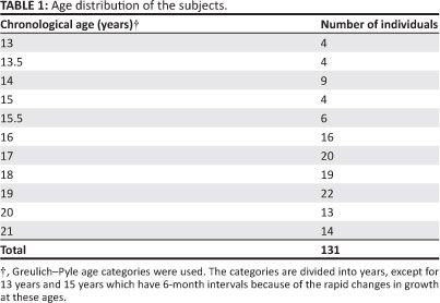
Table 1 shows the age distribution of the sample. From this table it is evident that the sizes of the younger age groups are smaller than the older age groups. Table 2 shows the estimated skeletal ages and the differences between skeletal age and chronological age. The mean skeletal age estimates are generally lower than their corresponding chronological ages, except for the older groups. The table was therefore divided into two areas of analysis. The first part of the table shows the 13- to 19-year age groups which are consistent with the Greulich-Pyle age categories with an upper limit for skeletal age of 19 years. The second part of the table shows skeletal age estimates for the 20- and 21-year age groups. This division is useful for the determination of age of skeletal maturation. The differences between skeletal age and chronological age ranged from 2.4 months to 8.4 months between the ages of 13 years and 18 years. However, for age groups 14, 16 and 17 years, the Greulich-Pyle method overestimated chronological age by 4.8, 3.6 and 6.0 months, respectively. A mean underestimation of 2.7 months was recorded for the 13- to 19-year age groups. At the chronological age of 19 years, skeletal age was underestimated by 1 year. As expected, this difference increased as chronological age increased from 19 years.
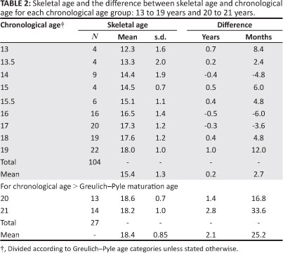
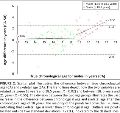
A positive correlation was found to exist between skeletal age and chronological age, as shown in Figure 1, a scatter plot of skeletal age against chronological age with the line of best fit indicated (R = 0.76). A Spearman rank-order correlation produced a value of 0.679 (significant at α = 0.05), indicating that the two parameters - chronological age and skeletal age - are both measuring an increase in age. However, skeletal age tended to underestimate chronological age. A Mann-Whitney test showed a significant difference between chronological age and skeletal age (p < 0.001) and the Kruskal-Wallis test showed those significant differences to be at chronological ages 19, 20 and 21 years. From Table 2 it can be seen that the 20- and 21-year-old groups had a mean skeletal age of 18.4 years and a mean difference in ages of 25.2 months. This result is not unexpected, as the Greulich-Pyle method identifies the attainment of maturity as 19 years. Thus individuals chronologically older than 19 years should have an estimated skeletal age of 19 years.

From Figure 2 it can be seen that the difference between chronological age and skeletal age is fairly consistent for individuals between 13 and 18 years old, with values within two standard deviations of the mean. It is also evident from Figure 2 that the Greulich-Pyle skeletal age estimation method is less accurate in older individuals. The change in gradient of the trend line between 19 years and 21 years, indicates the increasing difference between chronological age and skeletal age in this age range. Figure 2 also shows whether the skeletal age overestimated or underestimated chronological age, depending on whether the differences lie below or above the line y = 0, respectively. Skeletal age underestimated chronological age for approximately 74% of the sample and overestimated chronological age for approximately 26%. The y = 0 line is the line on which all points would lie if skeletal age and chronological age were identical at all ages.
Determination of age of maturation
As indicated by the number of points lying above the y = 0 line, the Greulich-Pyle method underestimated age in 57 of the 61 individuals between the ages of 18 and 21 years. Therefore skeletal maturation was still ongoing in these individuals at ages beyond the age of maturity of 19 years given by Greulich and Pyle. Further investigations took place to establish the age at which skeletal maturation was reached in the current subjects, the results of which are presented in Table 3.
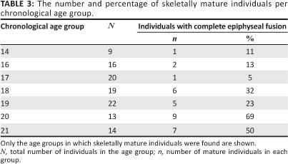
Table 3 shows that the individuals who were both chronologically and skeletally 19 years old, represent only 23% of the 19-year age group. Therefore 77% of 19-year-old individuals had not yet attained skeletal maturity. Of the total number of twenty-two 19-year-old individuals, two had a skeletal age of 15 years, one had a skeletal age of 16 years, five had a skeletal age of 17 years and six had a skeletal age of 18 years. For the 20-year age group, one individual had a skeletal age of 17 years and three had a skeletal age of 18 years. For the 21-year age group, only seven individuals had reached maturity while one individual recorded a skeletal age of 16 years, one of 17 years and five of 18 years. These findings confirm a delay in the skeletal maturation of this sample and account for the high level of variation in this age range.
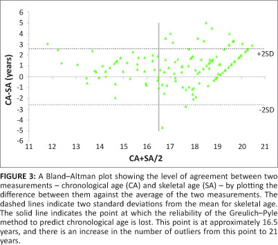
The Bland-Altman11 plot of the comparison between skeletal age and chronological age for the current sample is presented in Figure 3. The level of agreement between the two methods is shown by plotting the average of the two measurements (skeletal age and chronological age) against the difference between them. Also shown is the number of individuals for whom the difference in ages differed by more than two standard deviations. The vertical line shows the point at which the accuracy of the Greulich-Pyle method decreases as indicated by the increased number of points falling outside of two standard deviations.
Discussion
The precision, that is, the degree of similarity between measurements performed at different times on the same sample by the same or a different observer,12 of the Greulich-Pyle method was satisfactory. This is supported by the low inter- and intra-observer errors. The accuracy of the method, however, was unsatisfactory, as shown by the magnitude of the difference between chronological age and skeletal age.
Difference between chronological age and skeletal age
The overall results showed that skeletal ages determined using the Greulich-Pyle method5 were lower than the chronological ages for a large proportion of the sample. Our results are comparable to those published previously, in which it was found that the Greulich-Pyle method became inapplicable to the sample at 17 years of age, as indicated by the increased number of outliers or estimates falling outside of two standard deviations of the mean.13 For our current sample, the point at which progressively more outliers were observed with increasing age occurred at the chronological age of 16.5 years. These outliers were individuals whose chronological age was underestimated or overestimated by more than 2.5 years or two standard deviations. The majority of these individuals had not yet attained skeletal maturity, as shown in Table 3 by their skeletal ages being lower than their chronological ages in the 19-, 20- and 21-year age groups. The high incidence of individuals with differences between chronological age and skeletal age greater than two standard deviations is the main criticism for the Greulich-Pyle method.3,13 This high incidence occurs because the method assumes epiphyseal fusion in the hand and wrist is complete in male individuals by the age of 19 years.5
Delayed skeletal maturation
The underestimation of chronological age by the Greulich-Pyle method reported here can be interpreted as a delay in skeletal maturation in our subjects compared with Greulich and Pyle's reference population. These results are consistent with those of previous studies in which delays in skeletal development of between 1.5 months and 6 months were recorded when Greulich-Pyle standards were applied to populations of European descent.2,14 A discrepancy of up to 20 months was reported when estimating skeletal age in a Malawian sample.15 In the current sample the average difference between chronological age and skeletal age in individuals older than 19 years was 2.1 years (or 25.2 months). Such a difference would support the conclusion that there was a delay in skeletal development and thus attainment of skeletal maturation in our subjects.
Other studies on South African populations have tested the applicability of the Greulich-Pyle method for assessing age. One of these studies reported an underestimation of age of up to 1 year for the 'Negroid' sample.10 Another study also noted the increasing tendency for age to be underestimated in male individuals as chronological age increased, which we found in this study.16
In the few cases where age was overestimated, it is possible that these individuals were indeed developing at a faster rate than were the other individuals in our sample and those in Greulich and Pyle's reference population. Overestimations may also have been as a result of the position in which the hand was placed on the radiographic plate; because the subjects' hands were injured, they may have been unable
(physically or as a result of the pain) to place their hand in the appropriate position. The resulting image may have been distorted and the extent of epiphyseal fusion may thus have been misinterpreted (Phillips VM 2010, personal communication, 29 June). This possibility presents an important limitation to using pre-existing radiographs. Lastly, the overestimations observed may also be statistical artefacts resulting from the small sizes of each age group. In which case, this possible effect may have been reduced in the older age groups as these group sizes were larger than those of the younger age groups.
Possible causes of delay in skeletal development
Delayed skeletal maturity is not unique to the African context but is found in other populations as well. The question then arises as to the reason for this apparent delay. Factors affecting skeletal development range from biological origin, also referred to as 'race' or 'ethnicity', to secular trends in growth and socio-economic and health status.7,8,9
Although the current study was based on a single sample of African biological origin, the results are comparable to previous research on populations from North America, specifically in African Americans.7,8,17,18 The Greulich-Pyle method overestimated age in adolescent male African Americans in two studies.7,17 In this study, the same method underestimated age in male Africans, leading to the conclusion that our subjects were developmentally delayed compared with the African Americans in the other studies and therefore can be described as being 'accelerated', that is, their chronological age was in advance of their skeletal age.2 This finding provides a strong argument for biological origin as a factor affecting skeletal development. However, the design and aim of the current study did not include individuals of different biological origin and so such comparisons cannot be drawn.
Socio-economic status is reported to have only minimal effects on skeletal age,7 although some studies have found that growth and physiological development differed between population groups of different socio-economic status.19,20 In 1992, using height-weight measurements, it was found that the children of 'black' farm labourers in South Africa tended to weigh less and were shorter than their urban counterparts.19 Using menarche as an indicator of maturation, some researchers found that the age of menarche in middle-class 'Cape Coloured' girls was younger than that of 'white' girls and even younger than that of 'black' girls, but no further data on skeletal maturation was provided.21
The current sample was homogenous with respect to biological ancestry, so conclusions on the effect of ancestry on skeletal maturation are limited. In regard to socio-economic status, the sample consisted of individuals attending the Martin Singer Hand Clinic which draws patients from all sectors of society and with a wide range of income. However, in the current sample, there were more patients that received free or state-funded care than private patients, which limits comparisons based on socio-economic status.
Limitations and recommendations
The main limitations of this study were the small sample sizes of each age group and the reliance on pre-existing radiographs. Generating new radiographic images using more efficient high-resolution low radiation imaging, in addition to gathering data on genetic and geographic origin, health status, and socio-economic status on a larger sample would enable the testing of individual or combined effects of these factors on skeletal development.
Conclusion
The results of this study have shown that the current skeletal age estimation standards, formulated by Greulich and Pyle5 are not directly applicable to male South Africans of African biological origin. The Greulich-Pyle method, although precise, is not accurate for determining skeletal maturity, especially after the chronological age of 16.5 years. In our subjects, epiphyseal fusion of the hand and wrist was not complete by the chronological age of 19 years, suggesting that the onset of epiphyseal fusion occurs approximately 2 years later in male Africans. Moreover, whatever effect biological origin would have had on the rate of skeletal development, low socio-economic status and unfavourable environmental conditions are thought to have a much stronger effect on the rate of ossification of the bones of the hand and wrist.22
Although the difference recorded is within the accepted limits of error given by Greulich and Pyle5 by virtue of its consistency in the 13- to 19-year age groups, it would be advisable to formulate new standards in which the delay in development has been incorporated. New standards would be necessary for determining minimum adult age characterised by complete epiphyseal fusion. It is also recommended that, for the biologically diverse South African population, the average deviation from the Greulich-Pyle standard should be calculated for each age group.13 The age intervals given for each standard could then be adjusted by this value, thereby making the skeletal age estimation standards more applicable to, and more accurate in determining developmental age of a South African population.
Acknowledgements
We thank the Palaeontological Scientific Trust (PAST) for providing funding, Dr Michael Solomons and Sr Orie for granting permission to gather data from the Martin Singer Cape Hand Clinic, and Belinda Speed for performing the interobserver error analysis.
Competing interests
We declare that we have no financial or personal relationships which may have inappropriately influenced us in writing this paper.
Authors' contributions
A.G.M. made conceptual contributions to the project. K.D. designed the data collection methodology and performed the data analysis. K.D. wrote the manuscript and A.G.M. assisted with editing of the scientific content.
References
1. Garamendi PM, Landa MI, Ballesteros J, Solano MA. Reliability of the methods applied to assess age minority in living subjects around 18 years old: A survey on a Moroccan origin population. Forensic Sci Int. 2005;154(1):3-12. http://dx.doi.org/10.1016/j.forsciint.2004.08.018 [ Links ]
2. Schmidt S, Koch B, Schulz R, Reisinger W, Schmeling A. Comparative analysis of the applicability of the skeletal age determination methods of Greulich-Pyle and Thiemann-Nitz for forensic age estimation in living subjects. Int J Legal Med. 2007;121(4):193-296. http://dx.doi.org/10.1007/s00414-007-0165-7 [ Links ]
3. Schmidt S, Baumann U, Schulz R, Reisinger W, Schmeling A. Study of age dependence of epiphyseal ossification of the hand skeleton. Int J Legal Med. 2008;122(1):51-54. http://dx.doi.org/10.1007/s00414-007-0209-z [ Links ]
4. Lynnerup N, Belard E, Buch-Olsen K, Sejrsen b, Damgaard-Pedersen K. Intra- and interobserver error of the Greulich-Pyle method as used on a Danish forensic sample. Forensic Sci Int. 2008;179(2):242-247. http://dx.doi.org/10.1016/j.forsciint.2008.05.005 [ Links ]
5. Greulich WW, Pyle SI. Radiographic atlas of skeletal development of the hand and wrist. Stanford: Stanford University Press; 1959. [ Links ]
6. Malina RM. A consideration of factors underlying the selection of methods in the assessment of skeletal maturity. Am J Phys Anthropol. 1971;35(3):341-346. http://dx.doi.org/10.1002/ajpa.1330350308 [ Links ]
7. Loder RT, Estle DT, Morrison K, et al. Applicability of the Greulich and Pyle skeletal age standards to black and white children of today. Am J DisChild. 1993;147(12):1329-1333. [ Links ]
8. Zhang A, Sayre JW, Vachon L, Liu BJ, Huang HK. Racial differences in growth patterns of children assessed on the basis of bone age. Radiology. 2009;250:228-235. http://dx.doi.org/10.1148/radiol.2493080468 [ Links ]
9. Schmeling A, Schultz R, Danner B, Rösing FW. The impact of economic progress and modernization in medicine on the ossification of hand and wrist. Int J Legal Med. 2006;120(2):121-126. http://dx.doi.org/10.1007/s00414-005-0007-4 [ Links ]
10. Phillips VM, Thompson IOC. A correlation between dental age and bone age. In: Willems G, editor. Forensic odontology. Proceedings of the European IOFOS Millennium Meeting Leuven (Belgium). Leuven: Leuven University Press, 2000; p. 55-58. [ Links ]
11. Bland JM, Altman DG. Statistical methods for assessing agreement between two methods of clinical measurement. Lancet. 1986;327:307-310. http://dx.doi.org/10.1016/S0140-6736(86)90837-8 [ Links ]
12. Vignolo M, Milani S, Cerebello G, Coroli P, Di Battista E, Alcardi G. FELS, Greulich-Pyle, and Tanner-Whitehouse bone age assessments in a group of Italian children and adolescents. Am J Hum Biol. 1992;4(4):493-500. http://dx.doi.org/10.1002/ajhb.1310040408 [ Links ]
13. Van Rijn RR, Lequin MH, Thodberg HH. Automatic determination of Greulich and Pyle bone age in healthy Dutch children. Pediatr Radiol. 2009;39(6):591-597. http://dx.doi.org/10.1007/s00247-008-1090-8 [ Links ]
14. Groell R, Lindbichler F, Riepl T, Gherra L, Roposch A, Fotter R. The reliability of bone age determination in central European children using the Greulich and Pyle method. Br J Radiol. 1999;72:461-464. [ Links ]
15. Lewis CP, Lavy SBD, Harrison WJ. Delay in skeletal maturity in Malawian children. J Bone Joint Surg Br. 2002;84B:732-734. http://dx.doi.org/10.1302/0301-620X.84B5.12642 [ Links ]
16. Roff B. Greulich and Pyle (1959) skeletal age estimation using hand-wrist radiographs - does the method apply to South African children? A pilot study. Unpublished honours dissertation, Cape Town, University of Cape Town, 2008. [ Links ]
17. Ontell FK, Ivanovic M, Ablin DS, Barlow TW. Bone age in children of diverse ethnicity. Am J Roentgenol. 1996;167:1395-1398. [ Links ]
18. Mora S, Boechat MI, Pietka E, Huang HK, Gilsanz V. Skeletal age determinations in children of European and African descent: Applicability of the Greulich and Pyle standards. Pediatr Res. 2001;50(5):624-628. http://dx.doi.org/10.1203/00006450-200111000-00015 [ Links ]
19. Cameron N, Kgamphe JS, Leschner KF, Farrant PJ. Urban-rural differences in the growth of South African black children. Ann Hum Biol. 1992;19(1):23-33. http://dx.doi.org/10.1080/03014469200001892 [ Links ]
20. Henneberg M, Louw GJ. Average menarcheal age of higher socioeconomic status urban Cape Coloured girls assessed by means of status quo and recall methods. Am J Phys Anthropol. 1995;96(1):1-5. http://dx.doi.org/10.1002/ajpa.1330960102 [ Links ]
21. Simmons K., Greulich WW. Menarcheal age and the height, weight, and skeletal age of girls age 7 to 17 years. J Pediatr. 1943;22(5):518-548. http://dx.doi.org/10.1016/S0022-3476(43)80022-6 [ Links ]
22. Schmeling A, Reisinger W, Loreck D, Vendura K, Markus W, Gesrick G. Effects of ethnicity on skeletal maturation: Consequences for forensic age estimations. Int J Legal Med. 2000;113(5):253-258. http://dx.doi.org/10.1007/s004149900102 [ Links ]
 Correspondence to:
Kundisai Dembetembe
Private Bag X3
Observatory 7935, South Africa
Email: kundisai.dembetembe@yahoo.co.uk
Received: 06 Dec. 2011
Accepted: 23 Mar. 2012
Published: 10 Sept. 2012
© 2012. The Authors. Licensee: AOSIS OpenJournals. This work is licensed under the Creative Commons Attribution License.

