


Services on Demand
Article
 English (pdf)
 Article in xml format
 Article references
Indicators

Related links
-
 Cited by Google
 -
 Similars in Google
Share

 Permalink
Onderstepoort Journal of Veterinary Research
On-line version ISSN 2219-0635
Print version ISSN 0030-2465
Onderstepoort j. vet. res. vol.89 n.1 Pretoria 2022
http://dx.doi.org/10.4102/ojvr.v89i1.1968
ORIGINAL RESEARCH
Protective effects of methanolic leaf extracts of Monanthotaxis caffra against aflatoxin B1-induced hepatotoxicity in rats
Rhulani MakhuveleI, II; Kenn FoubertIII; Nina HermansIII; Luc PietersIII; Luc VerschaeveIV, V; Esam ElgorashiI, II
IToxicology and Ethnoveterinary Medicine, Agricultural Research Council-Onderstepoort Veterinary research, Onderstepoort, South Africa
IIDepartment of Paraclinical Sciences, Faculty of Veterinary Science, University of Pretoria, Onderstepoort, South Africa
IIIDepartment of Pharmaceutical Sciences, University of Antwerp, Antwerp, Belgium
IVDepartment of Risk and Health Impact Assessment, Sciensano, Brussels, Belgium
VDepartment of Biomedical Sciences, University of Antwerp, Antwerp, Belgium
ABSTRACT
Aflatoxins are potent hepatotoxic and carcinogenic secondary metabolites produced by toxigenic fungi. The present study investigated the protective effect of methanolic leaf extracts of Monanthotaxis caffra (MLEMC) against aflatoxin B1-induced toxicity in male Sprague-Dawley rats. The rats were randomly divided into 6 groups of 8 animals each. Five groups were administered orally for seven days with three different concentrations of MLEMC (100 mg/kg, 200 mg/kg and 300 mg/kg), curcumin (10 mg/kg) or vehicle (25% propylene glycol). The following day, these groups were administered 1 mg/kg b.w. of aflatoxin B1 (AFB1). The experiment was terminated three days after administration of AFB1. Group 6 represented untreated healthy control. Serum aspartate aminotransferase, alanine aminotransferase, alkaline phosphatase, lactate dehydrogenase, creatinine and liver histopathology were evaluated. Methanolic leaf extracts of M. caffra decreased the levels of aspartate aminotransferase, alanine aminotransferase, lactate dehydrogenase and creatinine in the sera of rats as compared with the AFB1 intoxicated group. Co-administration of MLEMC improved the histological characteristics of the hepatocytes in contrast to the AFB1 treated group, which had mild to severe hepatocellular injuries including bile duct proliferation, bile duct hyperplasia, lymphoplasmacytic infiltrate and fibrosis. Extracts of M. caffra were beneficial in mitigating the hepatotoxic effects of AFB1 in rats by reducing the levels of liver enzymes and preventing hepatic injury.
Keywords: mycotoxins; aflatoxins; Annonaceae; liver toxicity; liver enzymes; amelioration.
Introduction
Aflatoxins (AFs) are group of difuranocoumarins toxic secondary metabolites produced by Aspergillus flavus, Aspergillus parasiticus and Aspergillus nomius, which contaminate various foodstuff and feed. The most predominant major AFs that contaminate food and feed commodities include AFB1, AFB2, AFG1 and AFG2 and their hydroxylated metabolites, namely AFM1 (Ráduly et al. 2020). Aflatoxin B1 is the most potent hepatotoxin, mutagen, teratogen, immunosuppressive and carcinogen amongst other major groups of AFs in nature (Zarev et al. 2020). When ingested, it is bio-transformed in the liver primarily by CYP3A4 and CYP1A2 isoforms that are members of the cytochrome P450 (CYP) superfamily of drug metabolising enzymes and generate a highly reactive species, namely AFB1-exo-epoxide and other metabolites (Bedard & Massey 2006; Rushing & Selim 2019). These reactive species bind to the macromolecules causing toxicity and mutations, which lead to lipid peroxidation, necrosis, cell damage, cell death, deoxyribonucleic acid (DNA) lesions, carcinogenicity and other genetic diseases (Bedard & Massey 2006; Zarev et al. 2020).
Aflatoxin B1 deleteriously affects global public health as it is the major contributor to the worldwide occurrence of hepatocellular carcinoma (HCC). The toxin also works synergistically with hepatitis B and C viruses to significantly increase the risk of HCC far above either factor individually (Rushing & Selim 2019). Surgery and liver transplant offer limited treatment options as they are only useful in the treatment of early stages of HCC. Other non-surgical options such as chemo- and radio-therapies are only successful in patients with localised liver tumour (Darvesh, Aggarwal & Bishayee 2012). Therefore, research that focuses on the development of alternative preventative and therapeutic strategies may lead to valuable findings that may be effective in the control of HCC. Although the principal toxic target of AFB1 is the liver, several reports showed that exposure to AFB1 has adverse renal effects as it increases the levels of creatinine and urea in animal experiments (El-Mahalaway 2015; Yilmaz et al. 2018). Previous studies also revealed that oxidative stress resulting from AFB1 consumption leads to myocardial cell membrane destruction manifested by a marked increase in lactate dehydrogenase (LDH) activity (Mannaa et al. 2014).
The biosynthesis and activation of AFs can be attenuated using bioactive compounds present in herbal products. Plant extracts have been studied for their antifungal growth inhibition and mycotoxin detoxification. Several studies have been conducted on the protective effects of plant extracts and its bioactive compounds against AFB1-induced hepatotoxicity (Choi et al. 2010; Soni et al. 1997; Tang et al. 2007). Curcumin and ellagic acid are examples of phytochemicals isolated from plants and they inhibited hepatocarcinogenicity in rat and chicken models (Gowda et al. 2008; Soni et al. 1997). These compounds are known for their potent free radical scavenging ability and exert their anticarcinogenic effect primarily through prevention of oxidative stress (Darvesh et al. 2012). Furthermore, phytochemicals such as lycopene and quercetin have been reported to inhibit biotransformation of AFB1 toxicity by inducing detoxification enzymes (Choi et al. 2010; Tang et al. 2007).
Monanthotaxis caffra (Annonaceae) is a shrub occurring in evergreen forests of Eastern Cape, KwaZulu-Natal and Mpumalanga provinces of South Africa (National Research Council 2008). This plant species has been used in traditional medicine against tumours, microbial and parasitic infections in veterinary and human health (Mulholland et al. 2000; Okhale et al. 2016). Methanolic leaf extract of M. caffra has been also reported for its antigenotoxic properties against AFB1-induced genotoxicity (Makhuvele et al. 2018a). Furthermore, Crotepoxide, a known antitumour compound and other polyoxygenated cyclohexane derivative, 5,6-diacetoxy1-benzoyloxymethyl-1,3-cyclohexadiene were isolated from M. caffra (Makhuvele et al. 2018b; Mulholland et al. 2000). The in vivo hepatoprotective effects of M. caffra against mycotoxin-induced toxicity has never been explored. Therefore, this study aimed to investigate the protective effects of methanolic leaf extracts of M. caffra (MLEMC) against AFB1-induced hepatotoxicity on rats.
Materials and methods
Chemicals
Aflatoxin B1, dimethyl sulfoxide (DMSO) and curcumin were purchased from Sigma-Aldrich (St. Louis, United States). Methanol and acetonitrile were bought from Van Waters & Rogers Inc. (Radnor, USA) VWR and propylene glycol was from Merck (Darmstadt, Germany).
Preparation of methanolic leaf extract of M. caffra
Leaves of M. caffra (Annonaceae) were collected from Lowveld National Botanical Gardens (South Africa) in March 2015. The identities of the plants were confirmed by Mrs. E. Van Wyk, University of Pretoria, South Africa. A voucher (number: PRU 122761 for M. caffra) was deposited in the H.G.W.J. Schweickerdt Herbarium of the University of Pretoria. The plant material was dried in an oven set at 45 °C and thereafter, ground to a fine powder. The powdered plant material was stored in airtight glass container in the dark at room temperature until use.
The powdered leaf material of M. caffra (350 g) was added to 3500 mL of 80% methanol and extracted to exhaustion by maceration at room temperature. The plant extracts were filtered through Whatman No. 1 filter paper and concentrated to dryness under reduced pressure using Buchi rotary evaporator.
In vivo evaluation of hepatoprotective effect of methanolic leaf extracts of M. caffra
Animals
A total of 48 Sprague-Dawley male rats (7 weeks old), weighing between 150 g and 200 g, were obtained from South African Vaccine Producers (SAVP; Johannesburg, South Africa). The study was carried out using single gender as a way of reducing variation. During the experiment, the rats were kept in separate cages (2 per cage) under a controlled temperature of ±22 °C, and humidity at ±50% in a light and dark cycle of 12 h. The rats were fed with a conventional rodent diet and water, available ad libitum for the duration of the study. Rats were provided with enrichment including wooden sticks for gnawing, tissues and egg containers. All enrichment items and cages, water bottles and bedding were sterilised before use. Rats were acclimatised and closely monitored under laboratory conditions for five days prior to treatment.
Study design
The Sprague-Dawley rats were randomly divided into six groups of eight animals each (n = 8): Group A (healthy control, did not receive any treatment). Group B (negative control, received 25% propylene glycol), Group C (positive control, received curcumin dissolved in 25% propylene glycol (10 mg/kg body weight). The dose of curcumin was selected based upon previous studies where the dosage produced a marked hepatoprotective effects against aflatoxin B1 (Poapolathep et al. 2015). Group D, E and F rats were treated with 100 mg/kg, 200 mg/kg and 300 mg/kg body weight per day of MLEMC, respectively. The doses of the plant extracts were in line with those reported in literature (Sathya, Kokilavani & Ananta 2012). All treatments were administered once a day by oral gavage in the morning for 7 consecutive days. On day 8, all treated rats were administered 1 mg/kg b.w of AFB1 (Zarev et al. 2020), dissolved in reverse osmosis water by oral gavage, except the healthy control group. After three days, rats were sacrificed by using an isoflurane inhalation protocol for anaesthesia (Roustan, Perrin & Courbiere 2012).
Biochemical analysis
Blood samples were collected from the lateral tail vein from each rat on day 0 and by cardiac puncture on day 10 following anaesthesia with isoflurane prior to sacrifice for determination of serum biochemistry. The blood samples were centrifuged at 1500 × g, at 4 °C for 15 min and the serum was collected, then evaluated for serum enzyme level. Serum aspartate aminotransferase (AST), alanine aminotransferase (ALT), alkaline phosphatase (ALP), LDH and creatinine were measured using COBAS INTEGRA 400 kits from Roche following manufacturer's instructions.
Histopathological studies
The liver was excised and then fixed in 10% buffered formalin for histological analysis. The organ was sliced and processed according to routine histology tissue processing in an automated tissue processor. After tissue processing, the sections were cut into 5 µm - 6 µm and the slides were prepared and stained in an automated haematoxylin and eosin tissue stainer before histology examination. The slides were examined with a BX 63 Olympus electron microscope with Olympus cellSens dimension version 1.12 software.
Statistical methods
Data are presented as mean ± standard deviation. Differences between groups were determined using one-way analysis of variance (ANOVA). The standardised residuals were tested for deviations from normality using Dunnett's, t-test and Shapiro-Wilk's test. Least significant difference (LSD) test was used to determine statistical significance between the means of treated groups and the controls. The data were considered significant at p < 0.05.
Results
Effect of treatment on animal general conditions and body weight
Neither mortality nor clinical signs of illness and abnormalities were reported in all rats administered with MLEMC, negative (AFB1) and curcumin positive control treatments in this study. The administration of MLEMC did not affect the body weight, feed consumption and behavioural patterns of the rats at all tested concentration during 10 days of experiment. Based on these results, MLEMC can be considered safe for consumption by animals.
Effect of treatment on serum biochemistry
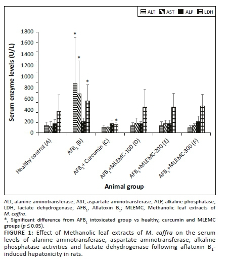
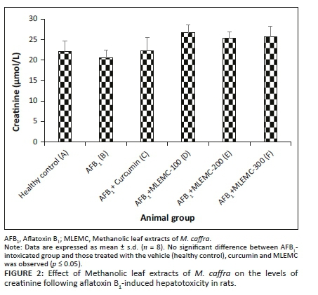
Effects of different concentrations of MLEMC on serum levels in rats induced with AFB1 are presented in Figure 1. The levels of ALT and AST significantly increased in AFB1-treated rats (Group B) in comparison to the healthy and curcumin positive control groups (p < 0.05). The levels of these enzymes markedly decreased in rats administered with AFB1+MLEMC at different concentrations and the positive control curcumin (p < 0.05). Furthermore, there were no significant differences observed in ALT and AST levels between the healthy and curcumin control group and the AFB1+MLEMC treated groups. No significant differences were observed in the levels of ALP and creatinine between the healthy, AFB1 intoxicating group and curcumin control group and those treated with different concentrations of MLEMC (Figure 1). The LDH levels of AFB1-treated rats were significantly higher when compared to the LDH levels of healthy and curcumin treated group and the AFB1+MLEMC treated groups, which were not statistically different from each other (Figure 2). Moreover, a significant decrease in the LDH level in rats treated with curcumin was observed (p < 0.05).
Effect of treatment on liver histopathology
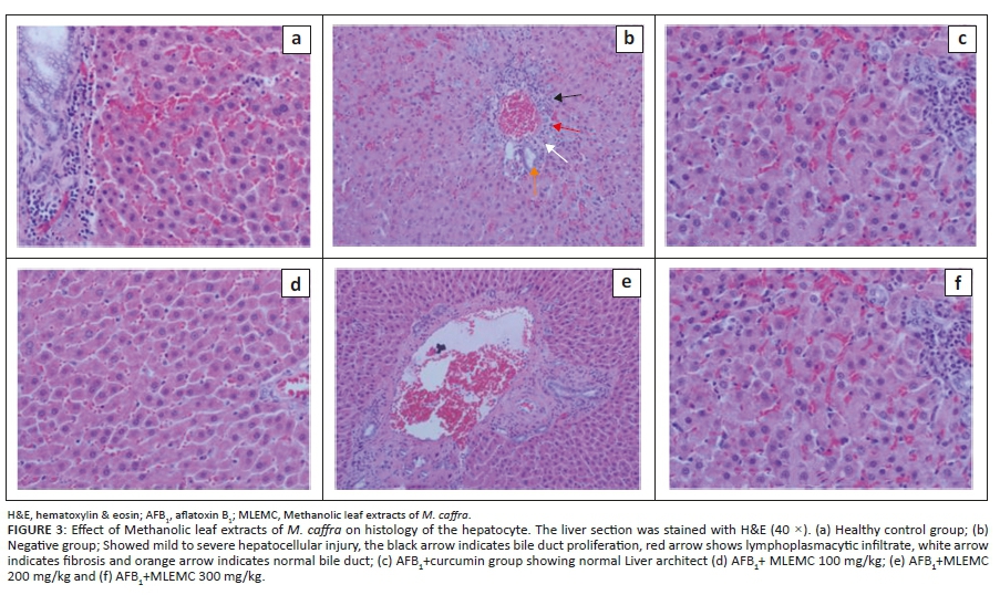
The effects of administration of MLEMC on liver histopathology are presented in Figure 3. Most rats in AFB1-intoxicated group (negative control) showed mild to severe hepatocellular degeneration with bile duct proliferation, hydropic degeneration, mild to moderate portal fibrosis in some of the larger tracts and portal lymphoplasmacytic infiltrates with the occasional neutrophil was observed in the periportal areas further extending into the periportal zone and disrupting the limiting plates, thus causing piecemeal necrosis. Scattered single cell hepatocellular necrosis was observed and the remaining hepatocytes had mildly granular and eosinophilic cytoplasm. The presence of phagocytic macrophages was also reported. These histological alterations were also observed in rats co-administered with AFB1+curcumin and AFB1+MLEMC but the injuries were substantially reduced to slight signs of sublethal non-specific hepatocellular injuries. Slight granular eosinophilic cytoplasms were also observed in all of the hepatocytes. Inflammation or necrosis were not observed and the portal tracts were within normal limits.
Discussion
Human and animals are exposed to different xenobiotic substance such as aflatoxins that cause deleterious effects on the biomolecules and cellular membrane by inducing oxidative damage (El-Agamy 2010). The liver is an essential organ of human and animals, which is involved in metabolism of xenobiotics (Colakoglu & Donmez 2012). Aflatoxin B1 is a well-known hepatotoxic agent. It induces cellular and tissue damage to the hepatocytes, thus resulting in aflatoxicosis, HCC, necrosis, cirrhosis, etc. (Shan 2020; Zarev et al. 2020). Aflatoxin B1 mediates these various deleterious effects through the induction of oxidative stress (El-Agamy 2010). Methanolic leaf extracts of M. caffra ameliorated AFB1-induced genotoxicity in in vitro antigenotoxic assays (Makhuvele et al. 2018b). Due to this antigenotoxic effect of MLEMC, we hypothesised that MLEMC will ameliorate AFB1-induced hepatotoxicity in rats. Therefore, we further investigated the protective effects of MLEMC against AFB1-induced hepatotoxicity in rats.
In our study, AFB1 intoxication significantly increased the serum levels of the liver enzymes AST and ALT in comparison with untreated control group, which implies that AFB1 caused cellular damage to the liver because of the release of these enzymes, which are normally located in the cytoplasm of the liver cells, in the blood stream. The higher standard deviation observed in the AFB1 intoxicated group were because of the two rats which did not respond to AFB1, their serum levels were not afffected by administration of AFB1. In general, AST and ALT are released into the blood stream when there is a liver damage or loss of cell membrane intergrity. These serum enzymes are considered as common biomarkers for the diagnosis of liver damage (Amacher 1998). Alanine aminotransferase and AST were normal in healthy control group whilst co-adminsteration of AFB1+curcumin and AFB1+MLEMC (100 mg/kg, 200 mg/kg and 300 mg/kg) normalised the serum enzymes ALT and AST. No significant difference was observed between the MLEMC and curcumin-treated groups in comparison with the healthy control group. This is a clear indication that MLEMC and curcumin ameliorated the toxic effect of the AFB1, by inhibiting AFB1-induced liver cell injury damage and consequently lowering the AST and ALT levels in the blood. Curcumin was used as a positive control in this study as it is renowned for its protective effects against AFB1-induced hepatocarcinogenesis and hepatotoxicity in rodents, broilers and ducklings by lowering the levels of serum markers and lipid peroxidation (Chuang et al. 2000; Gowda et al. 2008; Mathuria & Verma 2008). Methanolic leaf extracts of M. caffra demonstrated protective effect against AFB1-induced toxicity at all tested concentrations. However, the highest concentration of MLEMC showed high reduction comparably with the lower concentration, although no significant difference was observed.
An increase in LDH levels in rats intoxicated with AFB1 only was observed as compared with untreated control. Increased LDH activity is one of the major events involved in liver damage after AFB1 administration, as it is only released in the blood stream when the liver is injured (Choi et al. 2010; Nayak & Sashidhar 2010). However, this increase in LDH activity was not observed in the healthy control and in rats treated with different concentrations of AFB1+MLEMC. A significant decrease in the LDH level in rats treated with curcumin was observed, which is in line with literature reports (Nayak & Sashidhar 2010).
Creatinine, a major marker of kidney function, is the final metabolite of creatine conversion. Increased creatinine as an index of impaired kidney function because of chronic exposure to AFB1 was reported in chickens and rats (El-Mahalaway 2015; Valchev et al. 2014). In this study, significant changes were not observed in the serum level of creatinine in rats treated with a single dose of AFB1, which is in line with previous reports where nephrotoxicity was observed only in animals having chronic exposure to AFB1 (El-Mahalaway 2015; Valchev et al. 2014).
The result of biochemical analysis of the protective effects of MLEMC was also confirmed by the histopathological investigations. Aflatoxicosis is characterised by hydropic changes, vacuolar degeneration, bile duct proliferation and lymphoplasmacytic infiltration in exposed hepatocytes. However, the histopathological effects of AFB1 are directly proportional to the concentration and exposure time to AFB1 (Do & Choi 2007; Yaman, Yener & Celik 2016). Aflatoxin B1 intoxication caused mild to severe hepatocellular injury accompanied by bile duct proliferation, hydropic changes and lymphoplasmacytic infiltrate in the hepatocytes of exposed rats. This histological effect of AFB1 on rat hepatocytes has been reported in literature (Yaman et al. 2016). Minimal degree of the above-mentioned histological characteristics in rats exposed to co-administered AFB1+curcumin and AFB1+MLEMC were observed in this study, thus implying that curcumin and MLEMC had recuperative effects against AFB1-induced acute toxicity. Curcumin has been reported in literature to possess ameliorative effects on the histology of the liver and other organs (El-Agamy 2010; Gowda et al. 2008).
Members of the genus Monanthotaxis are known to contain essential oils (Parmena, Mgina & Joseph 2012), alkaloids, flavonoids and cyclohexane epoxides (Mulholland et al. 2000). Chemical analysis of the 80% extract of M. caffra revealed it is rich in cyclohexane epoxide derivatives including crotepoxide and 5,6-diacetoxy1-benzoyloxymethyl-1,3-cyclohexadiene and a mixture of two related benzoyloxy cyclohexidiene derivatives (Makhuvele et al. 2018b). Bioassay-guided fractionation of the extract using VITOTOX genotoxicity assay yielded the two antimutagenic compounds crotepoxide and 5,6-diacetoxy1-benzoyloxymethyl-1,3-cyclohexadiene. In addition to their antimutagenic effects against aflatoxin B1-induced mutagenicity in Ames and Vitotox assays, the antitumour or anticarcinogenic properties of these polyoxygenated cyclohexane derivatives have been reported in literature. Crotepoxide inhibited the expression of tumour necrosis factor (TNF) regulated gene products involved in anti-apoptosis such as Bcl-2, Bcl-XL, cyclin D1, Cox-2, Bax, Bid, c-Myc, MMP-9 and VEFG, etc. Furthermore, crotepoxide also inhibited the tumours by preventing the activation of genes that are involved in tumorigenesis at gene levels (Alonso-Amelot 2016). The compound also possessed antitumour properties against various carcinoma in rats and mice (Parmena et al. 2012; Shing & Tam 1996; Starks et al. 2012). The protective effects of these plant extracts against AFB1 hepatotoxicity may be due to the presence of the above-mentioned compounds, which are found in Annonaceae plant species.
Conclusion
The present study showed that pre-treatment of rats with MLEMC protected hepatocytes from AFB1-induced hepatotoxicity as evidenced by the normalisation of the serum enzyme levels of AST, ALT and LDH and the reduction of hepatocellular lesions. This effect was comparable to that produced by curcumin a known hepatoprotective agent against AFB1-induced toxicity. This hepatoprotective effects results from the synergistic effects of the compounds present in the MLEMC. Our results suggest that MLEMC can be considered as potential natural agent for prevention of AFB1-induced hepatotoxicity. Further studies on long time treatment of MLEMC against AFB1-induced hepatotoxicity in rats are recommended.
Acknowledgements
The authors would like to thank the Lowveld National Botanical Garden for permission to collect plant material for the study. They also thank Mrs. E. van Wyk for assisting with plant identification.
Competing interests
The authors declare that they have no financial or personal relationships that may have inappropriately influenced them in writing this article.
Authors' contributions
E.E., L.P., L.V. and N.H. designed the study. R.M., K.F. and E.E. performed the experiments and analysed data. R.M. drafted the manuscript. E.E., N.H., L.P. and L.V. revised the manuscript.
Ethical considerations
All animals received humane care in accordance with the guidelines of the University of Pretoria Animal Ethical Committee. The protocol (V073-15) was approved by the University of Pretoria Animal Ethics Committee.
Funding information
The study was supported by the National Research Foundation (grant number CPRR 87746 and NRF/FWO 87964), Fonds Wetenschappelijk Onderzoek (FWO) grant number G001014N, Flemish Interuniversity Council (VLIR) under grant, ZEIN2014Z184, and Agricultural Research Council.
Data availability
The raw data that support the findings of this study are available on request from the corresponding author, E.E.
Disclaimer
The views and opinions expressed in this article are those of the authors and do not reflect the official position or policy of the affiliation.
References
Alonso-Amelot, M.E., 2016, 'Multitargeted bioactive materials of plants in the Curcuma Genus and related compounds: Recent advances', in F.R. Atta-Ur-Rahman (ed.), Studies in natural products chemistry, vol. 47, pp. 111-200, Elsevier, Amsterdam. [ Links ]
Amacher, D.E., 1998, 'Serum transaminase elevations as indicators of hepatic injury following the administration of drugs', Regulatory Toxicology and Pharmacology 27(2), 119-130. https://doi.org/10.1006/rtph.1998.1201 [ Links ]
Bedard, L.L. & Massey, T.E., 2006, 'Aflatoxin B1-induced DNA damage and its repair', Cancer Letters 241(2), 174-183. https://doi.org/10.1016/j.canlet.2005.11.018 [ Links ]
Choi, K-C., Chung, W-T., Kwon, J-K., Yu, J-Y., Jang, Y-S., Park, S-M. et al., 2010, 'Inhibitory effects of quercetin on aflatoxin B1-induced hepatic damage in mice', Food & Chemical Toxicology 48(10), 2747-2753. https://doi.org/10.1016/j.fct.2010.07.001 [ Links ]
Chuang, S.E., Kuo, M.L., Hsu, C.H., Chen, C.R., Lin, J.K., Lai, G.M. et al., 2000, 'Curcumin-containing diet inhibits diethylnitrosamine-induced murine hepatocarcinogenesis', Carcinogenesis 21(2), 331-335. https://doi.org/10.1093/carcin/21.2.331 [ Links ]
Colakoglu, F. & Donmez, H.H., 2012, 'Effects of aflatoxin on liver and preotective effectiveness of esterified glucomannan in merino rams', The Scientific World Journal 2012, 462925. https://doi.org/10.1100/2012/462925 [ Links ]
Darvesh, A.S., Aggarwal, B.B. & Bishayee, A., 2012, 'Curcumin and liver cancer: A review', Current Pharmaceutical Biotechnology 13(1), 218-228. https://doi.org/10.2174/138920112798868791 [ Links ]
Do, J.H. & Choi, D-K., 2007, 'Aflatoxins: Detection, toxicity, and biosynthesis', Biotechnology and Bioprocess Engineering 12, 585-593. https://doi.org/10.1007/BF02931073 [ Links ]
El-Agamy, D.S., 2010, 'Comparative effects of curcumin and resveratrol on aflatoxin B1-induced liver injury in rats', Archives of Toxicology 84(5), 389-396. https://doi.org/10.1007/s00204-010-0511-2 [ Links ]
El-Mahalaway, A., 2015, 'Protective effect of curcumin against experimentally induced aflatoxicosis on the renal cortex of adult male albino rats: A histological and immunohistochemical study', International Journal of Clinical and Experimental Pathology 8(6), 6019-6030. [ Links ]
Gowda, N.K., Ledoux, D.R., Rottinghaus, G.E., Bermudez, A.J. & Chen, Y.C., 2008, 'Efficacy of tumeric (Curcuma longa), containing a known level of curcumin, and a hydrated sodium calcium aluminosilicate to ameliorate the adverse effects of aflatoxin in broiler chicks', Journal of Nutrition and Metabolism 87(6), 1125-1130. https://doi.org/10.3382/ps.2007-00313 [ Links ]
Makhuvele, R., Foubert, K., Apers, S., Pieters, L., Verschaeve, L. & Elgorashi, E., 2018b, 'Antimutagenic constituents from Monanthotaxis caffra (Sond.) Verdc', Journal of Pharmacy & Pharmacology 70(7), 976-984. https://doi.org/10.1111/jphp.12918 [ Links ]
Makhuvele, R., Matshoga, R.G., Anthonissen, R., Pieters, L., Verschaeve, L., and Elgorashi, E.E., 2018a, 'Genotoxicity and antigenotoxicity of selected South African indigenous plants', South African Journal of Botany 114, 89-99. https://doi.org/10.1016/j.sajb.2017.10.016 [ Links ]
Mannaa, F.A., Abdel-Wahhab, K.G. & Abdel-Wahhab, M.A., 2014, 'Prevention of cardiotoxicity of aflatoxin B1 via dietary supplementation of papaya fruit extracts in rats', Cytotechnology 66(2), 327-334. https://doi.org/10.1007/s10616-013-9579-x [ Links ]
Mathuria, N. & Verma, R.J., 2008, 'Ameliorative effect of curcumin on aflatoxin induced toxicity in serum of mice', Acta Poloniae Pharmaceutica 65(3), 339-343. https://doi.org/10.1016/j.fertnstert.2007.07.1300 [ Links ]
Mulholland, D., Naidoo, N., Hutchings, A., Lavaud, C. & Massiot, G., 2000, 'Crotepoxide, a cyclohexane diepoxide from Monanthotaxis caffra', Biochemical Systematics and Ecology 28(6), 595-597. https://doi.org/10.1016/s0305-1978(99)00099-x [ Links ]
National Research Council, 2008, Lost crops of Africa, Vol. III, Fruits, The National Academies Press, Washington, DC. [ Links ]
Nayak, S. & Sashidhar, R.B., 2010, 'Metabolic intervention of aflatoxin B1 toxicity by curcumin', Journal of Ethnopharmacology 127(3), 641-644. https://doi.org/10.1016/j.jep.2009.12.010 [ Links ]
Okhale, S.E., Akpan, E., Fatokun, O.T., Esievo, K.B. & Kunle, O.F., 2016, 'Annona senegalensis Persoon (Annonaceae): A review of its ethnomedicinal uses, biological activities and phytocompounds', Journal of Pharmacognosy and Phytochemistry 5(2), 211-219. [ Links ]
Parmena, D.S., Mgina, C.A. & Joseph, C.C., 2012, 'Composition of non volatile oils and antimicrobial activies of extracts from Mononthotaxis discolor and an undescribed Uvariondendron species', Tanzania Journal of Science 38(3), 221-231. [ Links ]
Poapolathep, S., Impsilp, K., Machii, K., Kumagai, S. & Poapolathep, A., 2015, 'The effects of curcumin on aflatoxin B1-induced toxicity in rats', Biocontrol Science 20(3), 171-177. https://doi.org/10.4265/bio.20.171 [ Links ]
Ráduly, Z., Szabó, L., Madar, A., Pócsi, I. & Csernoch, L., 2020, 'Toxicological and medical aspect of Aspergillus-derived mycotoxins entering the feed and food chain', Frontiers in Microbiology 10, 2908. https://doi.org/10.3389/fmicb.2019.02908 [ Links ]
Roustan, A., Perrin, J. & Courbiere, B., 2012, 'Reply: Mouse ethanasia by isoflurane inhalation: A controversial method for the animal welfare and a negative impact on oocyte quality', Laboratory Animals 46(4), 360. https://doi.org/10.1258/la.2012.012130 [ Links ]
Rushing, B.R. & Selim, M.I., 2019, 'Aflatoxin B1: A review on metabolism, toxicity, occurrence in food, occupational exposure, and detoxification methods' Food & Chemical Toxicology 124, 81-100. https://doi.org/10.1016/j.fct.2018.11.047 [ Links ]
Sathya, M., Kokilavani, R. & Ananta, T.K.S., 2012, 'Acute and subacute toxicity studies of ethanolic extract of Acalypha indica Linn in male winstar albino rats', Asian Journal of Pharmaceutical and clinical Research 5(Suppl 1), 97-100. [ Links ]
Shan, Y., 2019, 'The toxic efects of Aflatoxin B1: An update', in X.-D Long (ed.), Aflatoxin B1 occurrence, detection and toxicological effects, pp. 1-22, IntechOpen, London. [ Links ]
Shing, T.K.M. & Tam, K.W., 1996, 'First enantiospecific syntheses of crotepoxide and iso-crotepoxide from (-) -Quinic acid', Tetrahedron Asymmetry 7(2), 353-356. https://doi.org/10.1016/0957-4166(96)00004-3 [ Links ]
Soni, K.B., Lahiri, M., Chackradeo, P., Bhide, S.V. & Kuttan, R., 1997, 'Protective effect of food additives on aflatoxin-induced mutagenicity and hepatocarcinogenicity', Cancer Letters 115(2), 129-133. https://doi.org/10.1016/s0304-3835(97)04710-1 [ Links ]
Starks, C.M., Williams, R.B., Rice, S.M., Norman, V.L., Lawrence, J.A., Goering, M.G. et al., 2012, 'Polyoxygenated cyclohexene derivatives from Monanthotaxis congoensis', Phytochemistry 74, 185-189. https://doi.org/10.1016/j.phytochem.2011.10.014 [ Links ]
Tang, L., Guan, H., Ding, X. & Wang, J-S., 2007, 'Modulation of aflatoxin toxicity and biomarkers by lycopene in F344 rats', Toxicology and Applied Pharmacology 219(1), 10-17. https://doi.org/10.1016/j.taap.2006.12.001 [ Links ]
Valchev, I., Kanakov, D., Hristov, T.S., Lazarov, L., Binev, R., Grozeva, N. et al., 2014, 'Effects of experimental aflatoxicosis on renal function in broiler chickens', Bulgarian Journal of Veterinary Medicine 17(4), 314-324. [ Links ]
Yaman, T., Yener, Z. & Celik, I., 2016, 'Histopathological and biochemical investigations of protective role of honey in rats with experimental aflatoxicosis', BMC Complementary and Alternative Medicine 16, 232. https://doi.org/10.1186/s12906-016-1217-7 [ Links ]
Yilmaz, S., Kaya, E., Karaca, A. & Karatas, O., 2018, 'Aflatoxin B1 induced renal and cardiac damage in rats: Protective effect of lycopene', Research in Veterinary Science 119, 268-275. https://doi.org/10.1016/j.rvsc.2018.07.007 [ Links ]
Zarev, Y., Naessens, T., Theunis, M., Elgorashi, E., Apers, S., Ionkova, I. et al., 2020, 'In vitro antigenotoxicity, in silico ADME prediction and protective effects against aflatoxin B1 induced hepatotoxicity in rats of an Erythrina latissima stem bark extract', Food & Chemical Toxicology 135, 110768. https://doi.org/10.1016/j.fct.2019.110768 [ Links ]
 Correspondence:
Esam Elgorashi
elgorashie@arc.agric.za
Received: 17 Aug. 2021
Accepted: 25 Jan. 2022
Published: 23 Mar. 2022

