


Services on Demand
Article
 English (pdf)
 Article in xml format
 Article references
Indicators

Related links
-
 Cited by Google
 -
 Similars in Google
Share

 Permalink
Onderstepoort Journal of Veterinary Research
On-line version ISSN 2219-0635
Print version ISSN 0030-2465
Onderstepoort j. vet. res. vol.80 n.1 Pretoria Jan. 2013
RESEARCH COMMUNICATION
Concomitant fungal and Mycobacterium bovis infections in beef cattle in Kenya
Joseph N. KuriaI; Stephen M. GathogoII
IDepartment of Veterinary Pathology, Microbiology and Parasitology, University of Nairobi, Kenya
IIDepartment of Veterinary Services, Ministry of Livestock Development, Kenya
ABSTRACT
Bovine tuberculosis is an important zoonosis and accurate diagnosis is important for its surveillance. Post-mortem diagnosis may, however, be compromised by lesions caused by other pathogens. In an investigation on its prevalence in slaughter cattle in Kenya, Mycobacterium bovis and dimorphic fungi were inadvertently identified separately or concurrently in tuberculous lesions. Beef carcasses were inspected for lesions in two abattoirs in Nairobi. Tissues with lesions were collected and transported to the laboratory. Smears of lesions were stained by acid-fast procedure and examined microscopically. Lesions were cultured in Löwenstein-Jensen (LJ) and in BBL™ Mycobacterium growth indicator tubes (MGIT) media. Mycobacteria isolates in LJ medium were identified by DNA typing. Smears of BBLTM MGIT cultures were acid-fast stained and examined microscopically. Tissue sections were stained with periodic acid-Schiff reagent before examination. Of the 929 carcasses examined, 176 had granulomatous lesions. Dimorphic fungi were detected as acid-fast negative cells in 58 (32.9; 33.5%) of the lesion smears, either alone (29.0; 16.4%) or concurrently with acid-fast bacilli (29.0; 16.4%). The fungi were also detected in some BBLTM MGIT-culture smears and lesioned tissue sections. The fungi were identified, by means of cellular morphology, as Paracoccidioides brasiliensis and Blastomyces dermatitidis. A total of 64 isolates of mycobacteria were recovered in LJ medium, 19 of which were identified as M. bovis. The present report documents native P. brasiliensis infections outside the presumed endemic region and B. dermatitidis infections in a livestock animal. The findings further indicate the importance of dimorphic fungi as a differential diagnosis of bovine tuberculosis in the region.
Introduction
Bovine tuberculosis is caused by Mycobacterium bovis and manifests in cattle as granulomatous lesions mainly in the lungs and lymph nodes of the thorax (Liebana et al. 2008; Office International des Epizooties [OIE] 2009). Although post-mortem examination and culture are effective procedures for the diagnosis of bovine tuberculosis (OIE 2009), the sensitivity of the procedure is affected by the presence of non-tuberculous parasitic, bacterial or mycotic granulomas and bacterial abscesses, which may be indistinguishable macroscopically from tuberculous granuloma (Liebana et al. 2008; OIE 2009). A study in Mali cattle revealed that 72% of tuberculous lesions detected during standard meat inspections were due to pathogens other than M. bovis (Muller et al. 2009).
Dimorphic fungi, which include Blastomyces dermatitidis and Paracoccidioides brasiliensis, are known causative agents of endemic systemic mycoses. Although B. dermatitidis has a worldwide distribution, P. brasiliensis is geographically restricted to South and Central America (Chakrabarti & Shivaprakash 2005; McEwen et al. 1995; Restrepo 1985). Paracoccidioides brasiliensis is mainly a human pathogen, causing chronic granulomatous pulmonary or disseminated infection (Borges-Walmsley et al. 2002). However, infections have been reported in domestic and captive wild animals (Bagagli et al. 2003; Costa & Fara-Netto 1978; Costa et al. 1995; Ricci et al. 2004). Human paracoccidioidomycosis cases reported outside the endemic region are considered imported (Onda et al. 2011; Van Damme et al. 2006). Blastomyces dermatitidis primarily affects humans and dogs, causing a chronic suppurative or granulomatous respiratory infection that can disseminate to other tissues and organs, especially the skin (Chapman et al. 1997). However, infections have also been reported in cats and horses (Schmiedt et al. 2006). This report documents P. brasiliensis and B. dermatitidis infections in beef cattle in Kenya in association with granulomas grossly indistinguishable from those caused by M. bovis.
Materials and methods
Beef cattle carcasses were inspected for tuberculosis lesions in two abattoirs in Nairobi, Kenya, between July 2009 and November 2009. The animals slaughtered originated mainly from the nomadic pastoral communities in the arid and semi-arid northern areas of the country. Carcasses were randomly selected and inspected according to procedures established by legislation. The lungs, the pleura, abdominal organs and the lymph nodes of the head region, thoracic cavity and the mesentery were palpated, incised and examined. Samples of affected tissues were collected individually into sterile plastic bags, transported to the laboratory and preserved at -20 °C . The samples were then processed to be examined for acid-fast bacilli (AFB) and for Mycobacterium cultures according to standard procedures (OIE 2009; World Health Organization [WHO] 1998a, 1998b). Tissue samples were homogenised using Griffith tubes, decontaminated with 4% sodium hydroxide and neutralised with phosphate buffered solution. Smears were then prepared from the homogenates, stained according to the Ziehl-Neelsen (ZN) method and examined microscopically. Samples of the homogenates were cultured in Löwenstein-Jensen (LJ) medium and BBLTM MGITTM medium (Mycobacterium growth indicator tubes, Becton, Dickinson and Co, USA). Smears were prepared from the BBLTM MGITTM culture tubes and stained with the ZN stain. Mycobacteria isolated in LJ medium were identified by molecular, analysis using the genotype MTBC assay kit (HAIN Lifescience, Nehren, Germany), as described by Gathogo, Kuria and Ombui (2012). Briefly, a loopful of bacteria was collected from each AFB-positive LJ slant, suspended in 300 µL of purified water in a 1.5-mL microcentrifuge tube and incubated in boiling water for 20 min to inactivate the mycobacteria. This was followed by incubation in an ultrasonic bath to break the mycobacterial cell walls. DNA was extracted by centrifugation and amplified in a thermocycler. The amplification conditions consisted of 5 min of initial denaturation at 95 °C, 10 cycles of 30 s each at 95 °C and 2 min each at 58 °C, 20 cycles of 25 s each at 95 °C, 40 s at 53 °C and 40 s at 70 °C, and a final extension at 70 °C for 8 min. Hybridisation and detection were carried out using a semi-automated washing-and-shaking device (TwinCubator®, HAIN Lifescience, Nehren, Germany). Labelled hybridisation membrane strips were added into wells of the plastic reaction tray containing the amplicons and hybridisation reagents, after which plates were incubated. Colourimetric detection of hybridised amplicons was performed by addition of streptavidin-conjugated alkaline phosphatase and the appropriate substrate. After the final washing step, the strips were air dried, fixed on a data sheet and examined visually.
Subsequent to detection of fungi in the ZN preparations, histological examination of the lesions was carried out to confirm the presence of fungi. Formalin-fixed samples of the lesions were routinely processed for histopathology and embedded in paraffin wax. They were sectioned at 5 µm thickness, stained with the periodic acid-Schiff reagent and examined microscopically.
Results and discussion
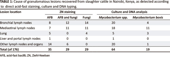
Of the 929 carcasses examined in total, 176 (19%) presented with tuberculous lesions. The majority of the lesions were localised (173/176; 98.29%) and were observed in bronchial lymph nodes (94), posterior mediastinal lymph nodes (94), lung parenchyma (14), liver parenchyma or portal lymph nodes (3), and in other lymph tissues (57). Fungi were detected as acid-fast negative cells in smears of 58 cases (32.9%), either alone (29; 16.4%) or concurrent with AFB (29; 16.4%). Mycobacterium spp. were detected from only 35 cases (19.8%). Of the 58 cases showing the presence of fungi, four presented with lesions in the lungs, whilst lesions in lymph nodes and other tissues and organs were seen in the remaining cases (Table 1). The fungi comprised two types: blastospores with multiple buds attached to the mother cell by a narrow neck, resembling a mariner's wheel and typical of P. brasiliensis, or large thick-walled cells with a single broad-based bud, identified as B. dermatitidis (Figure 1a and 1b). These fungi were also detected in some BBLTM MGIT cultures (Figure 1c and 1d) and B. dermatitidis cells were detected in some tissue sections (Figure 1e and 1f). Mycobacterium spp. were isolated from all 64 lesions that were AFB positive, with 19 isolates being identified as M. bovis.
The results of the investigation indicated that granulomatous lesions in the beef cattle carcasses examined were caused either separately by Mycobacterium spp. or dimorphic fungi, or concurrently by both bacterial and fungal infections. Bovine tuberculosis is endemic in many African countries (Ayele et al. 2004). In tropical and subtropical countries the disease is of particular importance as a large proportion of the population depends on livestock for their livelihood. Lack of control or eradication of the disease affects trade in animals and animal products (Biet et al. 2005). The zoonotic nature of bovine tuberculosis further puts the human populations at risk. Accurate diagnosis is therefore important in design and implementation of control programmes. In the present report dimorphic fungi were identified in tuberculous-like lesions as acid-fast negative cells. P. brasiliensis and B. dermatitidis are known to cause granulomatous lesions grossly similar to those caused by tuberculosis (Baily et al. 1991; Borges-Walmsley et al. 2002; Frean, Blumberg & Maureen 1993). In the presumed endemic region, paracoccidioidomycosis has also been found to occur concurrently with tuberculosis in 10% of human cases (Borges-Walmsley et al. 2002). In the present report, the fungi occurred concurrently with tuberculosis in 16.4% of the cases and separately in 17% of the cases. Clinically, dimorphic fungi mimic tuberculosis (Borges-Walmsley et al. 2002; Parvin et al. 2010) and chronic systemic mycoses caused by dimorphic fungi may therefore be confused with tuberculosis during ante-mortem examination of animals. Examination of acid-fast stained smears of post-mortem specimens can, however, provide a preliminary diagnosis of mycobacterial disease (OIE 2009).
Previous reports indicate that beyond the presumed endemic geographical region, paracoccidioidomycosis occurs only in patients who have previously resided in South or Central America (Miyaji & Kamei 2003; Shelbume & Hamil 2002). Detection of P. brasiliensis in cattle in the present report was an indication that this fungal infection does, however, occur outside the endemic area. It is noteworthy that climatic conditions similar to those in the presumed endemic regions of South and Central America occur in northern Kenya. Although B. dermatitidis infections are most common in dogs and humans in North America, some isolated cases have been reported in Africa, Israel, India and Bangladesh (Carman et al. 1989; Parvin et al. 2010; Rouhou et al. 2008).
In Africa, and especially South Africa, infections have previously been reported only in humans.
Conclusion
The present report documents native P. brasiliensis infections outside the reported endemic region and B. dermatitidis infection in a livestock animal. Dimorphic fungi should therefore be considered as an important differential diagnosis for tuberculosis during meat inspection in the region. In the present investigation, 16.4% of the cases were found to be caused exclusively by the fungi. Bovine tuberculosis is an important zoonosis and its diagnosis at slaughter requires condemnation of parts of or the whole carcass. Dimorphic fungi are not zoonotic and a diagnosis would therefore avert condemnation. However, observation of infection in cattle in the present report may also indicate infection in other animals, including humans, in the region.
Acknowledgments
We acknowledge the facilitation of the management of Kenya Meat Commission and Njiru abattoirs in sample collection, the National Tuberculosis Reference Laboratory for analytical facilities and the Department of Veterinary Services for partial funding of the study.
Competing interests
The authors declare that they have no financial or personal relationship(s) that may have inappropriately influenced them in writing this article.
Authors' contributions
J.N.K. (University of Nairobi) designed the project, S.M.G. (University of Nairobi) collected the samples and both authors contributed to sample analyses. J.N.K. wrote the manuscript.
References
Ayele, W.Y., Neill, S.D., Zinsstag, M.G. & Pavlik, I., 2004, 'Bovine tuberculosis: an old disease but a new threat to Africa', International Journal of Tuberculosis and Lung Disease 8, 924-937. PMid:12964713 [ Links ]
Bagagli, E., Franco, M., De Bosco, S., Hebeler-Barbosa, F., Trinca, L.A. & Montenegro M.R., 2003, 'High frequency of Paracoccidioides brasiliensis infection in armadillos (Dasypus novemcinctus): an ecological study', Medical Mycology 41, 217-23. PMid:12964713 [ Links ]
Baily, G.G., Robertson, V.J., Neill, P., Garrido, P. & Levy, L.F., 1991, 'Blastomycosis in Africa: Clinical features, diagnosis, and treatment', Clinical Infectious Diseases 13, 1005-1008. http://dx.doi.org/10.1093/clinids/13.5.1005, PMid:1962074 [ Links ]
Biet, F., Boschiroli, M.L., Thorel, M.F. & Guilloteau, L.A., 2005, 'Zoonotic aspects of Mycobacterium bovis and Mycobacterium avium-intracellular complex, (MAC)', Veterinary Research 3, 411-436. PMid:15845232 [ Links ]
Borges-Walmsley, M.I., Chen, D., Shu, X. & Walmsley A.R., 2002, 'The pathobiology of Paracoccidioides brasiliensis', Trends in Microbiology 10, 80-88. http://dx.doi.org/10.1016/S0966-842X(01)02292-2. [ Links ]
Carman, W.F., Frean, J.A., Crewe-Brown, H.H., Culligan, G.A. & Young, C.N., 1989, 'Blastomycosis in Africa: A review of known cases diagnosed between 1951 and 1987', Mycopathologia 107, 25-32. PMid:2682252 [ Links ]
Chakrabarti, A. & Shivaprakash, M.R., 2005, 'Microbiology of systemic fungal infections', Journal of Postgraduate Medicine 51, 16-20. PMid:16519250 [ Links ]
Chapman, S.W., Lin, A.C., Hendricks, K.A., Nolan, R.L., Currier, M.M., Morris, K.R. et al., 1997, 'Endemic blastomycosis in Mississippi: epidemiological and clinical studies', Seminars in Respiratory Infection 12, 219-228. PMid: 9313293 [ Links ]
Costa, E.O. & Fara-Netto, C., 1978, 'Contribution to the epidemiology of paracoccidioidomycosis and histoplasmosis in the state of Sao Paulo, Brazil: Paracoccidioidin and histoplasmin intradermal tests in domestic animals', Medical Mycology 16, 93-101. http://dx.doi.org/10.1080/00362177885380151 [ Links ]
Costa, E.O., Diniz, L.S., Netto, C.F., Arruda, C. & Dagli, M.L.J., 1995, 'Delayed hypersensitivity test with paracoccidioidin in captive Latin American wild mammals', Journal of Medical and Veterinary Mycology 33, 39-42. http://dx.doi.org/10.1080/02681219580000081 [ Links ]
Frean, J., Blumberg, L. & Maureen W., 1993, 'Disseminated blastomycosis masquerading as tuberculosis', Journal of Infection 26, 203-204. http://dx.doi.org/10.1016/0163-4453(93)93031-X [ Links ]
Gathogo, S.M., Kuria, J.K.N. & Ombui, J.N., 2012, 'Prevalence of bovine tuberculosis in slaughter cattle in Kenya: a postmortem, microbiological and DNA molecular study', Tropical Animal Health and Production 44, 1739-1744. http://dx.doi.org/10.1007/s11250-012-0131-3 [ Links ]
Liebana, E., Johnson, L., Gough, J., Durr, P., Jahans, K., Clifton-Hadley, R. et al. 2008, 'Pathology of naturally occurring bovine tuberculosis in England and Wales', The Veterinary Journal 176, 354-360. PMid:17728162 [ Links ]
McEwen, J.G., Garcia, A.M., Ortiz, B.L., Botero, S. & Restrepo, A., 1995, 'In search of the natural habitat of Paracoccidioides brasiliensis', Archives of Medical Research 26, 305-306. PMid:8580685 [ Links ]
Miyaji, M. & Kamei, K., 2003, 'Imported mycoses: an update', Journal of Infection and Chemotherapy 9,107-113. PMid:12825107 [ Links ]
Müller, B., Steiner, B., Bonfoh, B., Fané, A., Smith, N.H. & Zinsstag, J., 2008, 'Molecular characterisation of Mycobacterium bovis isolated from cattle slaughtered at the Bamako abattoir in Mali', BMC Veterinary Research 4, 26 http://dx.doi.org/10.1186/1746-6148-4-26, PMid:18637160 [ Links ]
Office International des Epizooties (OIE), 2009, 'Bovine tuberculosis', in Manual of diagnostic tests and vaccines for terrestrial animals. viewed 11 July 2013, from http://www.oie.int/fileadmin/Home/eng/Health_standards/tahm/2.04.07_BOVINE_TB.pdf [ Links ]
Onda, H., Komine, M., Murata, S. & Ohtsuki, M., 2011, 'Letter: Imported paracoccidioidomycosis in Japan', Dermatology Online Journal 17, 11. PMid:22233747 [ Links ]
Parvin, R., Amin, R., Mahbub, S., Hasnain, M., Arif, K.M., Miah, T. et al., 2010, 'Deep fungal infection - An emerging problem in Bangladesh', Journal of Medicine 11, 170-175. [ Links ]
Restrepo, A., 1985, 'The ecology of P. brasiliensis: a puzzle still unsolved', Journal of Medical and Veterinary Mycology 23, 323-334. http://dx.doi.org/10.1080/00362178585380481 [ Links ]
Ricci, G., Mota, F.T., Wakamatsu, A., Serafim, R.C., Borra R.C. & Franco M., 2004, 'Canine paracoccidioidomycosis', Medical Mycology 42, 379-383. PMid:15473365 [ Links ]
Rouhou, S.C., Racil, H., Ismail, O., Trabelsi, S., Zarrouk, M., Chaouch, N. et al., 2008, Pulmonary blastomycosis: a case from Africa', Scientific World Journal 8, 1098-1100. http://dx.doi.org/10.1100/tsw.2008.141, PMid:18979049 [ Links ]
Schmiedt, C., Kellum, H., Legendre, A.M., Gompf, R.E, Bright, J.M., Houle, C.D. et al., 2006, 'Cardiovascular involvement in eight dogs with Blastomyces dermatitidis infection', Journal of Veterinary Internal Medicine 20, 1351-1354. http://dx.doi.org/10.1892/0891-6640(2006)20[1351:CIIDWB]2.0.CO;2, PMid:17186849 [ Links ]
Shelbume, S.A. & Hamill, R.J., 2012, 'Paracoccidioidomycosis (South American blastomycosis)' in S.J. McPhee, M.A. Papadakis& M.W. Rabow (eds.), Current Medical Diagnosis and Treatment, 51st edn., p. 1497, MacGraw-Hill. [ Links ]
Van Damme, P.A., Bierenbroodspot, F., Telgtt, D.S., Kwakman, J.M., De Wilde, P.C. & Meis, J.F., 2006, 'A case of imported paracoccidioidomycosis: an awkward infection in the Netherlands', Medical Mycology 44, 13-18. PMid:16805088 [ Links ]
World Health Organization, 1998a, Laboratory Services in Tuberculosis Control Part II: Microscopy, World Health Organization, Geneva. [ Links ]
World Health Organization, 1998b, Laboratory Services in Tuberculosis Control. Part III: Culture, World Health Organization, Geneva. [ Links ]
 Correspondence:
Joseph Kuria
Email: jknkuria@uonbi.ac.ke
Postal address:
PO Box 29053, Nairobi 00625, Kenya
Received: 01 Mar. 2013
Accepted: 20 May 2013
Published: 31 July 2013

