


Servicios Personalizados
Articulo
 Inglés (pdf)
 Articulo en XML
 Referencias del artículo
Indicadores

Links relacionados
-
 Citado por Google
 -
 Similares en Google
Compartir

 Permalink
South African Dental Journal
versión On-line ISSN 0375-1562
versión impresa ISSN 0011-8516
S. Afr. dent. j. vol.78 no.8 Johannesburg sep. 2023
http://dx.doi.org/10.17159/sadj.v78i08.15675
RESEARCH
Radiographic assessment of developing maxillary canine ectopia and its association with dental anomalies in the mixed dentition
APG HudsonI; L JohanII; AMP HarrisIII; N MohamedIV
IBChD (Stell), HonsBSc (Med Sci) (Stell), MSc (Dent) (UWC). Senior Lecturer, Department of Orthodontics, Faculty of Dentistry, University of the Western Cape. ORCID: https://orcid.org/0000-0001-7971-0598
IIBDS (SRMC), MSc (Dent) (UWC). ORCID: https://orcid.org/0000-0002-6486-7645
IIIBChD (Stell), HonsBSc (Med Sci) (Stell), Dipl Ter Educ (Unisa), MChD Orthodontics (Stell), FFD(SA) Ortho, PhD (UWC .Professor, Department of Orthodontics, Faculty of Dentistry, University of the Western Cape. ORCID: https://orcid.org/0000-0001-6237-1200
IVBChD (Stell), BSc Hons (Paed Dent) (Stell), MSc (Paed Dent) (Stell), PhD (Comm Health) (Stell), MPhil HPE (Stell), Dip IPEH (UWC). Professor, Department of Paediatric Dentistry, Faculty of Dentistry, University of the Western Cape. ORCID: https://orcid.org/0000-0003-2184-2648
ABSTRACT
INTRODUCTION: Reciprocal associations have been found to exist between various dental anomalies.1-14 Maxillary canine ectopia may, however, occur in dentitions without any anomalies
AIMS AND OBJECTIVES: The aim of the study was to establish the prevalence of dental anomalies in a sample of panoramic radiographs. The objective was to establish whether associations exist between canine ectopia and the presence of one or more of a range of selected developmental dental anomalies. A cross-sectional study was carried out on 574 mixed dentition panoramic radiographs in patients with a dental age ranging from 8 to 12 years
RESULTS: Signs of potentially ectopic maxillary canines were evident in 85% of the radiographs and of these, 80.7% showed no evidence of the selected dental anomalies which were studied. The most prevalent association of potential canine ectopia and the anomalies studied was found with supernumerary teeth (6.5%), followed by infraocclusion of primary molars (4.5%). Peg-shaped lateral incisors showed a statistically significant association (p=0.043) with mesial overlap of the cusp tip of the maxillary canine and the root of the lateral incisor. Taurodontism was significantly associated with increased angulation of the developing canine (p=0.0049) and dilaceration showed a statistically significant association (p=0.03) with non- resorption of canines
CONCLUSION: In cases where dental anomalies are present, the developing canines should be carefully monitored both clinically and radiographically
Keywords: pre-eruptive canine ectopia, dental anomalies, interceptive orthodontics, mixed dentition, panoramic radiograph
Introduction
Pre-eruptive canine ectopia refers to erupting canines that show signs of moving in the wrong direction.1 When assessing the developing canines during the mixed dentition stage, important factors need to be taken into consideration.
• These have been identified by Hudson et al.1 as
• The presence of the canine bulges
• The presence of dental anomalies
• The late development of the dentition
• The size, position or absence of the lateral incisors
• The amount of space available in the arch and
• The mobility of the primary canines.
The early diagnosis of one or more developmental anomalies during the mixed dentition stage should be viewed as a potential early warning sign of possible canine ectopia.1 These include ectopic eruption of the first permanent molars, taurodontism, invaginations2, 3 4 and size and shape of the maxillary lateral incisor.5-12 The lateral incisors are thought to guide the canines into position, therefore, in cases where the root of the lateral incisor is smaller than normal or the lateral incisor is congenitally missing, ectopia may result. 13, 14
Significant reciprocal associations have been demonstrated between palatally displaced canines, small maxillary lateral incisors, infraocclusion of primary molars, missing second premolars and enamel hypoplasia.15 Several authors have suggested that infraocclusion of primary molars may cause ectopic eruption of the maxillary canines, because of the effect it has on the transeptal fibres.3,15, 16, 17 Late developing dentitions and in particular, late developing lateral incisors, may be more disruptive for canine eruption than missing lateral incisors.18, 19, 20 Studies have shown that aplasia of the second premolars may cause ectopic eruption of the permanent maxillary canines.3,15 However, since these premolars show a high variability in the initiation of calcification,21, 22, 23, 24 they should only be considered congenitally missing after the dental age of 7 years in order to avoid a false positive diagnosis.25 Some may erupt as late as a year after the eruption of the contralateral premolar.21 Aplasia of premolars could possibly create arch-length discrepancies, making it difficult for the maxillary canine to erupt into its normal position.26
Not much research has focused on the relationship between supernumerary teeth and ectopic maxillary canines. Baccetti15 found that supernumerary teeth did not have any association with palatally displaced maxillary canines. The presence of supernumerary teeth within the dental arch may cause delayed eruption of teeth, space issues, displacement of adjacent teeth and ectopic eruption of teeth.27,28 A mesiodens, for example, may prevent or delay eruption of the permanent maxillary central incisors which may cause ectopic eruption of a central incisor with a potential effect on the space remaining to accommodate canine eruption.29 Less frequently, a mesiodens may cause dilaceration or resorption of the permanent central incisor root, which may have an effect on the positioning of the canine.30, 31, 32
Bjerklin et al.3 suggested that there is a statistically significant association between ectopic first molars and ectopic maxillary canines. However, Baccetti15 found no association between ectopic molars and palatally displaced canines. Becktor et al.,2 showed that 23.3% of the 30 patients examined presented with ectopic eruption of the permanent first maxillary molar and root resorption of the second primary molar. This takes place prior to root resorption of the maxillary primary canines which is caused by ectopic maxillary permanent canines. Hence, the ectopic first molars could possibly be used as an early risk factor for the prediction of maxillary canine ectopia.
Methodology
The rationale for the study was to show whether or not there is a correlation between dental anomalies and canine ectopia in a South African context. An analytical, descriptive, cross-sectional, retrospective study was carried out to attempt to establish any relationship/ association between potential maxillary canine ectopia and the presence of a variety of selected developmental dental anomalies. Sequential, good quality mixed dentition panoramic radiographs (n = 574) from the UWC Paediatric Dentistry department at the Tygerberg Oral Health Centre were used. These radiographs were taken between 2011 and 2014 on patients with a dental age ranging from 8 to12 years. Patients with syndromes, cleft lip and palate, a history of previous extractions and those who received prior orthodontic treatment, were excluded.
The radiographic markers used for prediction of canine ectopia were:
• Rotated lateral incisors
• Non-resorption of primary canine roots
• Overlap between the developing permanent canine and the root of the lateral incisor
• Angulation of the developing canine greater than 300 to the mid-sagittal plane
• Enlarged developing maxillary canines
• Enlarged developing mandibular canines
Each of the radiographs exhibiting signs of potentially ectopic canines, were examined in order to determine the presence and/ or absence of the following developmental anomalies:
• Congenitally missing lateral incisors
• Aplasia of premolars
• Peg-shaped permanent maxillary lateral incisors (where the incisal width was less than the cervical width)
• I nfraocclusion of primary molars (diagnosed by a "step" in the occlusal plane)
• Supernumerary teeth
• Taurodontism
• Dilaceration of roots greater than 900
• Ectopic eruption of permanent first molars
The results obtained were coded accordingly and transferred to a Microsoft Excel spreadsheet.
Data processing and analysis
Pearson's correlation coefficient was used to determine the degree to which two variables were associated. For a correlation coefficient to be statistically significant, its absolute value must exceed 0.0834. This indicates an association. The Chi-square test of independence and Fisher exact test were also used to determine whether two categorical variables were dependent or independent. A p-value of <0.05 indicates that the variables have an association.
Results
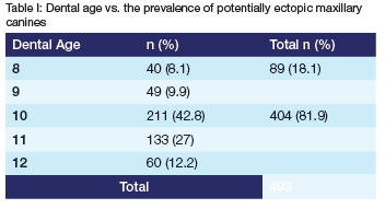
Of the 574 radiographs studied, 493 displayed potentially ectopic maxillary canines (Table I).
The anomalies detected are displayed in Table III. Roughly 80% of the cases presented with ectopia with no evidence of anomalies.
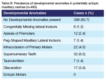
Of the 493 panoramic radiographs, 95 had anomalies present, 8 of which presented with more than 1 anomaly:
• 1 case presented with a congenitally missing lateral incisor, a peg-shaped lateral incisor and a supernumerary tooth.
• 2 cases presented with a congenitally missing incisor and a supernumerary tooth.
• 2 cases presented with infraocclusion and aplasia of a premolar.
• 1 case presented with infraocclusion and dilaceration.
• 1 case presented with infraocclusion and a supernumerary tooth.
• 1 case presented with a peg-shaped lateral incisor and a supernumerary tooth.
DISCUSSION
The findings of this study confirm the conclusion of Sorenson et al.4 that maxillary canine ectopia can occur in dentitions without any other dental deviations (Tables I, II and III). Other studies however suggest that the presence of one anomaly could predict another in the same case.15,33 The reciprocal associations found by Baccetti15 were not seen in this study. The low prevalence of developmental anomalies in this study (Table III) may be due to the genetic factors governing these various anomalies i.e. ethnicity/ hereditary factors.
No statistically significant associations (p > 0.05) were found to exist between congenitally missing lateral incisors and each of the radiographic markers when assessed separately (Table IV). This concurs with the findings of a study by Peck et al.10 which found no statistical significance in the relationship between missing maxillary lateral incisors and palatally displaced maxillary canines. Some studies have found that roughly 5% of congenitally missing maxillary lateral incisors occur with palatally displaced maxillary canines7,9, whilst others noted the frequent presence of palatally displaced canines.11,12 Nanda8 suggested that nonresorption of primary canines was likely to occur in cases with congenitally missing lateral incisors. These authors emphasized Broadbent's original 1941 observation, where the absence of a maxillary lateral incisor deprives the erupting permanent maxillary canine of the normal guidance provided by the root of the maxillary lateral incisor. This in turn leads to the high occurrence of palatally displaced canines. In the present study, the low number of cases with a congenitally missing maxillary lateral incisor may have accounted for the difference in the findings. Further investigations of congenitally missing maxillary lateral incisors using a bigger sample size may reveal a different association with maxillary canine ectopia.
The Chi-square independence test revealed no statistically significant associations between any of the potential markers of canine ectopia and aplasia of premolars (Table IV). The present study did not confirm the significant inverse relationship that Baccetti34 found between the maxillary lateral incisor rotation and aplasia of premolars. The difference in findings may be due to the low prevalence of aplasia of premolars within the selected sample.
A statistically significant association was found between peg-shaped maxillary lateral incisors (Table IV) and mesial overlap of the maxillary canine cusp tip over the root of the maxillary lateral incisor (p= 0.043).
The statistically significant association with mesial overlap supports the work of several authors who have reported the high incidence of peg-shaped maxillary lateral incisors In children with ectopic maxillary canines.6,9,10,15,35 All other radiographic markers showed no statistically significant association with peg-shaped maxillary lateral incisors. The significance of the mesial overlap may be understated because of the age limit on the study sample. Other studies have also demonstrated weak or no association between peg-shaped maxillary lateral incisors and the failure of eruption of the maxillary canine.36,37
A statistically significant association was found between peg-shaped maxillary lateral incisors (Table IV) and rotated maxillary lateral incisors (p= 0.01). The probability test showed that:
• There was a 0.74% chance for peg-shaped maxillary lateral incisors to occur when a rotated maxillary lateral incisor existed.
• There was a 33% chance for a rotated maxillary lateral incisor to occur in the presence of a peg-shaped maxillary lateral incisor.
Radiographically, the normal maxillary lateral incisor appears with the incisal edge of the crown parallel to the occlusal plane. The mesial and distal ridges are present and the V-shaped lingual fossa is visible. In the case of a rotation, only one of the ridges would be visible. The curvature of the cervical line is distinct in the direction of the incisal edge when the maxillary lateral incisor is rotated.38 The peg-shaped lateral presents with cervical margin broader than the crown tip with no ridges visible. Thus, radiographically, rotated maxillary lateral incisors could appear peg-shaped (depending on the severity of the rotation). This highlights a limitation of the present study in that it was a radiographic study without a clinical examination.
No statistically significant associations were found between infraocclusion of primary molars and the radiographic markers assessed. However, other studies showed significant associations between infraocclusion of primary molars and displaced maxillary canines.3,15,17 The difference in the results could possibly be due to the low prevalence of infraoccluded primary molars within the study sample.
No statistically significant association was found between supernumerary teeth and any of the radiographic markers. This study concurs with Baccetti's15 study, which found that the group with supernumerary teeth did not show any significant associations with palatally displaced maxillary canines. Gomes et al.39 also found that supernumerary teeth were common in his study of patients aged 9 to 10 years and noted that non-resorption of the primary maxillary canines occurred simultaneously.
Taurodontism showed no statistically significant association with potentially ectopic maxillary canines as determined by all the considered radiographic markers in Table II (p= 0.48). Nagpal et al.40 found a statistically significant relationship between maxillary canine ectopia and taurodontism. In the present study, however, taurodontism showed a statistically significant association (Table IV) with the angulation of the canine (p= 0.0049). When the angulation of the maxillary canine was greater than 300, the probability test found that:
• There was a 22% chance of taurodontism occurring.
• There was only a 9% chance of angulation of the maxillary canine being greater than 300 when taurodontism existed.
Dilaceration (Table IV) showed a statistically significant association with non-resorption of primary maxillary canines (p= 0.03). When non-resorbed primary maxillary canines existed, the probability test showed that:
• There was a 29% chance of dilaceration occurring.
• There was only a 2% chance for non-resorption of the primary maxillary canines to occur when dilaceration existed.
A total of three premolars and fourteen molars presented with dilacerations, whereas only four central incisors and two lateral incisors displayed dilaceration. This is consistent with studies conducted by Malcic et al.41 which recorded a 1.43% prevalence of dilaceration (900 or greater) of lateral incisors on panoramic radiographs. The question remains as to whether a dilaceration of 45o is normal or abnormal. No literature has specified the normal anatomical angle for dilaceration, which is why researchers use various criteria. This study found no statistically significant association between dilaceration and potentially ectopic maxillary canines as determined by the six radiographic markers (p= 0.24) (Table III).
In a sample size of 480 cases, Chohayeb42 reported that disto-labial dilaceration occurred in 52% of the maxillary lateral incisors. He disregarded angles less than 200 when recording dilaceration. Maxillary lateral incisors have a normal anatomical distal curvature43 for which, the exact degree of angulation is unknown. This result is therefore still questionable, because the normal anatomical curvature could have been 300 or more and these cases may have been included as an anomaly when Chohayeb42 was recording the prevalence of dilaceration, thus, bringing about the high prevalence of dilaceration of the maxillary lateral incisors in their study. The data reported by Chohayeb42 is not consistent with Malcic et al.'s41 results, where the prevalence of dilaceration for the lateral incisors was 1.43% in a sample size of 488 panoramic radiographs and 7% on periapical radiographs. Malcic et al.41 only recorded root dilacerations greater than or equal to 900, which is a strict criterion compared to Chohayeb's42 criteria. Hamasha et al.44 reported a prevalence of 1.2% for dilaceration in the maxillary lateral incisors in a sample size of 812 periapical radiographs. They also recorded all dilacerations of 900 and above. However, compared to Malcic et al.'s41 study, they found a lower prevalence for dilaceration using periapical radiographs. The present study used panoramic radiographs to identify the presence of root dilacerations. Hence, Hamasha et al.'s44 prevalence could not be compared with the present study.
The diagnosis of an ectopic first molar may be made clinically upon the eruption between a dental age of 5 to 7 years. Since the current study only used panoramic radiographs from dental age 8 years and above, the only way to determine if a first molar had ectopically erupted, was to identify the presence of resorption in the disto-buccal root of the second primary molar.2, 45 This study did not find any statistically significant associations with the various radiographic markers, thus supporting the findings of Baccetti's study.15 However, Bjerklin et al.3 found a statistically significant association between ectopic maxillary canines and ectopic molars.
CONCLUSION
When clinicians identify taurodontism prior to dental age 10, they should be aware of the possibility of angulation of the maxillary canines of greater than 300. This enables them to take interceptive measures (like the timeous extraction of the primary canines1) by monitoring the movement of the maxillary canine.
In the case of peg-shaped lateral incisors which can be clinically diagnosed on eruption at a dental age of 8 years, the overlap between the developing canines and lateral incisors should be monitored for signs of ectopia. The same holds true for supernumerary and infraoccluded teeth which demonstrated the strongest association with canine ectopia.
Should any root dilacerations of the lateral incisors or canines be identified prior to the dental age of 10 years, the resorption of the primary canine roots should be monitored for ectopia.
Author contributions
Dr A Hudson: Writing article 20%
Dr L Johan: Principal researcher 40%
Prof A Harris: Writing article, clinical input 20%
Prof N Mohamed: Writing article, editing 20%
Conflict of interest
None
REFERENCES
1. Hudson APG, Harris AMP, Mohamed N. Maxillary canine management in the pre-adolescent: A guideline for general practitioners. SADJ. 2010; 65 (8): 366-370. [ Links ]
2. Bektor KB, Steiniche K, Kjaer I. Association between ectopic eruption ectopic eruption of maxillary canines and first molars. Eur J Orthod. 2005; 27 (2): 186-189. [ Links ]
3. Bjerklin K, Kurol J, Valentin J. Ectopic eruption of maxillary first permanent molars and association with other tooth and developmental disturbances. Eur J Orthod. 1992; 14 (5): 369-375. [ Links ]
4. Sorenson BB, Artmann L, Larsen HJ, Kjaer I. Radiographic assessment of dental anomalies in patients with ectopic maxillary canines. Int J Paed Dent. 2009; 19 (2): 108- 114. [ Links ]
5. Shapira Y Kuftinec MM. Early diagnosis and interception of potential maxillary canine impaction. JADA. 1998; 129 (10): 1450-1454. [ Links ]
6. Becker A. Palatally impacted canines: In: Orthodontic treatment of impacted teeth. Martin Dunitz Ltd. 1998; Chapter 6, Pages 85-101. [ Links ]
7. Becker A, Smith P, Behar R. The incidence of anomalous maxillary lateral incisors in relation to palatally displaced cuspids. Angle Orthod. 1981; 51 (1): 24-29. [ Links ]
8. Nanda SK. The developmental basis of occlusion and malocclusion. Events in the life cycle of each permanent tooth. Quintessence Publishing Company, Chicago 1983; Pages 81-158. [ Links ]
9. Brin I, Becker A, Shalhav M. Position of the maxillary permanent canine in relation to anomalous or missing lateral incisors: a population study. Eur J Orthod. 1986; 8 (1):12-16. [ Links ]
10. Peck S, Peck L, Kataja M. Site specificity of tooth maxillary agenesis in subjects with canines malpositions. Angle Orthod. 1996; 66 (6): 473-476. [ Links ]
11. Miller BH. The influence of congenitally missing teeth on the eruption of the upper canine. Dent Pract Dent Rec. 1963; 13: 497-504. [ Links ]
12. Bass TB. Observations on the misplaced upper canine tooth. The Dental Practitioner and Dental Pract Dent Rec. 1967; 18: 25-33. [ Links ]
13. Bektor KB, Steiniche K, Kjaer I. Association between ectopic eruption ectopic eruption of maxillary canines and first molars. Eur J Orthod. 2005; 27 (2): 186-189. [ Links ]
14. Jacoby H. The etiology of maxillary canine impactions. Am J Orthod. 1983; 84 (2): 125-132. [ Links ]
15. Baccetti T. A controlled study of associated dental anomalies. Angle Orthod. 1998; 68 (3): 267-274. [ Links ]
16. Becker A. The effects of infraocclusion: Part 3. Dental arch length and the midline. Am J Orthod Dentofac Orthop. 1992; 102 (5): 427-433. [ Links ]
17. Shalish M, Peck S, Wasserstein A, Peck L. Increased occurrence of dental anomalies associated with infraocclusion of deciduous molars. Angle Orthod. 2010; 80 (3): 440-5. [ Links ]
18. Peck S, Peck L, Kataja M. The palatally displaced canine as a dental anomaly of genetic origin. Angle Orthod. 1994; 64 (4): 249-256. [ Links ]
19. Becker A, Chaushu S. Dental age in maxillary canine ectopia. Am J Orthod Dentofac Orthop. 2000; 117 (6): 657-662. [ Links ]
20. Leifert S and Jonas I. Dental anomalies as a micro symptom of palatal canine displacement. J Orofac Orthop. 2003; 64 (2): 108-120. [ Links ]
21. White SC, Pharaoh MJ. Oral radiology: Principles and interpretation. 6th edition. Mosby Elsevier 2010; Pages 295-304. [ Links ]
22. Wisth PJ, Thunold K, Böe OE. Frequency of hypodontia in relation to tooth size and dental arch width. Acta Odontol Scand. 1974; 32 (3): 201-206. [ Links ]
23. Nik-Hussein NN. Hypodontia in the permanent dentition: A study of its prevalence in Malaysian children. Aust Orthod J. 1989; 11 (2): 93-95. [ Links ]
24. Arte S, Pirinen S. Hypodontia. Orphanet Encyclopedia May 2004. http://www.orpha.net/data/patho/GP/UKhypodontia.pdf [ Links ]
25. Goya HA, Tanaka S, Maeda T, Akimoto Y. An orthopantomographic study of hypodontia in permanent teeth of Japanese pediatric patients. J Oral Sci. 2008; 50 (2): 143-150. [ Links ]
26. Kokich V. Early management of congenitally missing teeth. Semin Orthod. 2005; 11 (3): 146-151. [ Links ]
27. Peedikayil FC. Delayed tooth eruption. J Dent. 2011; 1 (4): 81-86. [ Links ]
28. Mitchell L and Bennett TG. Supernumerary teeth causing delayed eruption: A retrospective study. Br J Orthod. 1992; 19 (1): 41-46. [ Links ]
29. Moraes RS, Farinhas JA, Gleiser R, Primo LG. Delayed eruption of maxillary permanent central incisors as a consequence of mesiodens: a surgical re-treatment approach. J Clin Pediatr Dent. 2004; 28 (3): 195-198. [ Links ]
30. Gardiner J. Supernumerary teeth. Dent Pract Dent Rec. 1961; 12: 63-73. [ Links ]
31. Primosch RE. Anterior, teeth-assessment and surgical intervention in children. Pediatr Dent. 1981; 3 (2): 204-215. [ Links ]
32. Russell K.A., Folwarczna M.A. Mesiodens-diagnosis and management of a common supernumerary tooth. J Can Dent Assoc. 2003; 69 (6): 362-366. [ Links ]
33. Ooshima T, Ishida R, Mishima K and Sobue S. The prevalence of developmental anomalies of teeth and their association with tooth size in the primary and permanent dentitions of 1650 Japanese children. Int J Paediatr Dent. 1996; 6 (2): 87-94. [ Links ]
34. Baccetti T. Tooth rotation associated with aplasia of nonadjacent teeth. Angle Orthod. 1998; 68 (5): 471-474. [ Links ]
35. Zilberman Y, Cohen B, Becker A. Familial trends in palatal canines, anomalous lateral incisors, and related phenomena. Eur J Orthod. 1990; 12 (2): 135-139. [ Links ]
36. Mossey PA, Campbell HM, Luffingham JK. The palatal canine and the adjacent lateral incisor: A study of a West of Scotland population. Br J Orthod. 1994; 21 (2): 169-174. [ Links ]
37. Brenchley Z, Oliver RG. Morphology of anterior teeth associated with displaced canines. British J Orthod. 1997; 24 (1): 41-45. [ Links ]
38. Nelson SJ, Ash MM. The permanent canines: maxillary and mandibular. In: Wheeler's Dental Anatomy, Physiology and Occlusion. Saunders Elsevier Inc, China 2010 (9th ed); Pages 31, 107-111, 125. [ Links ]
39. Gomes CDO, Drummond SN, Jham BC, Abdo EN and Mesquita RA. A survey of 460 supernumerary teeth in Brazilian children and adolescents. Int J Paediatric Dent. 2008; 18 (2): 98-106. [ Links ]
40. Nagpal A, Pai KM, Sharma G. Palatal and labially impacted maxillary canine associated dental anomalies: A comparative study. J Contemp Dent Pract. 2009; 10 (4): 1-11. [ Links ]
41. Malcic A, Jukic S, Brzovic V Miletic I, Pelivan I, Anic I. Prevalence of root dilaceration in adult dental patients in Croatia. Oral Surg Oral Med Oral Pathol Oral Radiol Endod. 2006; 102 (1): 104-9. [ Links ]
42. Chohayeb AA. Dilaceration of permanent upper lateral incisors: Frequency, direction and endodontic treatment implications. Oral Surg Oral Med Oral Path. 1983; 55 (5): 519-20. [ Links ]
43. I ngle JI. Endodontics. 3rd edition. Lea and Febiger. Philadelphia, 1985. Pages 120121. [ Links ]
44. Hamasha AA, Al-Khateeb T, Darwazeh A. Prevalence of dilaceration in Jordanian adults. Int Endod J. 2002; 35 (11): 910-912. [ Links ]
45. Barberia-Leache E, Suarez-Clus MC, Seavedra-Ontiveros D. Ectopic eruption of the maxillary first permanent molar: Characteristics and Occurrence in growing children. Angle Orthod. 2005;75 (4): 610-615. [ Links ]
 Correspondence:
N. Mohamed
Department of Paediatric Dentistry, Faculty of Dentistry, University of the Western Cape
Private Bag X1, TYGERBERG 7505, South Africa
E-mail: namohamed@uwc.ac.za

