


Services on Demand
Article
 English (pdf)
 Article in xml format
 Article references
Indicators

Related links
-
 Cited by Google
 -
 Similars in Google
Share

 Permalink
South African Dental Journal
On-line version ISSN 0375-1562
Print version ISSN 0011-8516
S. Afr. dent. j. vol.78 n.3 Johannesburg Apr. 2023
RADIOLOGY CORNER
Case - Calcification of the epiglottis
L EbrahimI; S ShaikII
IBDS (WITS), PDD (UWC), Department of Oral and Maxillofacial Radiology, Faculty of Dentistry, University of the Western Cape, Tygerberg Oral Health Centre, South Africa. ORCID: 0009-0009-9780-2029
IIBChD, PDD, M.Sc(UWC), Department of Oral and Maxillofacial Pathology - Diagnostic Imaging, University of Pretoria. ORCID: 0000-0002-4898-3005
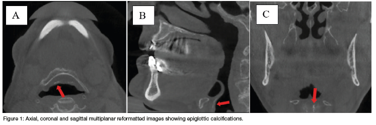
This 70-year-old male patient presented to the Department of Oral and Maxillofacial Radiology for CBCT imaging prior to implant therapy (Figure 1).
INTERPRETATION
The epiglottis play an important preventative role for preventing aspiration and assists in the coordination of swallowing.1 Epiglottic calcification presenting as single or multiple linear hyper-densities anterior to the airway space and posterior to the hyoid bone on CBCT imaging (red arrows). Calcification of the epiglottis has been rarely documented and is poorly understood. It is thought to be a normal physiological degenerative process and can also be a consequence of infection or trauma.12 CBCT imaging including the inferior border of the mandible and the hyoid bone may allow visualisation of this structure and careful interpretation of the images is therefore advised.
Calcification of the epiglottis may alter its morphology and function (elasticity), leading to symptoms such as dysphagia and/or dysphonia.1 Extra-osseous calcification is seen in patients with secondary hyperparathyroidism and renal diseases.1,3 The differential diagnosis for this presentation should include calcifications secondary to granulomatous diseases, calcifications secondary to radiotherapy and calcified tumours of the larynx.3 Thus, this patient may benefit from close monitoring or haematological tests (including biochemical serum testing for parathyroid hormone, vitamin D levels, calcium, or phosphate levels) to rule out systemic disease.4 Current imaging referrals include flexible fibreoptic laryngoscopy and further evaluation by pulmonologist. Advances have also been made in the field of contrast enhanced fluoroscopy.
The aetiology, clinical presentation and outcomes of epiglottic calcifications are poorly understood. Radiographic evaluation together with exclusion of other causes is advised, after ruling out common causes of dysphagia.4
AUTHORS DECLARATION
Funding
This research did not receive any specific grant from funding agencies in the public, commercial, or not-for-profit sectors.
Conflict of Interest
The authors declare that they have no conflict of interest.
Ethics approval
This study was approved by the University of the Western Cape, Faculty of Health Sciences Research Ethics Committee (Reference no.: BM21/03/06). All procedures followed the ethical standards of the Helsinki Declaration of 1975, as revised in 2008.
REFERENCES
1. Jeph, S., Aidi, M., Shah, A., Ly, T.T., Bronov, O. (2017) 'Calcification of the epiglottis presenting as foreign body sensation in the neck', Journal of Radiology Case Reports. EduRad, 11(6), pp. 1-5. doi: 10.3941/JRCR.V11I6.3093. [ Links ]
2. Ampanozi, G., Franckenberg, S., Schweitzer, W., Thali, M.J., Chatzaraki, V. (2021) 'Prevalence of calcified epiglottis in postmortem computed tomography. Is there a correlation to failed endotracheal intubation?', Dentomaxillofacial Radiology, 50(5). doi: 10.1259/dmfr.20200615. [ Links ]
3. URL: https://www.eurorad.org/case/14352 DOI: 10.1594/EURORAD/CASE.14352 ISSN: 1563-4086 [ Links ]
4. Punatar S, Song D, Zaman A, Jiao B, Spyratos T. September 2022 Medical Image of the Month: Epiglottic Calcification. Southwest J Pulm Crit Care Sleep. 2022;25(3):41-42. doi: https://doi.org/10.13175/swjpccs031-22 [ Links ]
 Correspondence:
Dr L Ebrahim
Department of Oral and Maxillofacial Radiology, Faculty of Dentistry, University of the Western Cape
Tel: +27 64 532 6428

