


Serviços Personalizados
Artigo
 Inglês (pdf)
 Artigo em XML
 Referências do artigo
Indicadores

Links relacionados
-
 Citado por Google
 -
 Similares em Google
Compartilhar

 Permalink
South African Dental Journal
versão On-line ISSN 0375-1562
versão impressa ISSN 0011-8516
S. Afr. dent. j. vol.78 no.2 Johannesburg Mar. 2023
RADIOLOGY CORNER
Oral and Maxillofacial Radiology
J WaltersI; S IndermunII
IBChD PDD (MFR) PGD (OS) MSc (MFR); Department of Oral and Maxillofacial Radiology, Faculty of Dentistry, University of the Western Cape, Tygerberg Oral Health Centre, Francie Van Zijl Drive, Cape Town 7505, South Africa. ORCiD: 0000-0002-0593-6890
IIBChD PDD (MFR) MSc (MFR); Department of Oral and Maxillofacial Radiology, Faculty of Dentistry, University of the Western Cape, Tygerberg Oral Health Centre, Francie Van Zijl Drive, Cape Town 7505, South Africa. ORCiD: 0000-0001-6954-0281
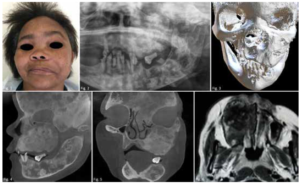
This 40-year-old female presented for extractions of mobile teeth. A bony hard mass affected the left side of the face. She reported being blind and deaf on the affected side. What is your diagnosis?
INTERPRETATION
Clinically (Figure 1) a unilateral swelling, proptosis and obliteration of the nasolabial fold was noted. Intraoral examination revealed normal-appearing overlying mucosa. A pantomograph (Figure 2) demonstrates a mixed diffuse expansile lesion and thinning of the cortices affecting both jaws. 3D reconstruction (Figure 3) overview the lesions' extent. CBCT interpretation (Figures 4 and 5) indicated engrossment of the frontal, parietal, temporal, sphenoid, ethmoid, maxillary, palatine, zygomatic, and mastoid bones. T1-weighted gadolinium-enhanced MRI image (Figure 6) of a patient with a similar lesion in the right maxilla demonstrates a heterogeneous appearance. Fibrous dysplasia (FD) is an uncommon benign non-neoplastic developmental bone disease of fibro-osseous origin. In accordance with which normal medullary bone is replaced by fibrous tissue. Variants are defined as monostotic (MFD), affecting one bone, polystotic (PFD) multiple, and craniofacial includes the head-and-neck only. Association with McCune-Albright syndrome (MAS) is well documented.1 The mean age at the time of diagnosis is 25 years. Onset of PFD ranges from 3 months old in precocious puberty to late 60s in adults. Sex prevalence expressed (male:female) as 1.6:1 (MFD), 1.2:1 (PFD), and 9.4:1 in craniofacial.2 The femur, jaws, skull, and ribs are typically affected. Predilection for the maxilla is approximately double compared to the mandible. Affected sites present painless, progressively enlarging, bony lesions with marked unilateral tendency. Facial asymmetry, malocclusion, and teeth spacing may occur in the jaws. Pigmented skin lesions (café au lait macules) can be observed in MAS.3 Radiographic presentations consist of three distinct patterns. Pagetoid features mixed densities; sclerotic as homogenous "ground glass"; and cyst-like with oval lesions accompanied by sclerotic borders. Indiscernible merging with adjacent bone occurs as lesions mature. Radiation treatment is contraindicated due to documented sarcomatous changes. Differential conditions include ossifying fibroma, Paget disease, florid osseous dysplasia, and osteopetrosis.4
REFERENCES
1. Farah C, Balasubramaniam R, McCullough M. Contemporary Oral Medicine: A Comprehensive Approach to Clinical Practice, 1st ed. Cham: Springer Nature Switzerland AG, 2019: 612-613. [ Links ]
2. Reichart P and Philipsen HP. Odontogenic Tumors and Allied Lesions, 1st ed. Surrey: Quintessence, 2004: 281-291. [ Links ]
3. Larheim TA and Westesson P-L. Maxillofacial Imaging, 2nd ed. Verlag Berlin Heidelberg: Springer, 2008: 81-82. [ Links ]
4. Koch BL, Hamilton BE, Hudgins BA, Harnsberger HR. Diagnostic Imaging: Head and Neck, 3rd ed. Philadelphia: Elsevier, 2017: 922-924. [ Links ]
 Correspondence:
Jaco Walters
Address: Department of Oral and Maxillofacial Radiology, Faculty of Dentistry, University of the Western Cape.
E-mail: jawalters@uwc.ac.za; Tel: +27 21 937 3078/3045
Authors contribution:
Jaco Walters: 70%
Suvarna Indermun: 30%

