


Services on Demand
Article
 English (pdf)
 Article in xml format
 Article references
Indicators

Related links
-
 Cited by Google
 -
 Similars in Google
Share

 Permalink
South African Dental Journal
On-line version ISSN 0375-1562
Print version ISSN 0011-8516
S. Afr. dent. j. vol.78 n.1 Johannesburg Feb. 2023
RESEARCH
The prevalence and associations of radiographic diagnostic signs indicating possible pre-emptive canine ectopia: The results of a mixed dentition radiographic survey
APG HudsonI; L JohanII; AMP HarrisIII; N MohamedIV
IBChD (Stell), HonsBSc (Med Sci) (Stell), MSc (Dent) (UWC); Senior Lecturer, Department of Orthodontics, Faculty of Dentistry, University of the Western Cape. ORCID: https://orcid.org/0000-0001-7971-0598.
IIBDS (SRMC), MSc (Dent) (UWC). ORCID: https://orcid.org/0000-0002-6486-7645
IIIBChD (Stell), HonsBSc (Med Sci) (Stell), Dipl Ter Educ (Unisa), MChD Orthodontics (Stell), FFD(SA) Ortho, PhD (UWC); Professor, Department of Orthodontics, Faculty of Dentistry, University of the Western Cape. ORCID: https://orcid.org/0000-0001-6237-1200
IVProf N Mohamed: BChD (Stell), BSc Hons (Paed Dent) (Stell), MSc (Paed Dent) (Stell), PhD (comm Health) (Stell), MPhil HPE(Stell); Professor, Department of Paediatric Dentistry, Faculty of Dentistry, University of the Western Cape. ORCID: https://orcid.org/0000-0003-2184-2648
ABSTRACT
Maxillary canine ectopia is an anomaly of the mixed dentition which can and should be diagnosed early and treated interceptively wherever possible. Various radiographic markers have been associated with canine ectopia, and these are significant aids to a thorough clinical examination, in order to diagnose ectopia.
METHODOLOGY: A cross sectional study was carried out on a sample of 465 mixed dentition panoramic radiographs in order to establish the prevalence of maxillary canine ectopia according to a set of radiographic markers. The sample of radiographs included patients with dental ages between 10 and 12 years of age
RESULTS: 404 radiographs displayed signs of canine ectopia according to the markers studied. Non- resorption of the root of the primary canine was the most common marker (63%) found. This was followed by overlap in 25.2% of cases, whilst increased angulation of the developing canine was the least prevalent (4.7%). Non-resorption showed a statistically significant association with distal overlap and overlap over the pulp chamber. Increased angulation was significantly associated with non-resorption in all degrees of overlap. Unilateral increased size of the mandibular canine showed a significant association with cases displaying a mesial overlap (p= 0.027
CONCLUSION: Dental age is an important aspect of predicting canine ectopia. Non-resorption of the roots of the primary canine must be viewed with caution at the dental age of 10 years. Enlargement of the mandibular canine maybe viewed as a potential early warning sign for maxillary canine ectopia
Keywords: Dental age; Nolla stage 9, Canine ectopia; Maxillary canines; Interceptive orthodontics, Mixed dentition, Panoramic radiographs, non-resorption of primary canine roots
INTRODUCTION
It is not uncommon to encounter maxillary canine ectopia when managing paediatric patients.1 Maxillary canine ectopia may present either pre-eruptively or post-eruptively. Pre-eruptive ectopia occurs due to the tooth germ being displaced, which then causes the tooth to erupt along the wrong path.2 Post-eruptive ectopia refers to a tooth that has erupted into the mouth but is out of its normal position.3 There are important considerations that need to be accounted for when assessing the developing canines during the mixed dentition. The most critical of these is whether or not the buccal bulge resulting from the developing canine is present. This should be clinically palpable from a dental age of 10 years. In the absence of this buccal bulge, a radiographic investigation is considered to be the preferred practice in order to verify whether the developing canine is ectopic or not.1 These have been identified as:1
• The amount of overlap between the crown tip of the developing canine and the root of the lateral incisor
• The angulation of the long axis of the developing canine to the mid sagittal plane
• Resorption or non-resorption of the roots of the primary canines and
• The size of the developing canine when compared to the contralateral canine.
On a pantomogram, a visible overlap between the cusp tip of the erupting maxillary canine and the permanent maxillary lateral incisor root may be considered normal, prior to the maxillary lateral incisor reaching Nolla's root development stage 9. At this stage, the lateral incisor roots have developed to full length but still have open apices, and half the root of the permanent maxillary canine has formed but active eruption has not yet started.4,5 Nolla stage 9 coincides with a dental age of 10 years.4 However, after Nolla stage 9, the degree of overlap of the permanent maxillary canine cusp tip and maxillary lateral incisor root is considered to be a significantly accurate indicator of potential pre-eruptive maxillary canine ectopia.6,7
Ericson and Kurol (1988a)8 evaluated the degree of overlap of the permanent maxillary canine cusp tip with the root of the maxillary lateral incisor. The amount of overlap between lateral incisor root and canine tip was grouped as follows:
• Group 1 displayed no overlap,
• Group 2 displayed an overlap where the cusp tip was located distal to the pulp chamber of the lateral incisor.
• Group 3 displayed an overlap where the cusp tip was located directly over the pulp chamber of the lateral incisor.
• Group 4 applied to all instances where the overlap of the cusp tip was located mesial to the pulp chamber of the lateral incisor.
Warford et al., (2003)6 used this method to gauge maxillary canine displacement and predict the non-eruption of the maxillary canine and they found that groups 3 and 4 had the highest odds of non-eruption of the maxillary canine. Lindauer et al., (1992)9 found that 78% of cases with non-erupted maxillary canines exhibited an overlap as described in groups 2, 3 and 4, above. The amount of overlap after the dental age of 10 determines the prognosis of the interceptive treatment. A positive outcome of interceptive treatment decreases if the erupting maxillary canine overlaps more than half of the root of the maxillary lateral incisor at dental age 11.10,11 A clinical study by Ericson and Kurol (1988)10 showed that 91% of cases respond favourably to interceptive treatment when the canine crown-lateral root overlap is distal to the midline of the lateral incisor. This success rate drops significantly to 64% should the canine overlap be mesial to the midline of the lateral incisor.
Baccetti et al., (2008)12 modified Ericson and Kurol's classification (1988a)10 and measured the medial crown position of the maxillary canine through sectors 1 to 5:
• Sector 1 corresponded to the primary maxillary canine (present or absent).
• Sector 2 was the area from the distal aspect of the maxillary lateral incisor to the midline of the maxillary lateral incisor.
• Sector 3 was the area from the midline of the maxillary lateral incisor to the distal aspect of the maxillary central incisor.
• Sector 4 corresponded to the area from the distal side of the maxillary central incisor to the midline of the central incisor.
• Sector 5 was designated to the area from the midline of the central incisor to the midline of the maxillary arch.
This modification showed a high rate of reproducibility (0.94) with both methods giving accurate results.12
Angulation of the maxillary canines to the mid-sagittal plane appears to be less significant than the amount of overlap between the cusp tip of the maxillary canine and the root of the maxillary lateral incisor.6 A favourable inclination for the maxillary canine in the arch is no more than 30° to the mid-sagittal plane.11 The angle is measured between the long axis of the maxillary canine and the midline.8 Landmarks on the panoramic radiograph such as the intermaxillary suture, anterior nasal spine, nasal septum and internasal suture, demarcates the midline.13 When the developing maxillary canine (after the dental age of 10 years) has an angulation greater than 30°, it suggests that the maxillary canine has a greater tendency to become impacted.11
External root resorption of the primary canines associated with the erupting permanent canines occurs in the apical area of the root, causing a smooth resorption pattern which results in blunting of the root apex.4,5 When external root resorption involves the lateral aspect of roots, it causes an irregular resorption pattern where one side is resorbed more than the other.14 This can occur unilaterally or bilaterally.15 Resorption of primary maxillary canines should have begun by dental age 10 and be completed by dental age 12.16
Lappin (1951)17 put forward non-resorption of the root of the primary maxillary canine as a possible cause for maxillary canine ectopia. In support of Lappin's (1951)17 theory, various studies have shown the subsequent eruption of the displaced maxillary canines, following the extraction of non-resorbed primary maxillary canines.18,8,9,11
Ericson et a!,, (2002)19 however, suggested that root resorption of the primary maxillary canines was merely a consequence of maxillary canine ectopia rather than a cause of it. As part of the eruptive mechanism of the permanent maxillary canine, the active pressure exerted causes various cellular changes which, together with the actual physical contact between the adjacent primary teeth and the permanent maxillary canine, brings about root resorption of the primary maxillary canine. Becker (1998)2 suggested that the erupting permanent maxillary canine provides the stimulus for the resorption of the roots of the primary maxillary canine. A portion of the root far from the permanent canine may be unaffected by this process, thus bringing about non-resorption of the primary maxillary canine.
An enlarged radiographic image of a maxillary/ mandibular canine in comparison to its opposite number and surrounding teeth, indicates palatal positioning of the tooth on the panoramic radiograph.20,21,5 Palatally displaced maxillary canines have been studied by several authors, where the palatally positioned maxillary canines were verified both clinically and radiographically.22,23,24,25,26,27,8,15
A mandibular canine that is situated lingually may be as a result of spontaneous early loss of a primary mandibular canine, an unfavourable sequence of permanent tooth eruption in the mandible or eruption anomalies. These may be determined radiographically and clinically where an early warning sign may be crowded mandibular incisors.28
This lingual ectopia of mandibular canines may cause a lingual collapse of the mandibular incisors which can impact on the maxillary incisors by diminishing the space available for normal eruption of the maxillary canines, thus forcing them into an ectopic position.28
METHODOLOGY
An analytical, descriptive, cross-sectional study was carried out to establish the radiographic prevalence of potential maxillary canine ectopia visible on mixed dentition panoramic radiographs between the dental ages of 10 and 12 years as described by Proffit et al., (2007).4
A sequential sample consisting of 465 radiographs from UWC's Paediatric Dentistry department at the Tygerberg campus, were used for this study. The radiographs were taken between 2011 and 2014. The data was recorded according to the presence of one or more of the four canine ectopia prediction markers as described by Hudson et al., (2010).1 These markers have been identified as:
• The amount of overlap between the crown tip of the developing canine and the root of the lateral incisor.
• The angulation of the developing canine to the mid sagittal plane.
• Resorption or non-resorption of the roots of the primary canines.
• The size of the developing canine when compared to the contralateral canine. The size of both the maxillary and mandibular canines were assessed in order to establish the probability of any relationship between mandibular canine ectopia and maxillary canine ectopia.
Inclusion criteria
1. Patients with no previous history of orthodontic treatment.
2. Only good quality dental panoramic radiographs were used.
Exclusion criteria
1. Patients with cleft lip and palate.
2. Patients with syndromes.
3. Panoramic radiographs with only primary or only permanent dentitions.
Data processing and analysis
Pearson's correlation coefficient was used to determine the degree to which two variables were associated. For a correlation coefficient to show a statistically significant association, its absolute value must exceed 0.0834. The Chi-square test of independence and Fishers exact test were also used to determine whether two categorical variables were dependent or independent. A p-value of less than 0.05 indicates that the variables have a statistically significant association.
RESULTS
Of the 465 mixed dentition panoramic radiographs of children between dental ages 10-12 years, 404 displayed potentially ectopic maxillary canines according to the markers studied.
Non-resorption of the primary maxillary canines showed a statistically significant association with overlap:
• Distal overlap (p < 0.001)
• Overlap over the pulp chamber (p = 0.003)
The probability test showed that:
• Non-resorption of the primary canines was 63% more likely to occur when distal overlap was present.
• There was a 76% chance of non-resorption in cases where the overlap was over the pulp chamber.
• In cases where non-resorption of the primary canine existed, there was 19.8% chance for distal overlap or only a 6% chance for overlap over the pulp chamber to occur.
With angulation greater than 300 as the primary marker, a statistically significant association was found with distal overlap (p < 0.001). The probability test showed that:
• Maxillary canines angulated greater than 300 were 39% more likely to cause the maxillary canine cusp tip to have a distal overlap with the root of the maxillary lateral incisor.
• In cases where there was an existing distal overlap, angulation of the maxillary canine had an 11% chance that the angulation would become greater than 300.
Angulation greater than 300 also showed a statistically significant association with overlap over the pulp chamber of the maxillary lateral incisor root (p= 0.014). The probability test showed that:
• There was a 13% chance for the maxillary canine cusp tip to overlap the pulp chamber of the root of the maxillary lateral incisor.
• When an overlap existed over the pulp chamber, the probability for the maxillary canines to have an angulation greater than 300 was 14%.
Maxillary canines angulated greater than 300 also showed a statistically significant association (p= 0.015) with an overlap that was mesial to the pulp chamber. The probability test showed that:
• There was a small chance (8.7%) for the maxillary canine cusp tip to be positioned mesial to the maxillary lateral incisor root.
• The probability of the maxillary canine to have an angulation greater than 300 doubled to 18% when a mesial overlap existed.
With increased angulation as the primary marker, a statistically significant association with non-resorption of primary maxillary canines (p = 0.004) was found. The probability test showed that:
• Non-resorption of the primary canines was 74% more likely to occur when the maxillary canines had an angulation greater than 300.
• When non-resorption of the primary canine existed, there was only a 7% chance for the angulation of the maxillary canines to be greater than 300.
When maxillary canine enlargement was the primary marker, non-resorbed primary maxillary canines occurred 48.1% of the time and 57.1% of the time when enlarged mandibular canines was the primary marker (Table 5). Maxillary canine enlargement (p = 0.32) and mandibular canine enlargement (p = 0.65) did not show a statistically significant association with non-resorption of primary maxillary canines.
Mandibular canine enlargement as the primary marker showed no statistically significant association with distal overlap or overlap over the pulp chamber of the root of the maxillary lateral incisor (p > 0.05). However, there was a statistically significant association with those cases displaying a mesial overlap (p= 0.027). The probability test showed:
• A 6% chance of a mesial overlap in cases with enlarged mandiular canines.
• It was 27% more likely for the mandibular canine to be lingually displaced when the maxillary canine displayed a mesial overlap.
DISCUSSION
This study showed that radiographic evidence of potentially ectopic maxillary canines seems to become less prevalent as the dental age increases from 10 to 12 years (Tables 1 and Table 3). The timing of normal eruption of the maxillary canine should coincide with a dental age of 12 years.4
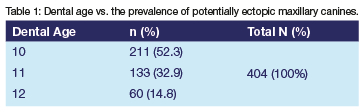
Thilander and Jakobsson (1968)29 examined dental casts and radiographs and recorded an ectopia prevalence of 37% for unerupted maxillary canines at the initial examination in cases with a mean chronological age of 11.5 years. In the present study, the prevalence of potentially ectopic maxillary canines at dental age 12 was 14.8% (Table 1). The differences in the findings may be because Thilander and Jakobsson (1968)29 had access to the models and did not use all the radiographic markers used in this study, but more importantly, the present study used dental age and not chronological age. Studies have shown that dental age can differ by between 4 to 5 years from the actual chronological age.30,31 Davidson and Rodd (2001)32 found that the difference between dental age and chronological age was most evident in 8 to 12-year-old children.
If a clinical examination had been conducted in the present study and it was found that the maxillary canine buccal bulge was palpable and/ or the primary canine was mobile, the 404 radiographs showing potential ectopia may have been judged to be displaying normal canine development.
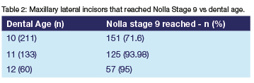
Any overlap of the permanent maxillary canine is to be considered normal prior to the permanent maxillary lateral incisor reaching Nolla Stage 9.7 Table 2 shows the dental age distribution of the maxillary lateral incisor at Nolla Stage 9. In three cases at dental age 12, the maxillary lateral incisors did not reach Nolla stage 9 but were close to reaching this stage (Table 2).
Using the Erickson and Kurol (1988a)8 overlap classification, 25% of the potentially ectopic maxillary canine cases presented with an overlap, the prevalence of which is shown in each age group in Table 3. Chalakkal et al., (201 1)33 found that overlap displayed a prevalence of 73%. The difference seen was due to variations in criteria in the sample selection. Children between the chronological ages of 10 and 12 years were selected. These cases were clinically examined and only cases with unilaterally palpable maxillary canine bulges were included in the study. In the present study, 2.6% of the potentially ectopic maxillary canines overlapped the root of the lateral incisor mesial to the pulp chamber (Table 4). Chalakkal et al., (2011)33 found that 30% of the maxillary canines were positioned mesial to the root of the maxillary lateral incisors between the chronological ages of 10 and 12 years. In this study non-resorption of primary canines did not show a statistically significant association with mesial overlap (p = 0.21). This may have resulted due to the dental age at which mesial overlap is being identified. If non-resorption of primary canines and mesial overlap were studied in an older age group, the results may have been different. Mesial overlap is perhaps an extreme situation at dental ages 8-12 years but this needs to be further investigated.
Proffit et al., (2007)4 and Duterloo (1991)5 suggested that resorption of the apical third of the root of the primary maxillary canines should have taken place at dental age 10. The high prevalence (83%) of non-resorption of primary canines at dental age 10 (Table 3) suggests one of two conclusions:
• A potentially ectopic maxillary canine is present and has not resorbed the root of the primary canine.
• The minor resorption of the primary maxillary canine was not clearly visible from the panoramic radiograph.
Between the dental ages of 10 and 12 years, there is a decline in the prevalence of non-resorbed primary canines (Table 3). As the permanent maxillary canine actively erupts during dental age groups of 10 to 12 years, the resorption of the root of the primary maxillary canine should occur at the same time.4,5 Duterloo (1991)5 also stated that resorption of the primary maxillary canine should take place at this age. It is therefore normal to find a decline in the number of non-resorbed primary maxillary canines from dental age 10 to 12 years as shown in Table 3.
The present study had one marker (non-resorption of primary maxillary canines) in common with the study conducted by Thilander and Jakobsson (1968).29 The present study showed a prevalence of non-resorption of 16.7% at dental age 12 years (Table 3) compared to Thilander and Jakobsson (1968)29 who recorded non-resorption of primary maxillary canines as 67% at chronological age of 12 years.
The fundamental problem is that early stages of root resorption are difficult to detect on a panoramic radiograph as it is two-dimensional and root resorption in the 3rd dimension may only be identified at a later stage or not at all.21,34 Furthermore, the fact that overlap, particularly of the first premolars and the root of the primary canine at dental age of 10 years, is a complicating factor.35
This study showed 25.9% of the cases displayed an overlap (all types), when non-resorbed primary maxillary canines was viewed as the primary marker while 65% had non-resorption of primary canines when overlap (all types) was viewed as the primary marker (Table 5). The probability test findings suggest that non-resorption of the primary canines is more a consequence of potentially ectopic maxillary canines rather than a cause, thus concurring with the work of Ericson et al., 19.
Warford et al., (2003)6 found a degree of overlap to be a significant predictor of maxillary canine impaction when compared to angulation of the maxillary canine. This was only possible because they had the impaction status of the maxillary canines, allowing them to run a logistic regression test between the two predictive markers. Since the present study could not determine the impaction status of the maxillary canines, their statement could not be verified. However, as the severity of overlap increased, potentially ectopic maxillary canines became less prevalent (Table 3). This suggests that an absence of the buccal canine bulge upon clinical examination (dental age > 10 years) and identifying the degree of overlap on the panoramic radiograph could act as a good predictor of ectopic maxillary canines.
Although increased angulation showed a statistically significant association with overlap (all types), the statistical results above suggest that the marker did not add significantly to the prediction of ectopic maxillary canines when compared to overlap as a marker. Most of the maxillary canines positioned over the pulp chamber or mesial to the root of the maxillary lateral incisor will become impacted. Hence, the small increase that angulation contributes is not clinically significant. Angulation would only have significance in predicting impaction for the maxillary canines positioned distal to the root of the maxillary lateral incisor, confirming the work of Warford et al., (2003).6
When non-resorption of the primary maxillary canine was the primary marker 6.6% of the cases had angulated maxillary canines greater than 300. When angulated maxillary canines greater than 300 was the primary marker (Table 5), 89.5% of the cases displayed non-resorption of primary canines. The probability tests in this study suggest that non-resorption of the primary maxillary canines is caused by an ectopically erupting maxillary canine and is not a cause of it thus confirming the work of Warford.
Maxillary canine enlargement was detected in 12.9% (52/ 404) of the radiographs studied (Table 3). When overlap was the primary marker, enlargement occurred in 15.5% of the cases. When enlargement was the primary marker overlap (all types) occurred 30.8% of the cases (Table 5). Maxillary canine enlargement resulted in no statistically significant association with distal overlap (p= 0.21), overlap over the pulp chamber (p= 0.64), and/ or mesial overlap (p= 0.45).
Other studies are yet to examine the relationship between these two radiographic markers. This study however, suggests that when the maxillary canine is palatally displaced and the maxillary canine bulge is not palpable by dental age 10, the maxillary canine is likely to overlap the root of the adjacent maxillary lateral incisor to some extent. As the dental age of the patient increases, the extent of the overlap may worsen.
This result of this study suggests that if there is an ectopically positioned mandibular canine, there is a chance that an ectopic maxillary canine also exists (Table 5). Further investigations are needed to reveal any clear link between mandibular canine ectopia and maxillary canine ectopia. Both maxillary canine enlargement and mandibular canine enlargement were identified in approximately 9% of cases when non-resorbed primary maxillary canine was the primary marker (Table 5). Non-resorbed primary maxillary canines occurred 48.1% of the time when maxillary canine enlargement was the primary marker and 57.1% of the time when enlarged mandibular canine was the primary marker (Table 5). Maxillary canine enlargement (p = 0.32) and mandibular canine enlargement (p = 0.65) did not show a statistically significant association with non-resorption of primary maxillary canines. This result could not be compared since no other studies have examined the relationship of these anomalies.
CONCLUSIONS
Dental age is an important aspect in the diagnosis of canine ectopia, as there is little correlation between dental and chronologic age.
From the dental age of 10 years, regular thorough clinical examinations, buccal bulge palpations and primary canine mobility assessments are vital to the monitoring of the developing canine. In patients with dental age greater than 10 years, the absence of the buccal canine bulge on clinical examination and identifying the degree of overlap on the panoramic radiograph, could be valuable indicators of maxillary canine ectopia.
Non-resorption of the apical third of the root of the maxillary canines at dental age 10 years should be interpreted with caution, as the early stages of root resorption can be difficult to detect on panoramic radiographs and overlap may be present between the first premolar and the root of the primary canine. The findings of this study suggest that non-resorption of the primary canines may more likely be a consequence of potentially ectopic maxillary canines rather than the cause of it.
Canines positioned over the pulp chamber or mesial to the root of the maxillary lateral incisor will most likely become impacted. Angulation would only be a significant predictor of ectopia for canines positioned distal to the root of the maxillary lateral incisor.
Clinicians who detect an enlarged mandibular canine on a panoramic radiograph i.e. a lingually displaced mandibular canine, should be aware of the possibility for the maxillary canine to also become ectopic as it should erupt a year after the mandibular canine. Since mandibular canines develop earlier than the maxillary canines, clinicians can take timeous interceptive measures if needs be.
REFERENCES
1. Hudson APG, Harris AMP, Mohamed N. Maxillary canine management in the preadolescent: A guide for general practitioners. SADJ. 2010; 65(8): 366-370. [ Links ]
2. Becker A. Palatally impacted canines: In: Orthodontic treatment of impacted teeth. Martin Dunitz Ltd, 1998: 85-101. [ Links ]
3. Nikiforuk G. Ectopic Eruption: Discussion and clinical report. J Ont Dent Assoc. 1948; 25:243-246. [ Links ]
4. Proffit WR, Fields HW and Sarver DM. The later stages of development. In: Contemporary Orthodontics. Mosby Elsevier, 2007: 63-94. [ Links ]
5. Duterloo HS. Development and chronology of the dentition and Abnormalities of dentitional development. In: An atlas of dentition in childhood, orthodontic diagnosis and panoramic radiology. Wolfe Publishing Limited, 1991: 69-96, 129-196. [ Links ]
6. Warford JH, Grandhi RK, Tira DE. Prediction of maxillary canine impaction using sectors and angular measurement. Am J Orthod Dentofac Orthop. 2003; 124(6): 651655. [ Links ]
7. Fernandez E, Bravo LA, Canteras M. Eruption of the permanent upper canine: A radiological study. Am J Orthod Dentofac Orthop. 1998; 113(4): 414-420. [ Links ]
8. Ericson S, Kurol J. Early treatment of palatally erupting maxillary canines by extraction of the primary canines. Eur J Orthod. 1988a; 10(4): 283-295. [ Links ]
9. Lindauer SJ, Rubenstein LK, Hang WM, Andersen WC, Isaacson RJ. Canine impaction identified early with panoramic radiographs. JADA. 1992; 123(3): 91-7. [ Links ]
10. Ericson S, Kurol J. Resorption of maxillary lateral incisors caused by ectopic eruption of the canines. A clinical and radiographic analysis of predisposing factors. Am J Orthod Dentofac Orthop. 1988b; 94(6): 503-13. [ Links ]
11. Power SM, Short MB. An investigation into the response of palatally displaced canines to the removal of deciduous canines and an assessment of factors contributing to favourable eruption. Br J Orthod. 1993; 20(3): 217- 23. [ Links ]
12. Baccetti T, Leonardi M, Armi P. A randomized clinical study of two interceptive approaches to palatally displaced canines. Eur J Orthod. 2008; 30(4): 381-385. [ Links ]
13. Sajnani AK, King NM. Early prediction of maxillary canine impaction from panoramic radiographs. Am J Orthod Dentofac Orthop. 2012; 142(1): 45-51. [ Links ]
14. White SC, Pharaoh MJ. Oral radiology: Principles and interpretation. 6th edition. Mosby Elsevier, 2010; 295-304. [ Links ]
15. Peck S, Peck L, Kataja M. The palatally displaced canine as a dental anomaly of genetic origin. Ang Orthod. 1994; 64(4): 249-256. [ Links ]
16. Van der Linden PGM, Duterloo HS. Development of the Human Dentition: An Atlas. Harper and Row, 1976: 75-212. [ Links ]
17. Lappin M. Practical management of the impacted maxillary cuspid. Am J Orthod Dentofac Orthop. 1951; 37(10): 769-78. [ Links ]
18. Howard RD. The unerupted incisor. A study of the postoperative eruptive history of incisors delayed in their eruption by supernumerary teeth. Dent Pract Dent Rec. 1967; 17(9): 332-41. [ Links ]
19. Ericson S, Bjerklin K, Falahat B. Does the canine dental follicle cause resorption of permanent incisor roots? A computed tomographic study of erupting maxillary canines. Angle Orthod. 2002; 72(2): 95-104. [ Links ]
20. Hudson APG, Harris AMP, Mohamed N. The mixed dentition pantomogram: A valuable dental development assessment tool for the dentist. SADJ. 2009; 64(10): 480-483. [ Links ]
21. Mason C, Papadakou P, Roberts GJ. The radiographic localization of impacted maxillary canines: a comparison of methods. Eur J Orthod. 2001; 23(1): 25-34. [ Links ]
22. Becker A, Smith P, Behar R. The incidence of anomalous maxillary lateral incisors in relation to palatally displaced cuspids. Angle Orthod. 1981; 51(1): 24-29. [ Links ]
23. Becker A, Zilbermann Y Tsur B. Root length of lateral incisors adjacent to palatally displaced maxillary cuspids. Angle Orthod. 1984; 54(3): 218-225. [ Links ]
24. Baccetti T.A controlled study of associated dental anomalies. Angle Orthod. 1998a; 68(3): 267-274. [ Links ]
25. Nagpal A, Pai KM, Sharma G. Palatal and labially impacted maxillary canine associated dental anomalies: A comparative study. J Contemp Dent Pract. 2009; 10(4): 1-11. [ Links ]
26. Shapira Y, Kuftinec MM. Early diagnosis and interception of potential maxillary canine impaction. JADA. 1998; 129(10): 1450-1454. [ Links ]
27. Liuk IW, Olive RJ, Griffin M, Monsour P Associations between palatally displaced canines and maxillary lateral incisors. Am J Orthod Dentofac Orthop. 2013; 143(5): 622632. [ Links ]
28. Hudson APG, Harris AMP, Mohamed N. Early identification and management of mandibular canine ectopia. SADJ. 2011; 66(10): 462-467. [ Links ]
29. Thilander B, Jakobsson SO. Local factors in impaction of maxillary canines. Acta Odontol Scand. 1968; 26(2): 145-168. [ Links ]
30. Hurme VO. Ranges in normalcy in the eruption of permanent teeth. J Dent Child. 1949; 16(2): 11-5. [ Links ]
31. Taranger J, Lichtenstein H, Svennberg-Redegren I. Dental development from birth to 16 years. Acta Paediatrica. 1976; 65: 83-97. [ Links ]
32. Davidson LE, Rodd HD. Interrelationship between dental age and chronological age in Somali children. Community Dent Health. 2001; 18(1): 27-30. [ Links ]
33. Chalakkal P, Thomas AM and Chopra S. Displacement, location, and angulation of unerupted permanent maxillary canines and absence of canine bulge in children. Am J Orthod Dentofac Orthop. 2011; 139(3): 345-350. [ Links ]
34. Rimes RJ, Mitchell CNT, Willmot DR. Maxillary incisor root resorption in relation to the ectopic canine: A review of 26 patients. Eur J Orthod. 1997; 19(1): 79-84. [ Links ]
35. Falahat B, Ericson S, D'Amico RM, Bjerklin K. Incisor root resorption due to ectopic maxillary canines: A long-term radiographic follow-up. Angle Orthod. 2008; 78(5): 778785. [ Links ]
 Correspondence:
N Mohamed
Department of Paediatric Dentistry, Faculty of Dentistry, University of the Western Cape
Private Bag X1, TYGERBERG 7505, South Africa
E-mail: namohamed@uwc.ac.za
Author contributions:
Dr A Hudson: Writing article 20%
Dr L Johan: Principal researcher 40%
Prof A Harris: Writing article, clinical input 20%
Prof N Mohamed: Writing article, editing 20%
Conflict of interest: None

