


Services on Demand
Article
 English (pdf)
 Article in xml format
 Article references
Indicators

Related links
-
 Cited by Google
 -
 Similars in Google
Share

 Permalink
South African Dental Journal
On-line version ISSN 0375-1562
Print version ISSN 0011-8516
S. Afr. dent. j. vol.77 n.10 Johannesburg Nov. 2022
http://dx.doi.org/10.17159/2519-0105/2022/v77no10a9
RADIOLOGY CASE
C SmitI; L RobinsonII
IBChD, MSc (Maxillofacial and Oral Radiology). Department of Oral and Maxillofacial Pathology, University of Pretoria. ORCID: 0000-0003-4047-6356
IIBChD, PDD (MaxillofacialRadiology), PDD (Forensic Odontology), MChD (Oral Path), FC Path (SA) Oral Path. Department of Oral and Maxillofacial Pathology, University of Pretoria. ORCID: 0000-0002-0549-7824
CASE
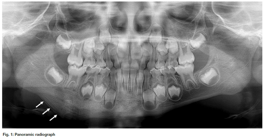
A 5-year-old healthy female patient presented with a one-year history of a slow-growing swelling of the right mandible. The patient reported that the swelling was slightly tender. Intraoral examination revealed a grossly carious lower right primary molar (tooth 85). A panoramic radiograph showed bony expansion of the inferior mandibular border with a lamellated or 'onion-skin' appearance. The trabecular bone in the vicinity had a sclerotic appearance. What is your diagnostic hypothesis?
INTERPRETATION
Chronic osteomyelitis with an associated periosteal reaction has been described under a multitude of different terms including Garrè's osteomyelitis, proliferative periostitis and periostitis ossificans.1 Carl Garrè's publication of periosteal
new bone formation in a so-called 'onion-skin' pattern has forever linked his name to this condition.1-3 Interestingly, Wood et al found that Garrè did not actually describe this type of osteomyelitis in his original publication.4 The term Garrè's osteomyelitis has however remained and is now synonymous with the more widely accepted term chronic osteomyelitis with proliferative periostitis.
Chronic osteomyelitis with proliferative periostitis usually affects young patients with a mean age of 13 years and a near equal gender predilection.1,2 Most cases arise in the molar-premolar region of the mandible, involving the lower border or buccal aspect in most instances.1,2 Common causative factors of this condition include dental caries with associated periapical inflammation, periodontal infection, fractures, and other nonodontogenic infections of the jaw bones.1 The new subperiosteal bone formation represents a bony reaction to persistent low-grade inflammation in the region. The reason for the propensity of this condition in young patients is likely related to the ease with which the periosteum may be separated from the bone. Additionally, this age group has a greater susceptibility to caries in the region of involvement.2
Radiographic examination shows bony laminations parallel to each other and to the cortical surface of the involved bone. The cortical bone is usually thickened and the adjacent jawbone usually appears normal.1-3 Appropriate imaging may show an associated soft tissue swelling, resulting in facial asymmetry.1,3 The histopatho-logical features are distinct, characterised by periosteal histopathologic layering of vital bone parallel to each other and the inferior surface of the bone.1
The radiographic differential diagnoses list includes both benign and malignant conditions including osteosarco-ma, Ewing's sarcoma, infantile cortical hyperostosis, callus formation, bony exostosis, and osteomas.1,3 Signs of malignant bony changes should be viewed with caution and necessitates an appropriate biopsy.2
Removal of the causative agent results in resolution of the infection with the eventual remodeling of the bone.1, 2 Surgical recontouring may be performed in cases without spontaneous regression.3
Authors declaration Funding
This research did not receive any specific grant from funding agencies in the public, commercial, or not-for-profit sectors.
Conflict of Interest
The authors declare that they have no conflict of interest.
Ethics approval
This study was approved by the University of Pretoria, Faculty of Health Sciences Research Ethics Committee (Reference no.: 587/2022). All procedures followed the ethical standards of the Helsinki Declaration of 1975, as revised in 2008.
REFERENCES
1. Tong AC, Ng IO, Yeung KA. Osteomyelitis with proliferative periostitis: an unusual case. Oral Surgery, Oral Medicine, Oral Pathology, Oral Radiology, and Endodontology. 2006 Nov 1;102(5):14-19. [ Links ]
2. Nortje CJ, Wood RE, Grotepass F. Periostitis ossificans versus Garrè's osteomyelitis: Part II: Radiologic analysis of 93 cases in the jaws. Oral surgery, oral medicine, oral pathology. 1988 Aug 1;66(2):249-260. [ Links ]
3. Belli E, Matteini C, Andreano T. Sclerosing osteomyelitis of Garré periostitis ossificans. Journal of Craniofacial Surgery. 2002 Nov 1;13(6):765-768. [ Links ]
4. Wood RE, Nortjé CJ, Grotepass F, Schmidt S, Harris AM. Periostitis ossificans versus Garrè's osteomyelitis. Part I. What did Garrè really say?. Oral surgery, oral medicine, oral pathology. 1988 Jun 1;65(6):773-777. [ Links ]
 Correspondence:
Chané Smit
Department of Oral and Maxillofacial Pathology
University of Pretoria
Tel +27 (0)12 319 2311
Email: chane.smit@up.ac.za
Authors contribution:
Chané Smit: 50%
Liam Robinson: 50%

