


Serviços Personalizados
Artigo
 Inglês (pdf)
 Artigo em XML
 Referências do artigo
Indicadores

Links relacionados
-
 Citado por Google
 -
 Similares em Google
Compartilhar

 Permalink
South African Dental Journal
versão On-line ISSN 0375-1562
versão impressa ISSN 0011-8516
S. Afr. dent. j. vol.77 no.6 Johannesburg Jul. 2022
http://dx.doi.org/10.17159/2519-0105/2022/v77no6a1
RESEARCH
Determination of the influence of body mass percentile on mandibular canine calcification stages among 5-17 years old Northern Nigerian children
Osaronse Anthony AghimienI; Osasumwen Aghimien-OsaronseII
IBDS, FWACS), Federal Medical Centre, Keffi, Nasarawa State. Nigeria. ORCID number: 0000-0002-6737-7959
IIBDS), University of Benin Teaching Hospital, Benin City, Edo State. Nigeria
ABSTRACT
BACKGROUND: Dental calcification is a biological phenomenon used to estimate the maturation status of growing children. The effect of body mass index percentile (BMI-percentile) on this process appears contentious among researchers
AIMS AND OBJECTIVES: To determine the predictive effect of body mass percentile on mandibular canine calcification.
DESIGN: A prospective descriptive cross-sectional study
METHODS: This was a prospective cross sectional descriptive study comprising of eighty four participants (5-17 years) who visited the Child Health Dental Clinic of Federal Medical Centre, Keffi, Nigeria between January and September, 2021. Mandibular canine calcifications of the study participants were staged using the Demirjian method while the World Health Organisation growth chart specific for age and gender was used to classify the BMI-percentile. The effect of BMI-percentile on the mandibular canine calcifications was determined using multinomial logistic regression
RESULTS: Chronological age had a significant predictive effect on the mandibular canine calcification (P=0.002) as against gender and BMI-percentile. A one-percentile increase in the BMI-percentile increases the likelihood of healthy children of having to present in stage D by 3.454 compared to obese children, but this effect was not statistically significant (P= 1.000
CONCLUSIONS: Obese children have a tendency of having advanced mandibular canine calcification than healthy children. Female participants were likely to be in advanced mandibular canine calcification stage. Early intervention is therefore suggested for obese children
Key words: Body mass percentile, mandibular canine, calcification.
INTRODUCTION
In 2006, the World Health Organisation (WHO) released standard growth charts for describing growth of healthy children in ideal conditions.1 These charts were used to assess the nutritional status of children globally. Children and adolescents were categorized as either underweight, healthy, overweight or obese using body mass index percentile (BMI-percentile) scores of <5%, 5%-85%, 85%-95% and >95% respectively.
The prevalence of obesity is on the rise globally with increasing trend in developing countries like Nigeria.2-4 Continuous increase in the BMI-percentile of an individual can affect their general wellbeing by; predisposing them to increased blood pressure, coronary heart disease and increased risk of diabetic mellitus. It can also affect their dental development.5
Dental development is a biological phenomenon that is used to estimate the maturation of growing children. Orthodontists and paedodontists consider dental development as a crucial parameter that is usually evaluated before commencing treatment on their patients. Apart from dental eruption age, the dental formation stages (calcification/mineralization) of the teeth are more confidently used to estimate an individual's dental age because it has been reported to follow a more independent process and can be evaluated at any stage of its developmental process.6,7 The effects of changes in BMI-percentile on dental development have been studied by several researchers.8-17 Changes in the BMI-percentiles have been reported to either cause acceleration or a delay in tooth eruption.8-11 Advancement in dental age among overweight and obese children as against normal healthy individuals have also been reported in different population.12-17 Recently, it was reported that black South Africans overweight children aged 5-20 years were observed to be significantly advanced in their dental development when compared to underweight children.18 If nutritional status as classified by the BMI-percentile have influences on dental development, it cannot be said to absolutely follow an independent process of growth.18 The effect of BMI-percentile on dental maturation have been observed to be dichotomous among researchers.8-22 While some researchers observed BMI-percentile to have a statistically significant explanatory effect on dental maturation,18-20 some others have reported non-statistically significant explanatory effects.21,22
The effect of increased BMI-percentile on orthodontic tooth movement is still considered controversial among researchers.23
There is no doubt that fluctuation in BMI-percentile can influence dental development. However, there appears to be scarcity of researches on the influence of BMI-percentile on dental development among Nigerians, despite the increasing prevalence of overweight and obesity among the Nigerian population.24 This study was conducted to determine the influence of BMI-percentile on the developmental stages of the right mandibular canine in Northern Nigerian children using the Demirjian method.25 It was hypothesized that there was no statistically significant correlation between BMI-percentile and the developmental stages of the right mandibular canine. Secondly, that BMI-percentile has no statistically significant explanatory effect on the developmental stages of the right mandibular canine. The outcome of this study will guide clinicians especially those in the fields of Orthodontists and paedodontists on proper timing of implementation of treatment procedures.
MATERIALS AND METHODS
Study population
This was a prospective cross-sectional study. It included 84 children and adolescents that visited the Child Health Dental Clinic, Federal Medical Centre, Keffi, Nigeria (between January 2021 and September, 2021). Data were extracted from records of patients that visited for routine treatment.
Ethical consideration
Ethical approval was obtained from the institution's Health Research Ethical Committee (FMC/KF/ HREC/2571/21) prior to collection of the data, informed consent was obtained from parents/guardians of the participants while verbal consent was obtained from the children.
Inclusion and exclusion criteria.
Participants recruited for the study included patients between aged 5 to 17 years with no previous orthodontic treatment and the absence of congenital or developmental defects. Participants with serious childhood illness, multiple extractions, congenital anomalies affecting the teeth in the form of supernumeraries and; dilacerations as well as those who refused consent and assent were not recruited for the study. Participants with distorted perl-apical radiographs were also excluded from the study
Data collection
Digital peri-apical radiographs of the mandibular canine were obtained using the bisecting angle technique. The Demirjian method25 was used to grade the developmental stages of the right mandibular canine calcification.
Mandibular canine calcification stages using the Demirjian method
• Stage D: Crown formation is complete down to CEJ. Superior border of pulp chamber is curved and concave towards the cervical region. The beginning of root formation is in the form of a spicule.
• Stage E: The walls of the pulp chamber form straight lines. The root length is less than the crown height.
• Stage F: The walls of the pulp chamber form a more or less isosceles triangle, with the apex ending in a funnel shape. The root length is equal to or greater than the crown height.
• Stage G: The walls of the root canal are parallel and its apical end is still partially open.
• Stage H: The apical end of the root canal is completely close. The periodontal membrane has a uniform width around the root and the apex.
Measurement of body mass index (BMI) and body mass index-percentile
Weight (in kilogram) and height (in meters) were measured using a stadiometer with a weighing scale at the base. Body mass index was calculated by dividing the weight in kilogram (kg) by the square of the height in meters (m2).
BMI percentile for each participants was obtained from BMI scores with reference to the growth charts specific for age and gender. The WHO growth chart specific for age (2-20 years) and gender was used for this participants. A BMI-percentile less than 5%, between 5%-85%, between 85%-95% and greater than 95% were scored as either underweight, healthy, overweight or obese respectively.
Description of equipment and machines
The Carestream peri-apical x-ray machine model CS2100 with a standard wall-mounted unit was used to take radiographs in for this study. It has exposure dose of 60KV-7mA at a distance of 20cm from the x ray tube focal spot to the skin. Carestream digital sensor, RVG 142 size 1 (24mm x 40mm) was used to obtain the peri-apical radiographs of the study participants.
Intra-investigator reliability
Before the commencement of the study, a reliability survey was conducted to assess the level of intra-investigator error of the mandibular calcification stages. Eight (8) digital peri-apical radiographs of the participants were randomly selected and assessed at two different sections of 2 weeks interval to determine intra-class reliability. Intra-class coefficient (ICC) showed excellent intra-investigator reliability to be 0.948 for the mandibular canine calcification stages, p<0.001.
Data analysis
Data collected were coded and entered into the computer system and analyzed using Statistical Package for Social Sciences (SPSS) version 22.
Unpaired t-test was used to compare the mean of the chronological age, height, weight and Body Mass Index (BMI) according to gender. The mean chronological age and mean BMI of the various groups of BMI-percentile and the various stages of mandibular canine calcifications were compared according to gender using the unpaired t-test. Spearman's correlation were used to determine the association of BMI-percentile and the mandibular canine calcifications stages. Multinomial logistic regression was thereafter used to determine the effect of some explanatory independent variables (chronological age, gender and BMI percentile) on the dependent variable (mandibular canine calcifications stages) with stage F of the mandibular canine calcifications stages as the reference category.
RESULTS
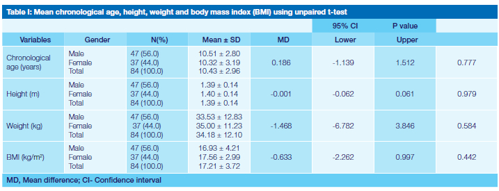
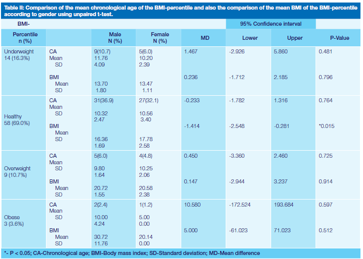
Table I shows the comparison of the mean values of chronological age, height, weight and Body Mass Index (BMI) according to gender using unpaired t-test. Female participants had higher heights (1.40 ± 0.14 mm), larger weights (35.00 ± 11.23 kg) and BMI (17.56 ± 2.99 kg/ m2). However, but they were not statistically significant, with p values of 0.979, 0.584 and 0.442 respectively.The mean BMI of the healthy females was significantly higher than the males (p=0.015). Overweight and obese males participants (20.72 ± 1.55 kg/m2 and 30.72 ± 11.76 kg/m2 respectively) had higher mean BMI when compared to females (20.58 ± 2.38 kg/m2 and 20.14 kg/m2 respectively) but no statistical significance was observed. The mean ages of obese participants was observed to be 10 years for males and 5 years for female participants, p=0.597 (see table II).
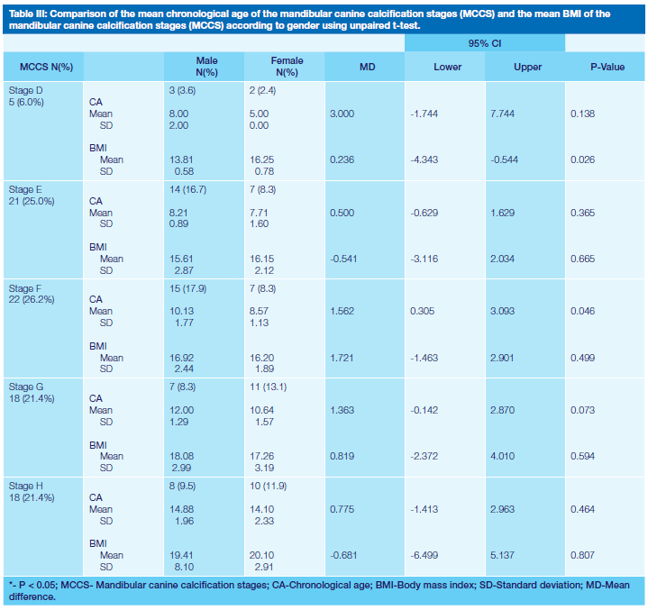
Table III shows that Stage F of mandibular canine calcification was mostly represented among the various stages of the tooth development with 26.2% while stage D was least represented with 6.0%. The mandibular canine calcification stages among the male participants were majorly categorized as stage F (17.9%) while stage G had the highest representation among females (13.1%). The mean chronological age of the various developmental stages of the mandibular canine were consistently earlier among females than in males. Statistical significant difference was only observed at stage F were the mean chronological age was 8.57 years for females and 10.13 years for males, (p=0.046). Mean BMI for each stage were lower among the males compared to females in stages D, E, G and H but higher in stage F. Statistical difference was only observed in stage D, (p = 0.026). As the mandibular canine calcification stages advanced from stage to stage, the mean BMI also steadily increased among the males.
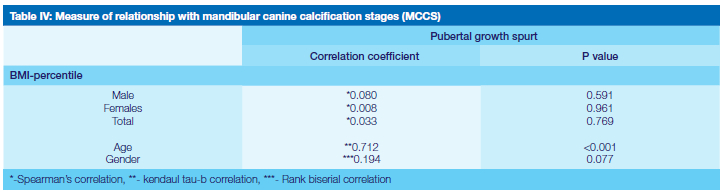
Table IV shows that the average correlation of the mandibular canine calcification stages with the BMI-percentile was positive but weak and not statistically significant, (p = 0.769). Table IV also shows that age has significant (p <0.001) positive and moderate correlation while gender had weak correlation with the mandibular canine calcification stages (p = 0.077).
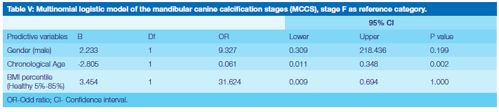
Multinomial logistic regression was used to determine the influence of age, gender and BMI percentile on the dependent variable (mandibular canine calcification stages) with stage F of the mandibular canine calcification stages as the reference category as shown in Table V. There was an increase in the likelihood of males to present with stage D of mandibular canine calcification by 2.233 than females. This shows that females are more likely to be in advanced stage F. However the effect of gender on the canine calcification was observed not to statistically significant (p = 0.199).
Furthermore, a one-percentile increase in the BMI-percentile increased the likelihood of healthy children to present in stage D by 3.454 than in stage F as against obese children. This revealed that obese children tend to have advance mandibular canine development than healthy children. This effect was also not statically significant (p= 1.000).
An increase in chronological age by one year was observed to have significantly (P = 0.002) decreased the likelihood of presenting with stage d of mandibular canine calcification by 2.805. Thus showing that an increase in chronological age is accompanied by increase in developmental stages of the mandibular canine calcification stages.
This result shows that gender and BMI-percentile were not statistical significant predictors of mandibular canine calcification stages.
DISCUSSION
In this current study, it was observed that the correlation of the mandibular canine calcification with the BMI-percentile was positive but weak irrespective of gender. This findings is consistent with results of a previous study conducted in an Iranian population.14 While the present study recorded a non-statistically significant correlation among males and females, Anbiaee et al14 only reported a non-statistically significant correlation among the males.
The present study also confirms that age has a significant predicting effect on the calcification stages of mandibular canine. These findings corroborate previous observations. Hedayati & Khalafinejad in a study conducted among an Iranian population observed that gender was not a statistical significant predictor of mandibular canine calcification.15 The findings in the present study corroborate the observation made by the authors.15
Findings in the current study differ from previous reports made by Mack et al19 and Erhamza et al22 where it was observed that gender played a significant predicting role in determining the outcome of the dental development.
Although this present study shows that an increase in the BMI-percentile increases the tendency towards advanced dental calcification, the effect was however not statistically significant. There is general consensus among several authors that there is a tendency towards advanced dental development when there is an increase in the BMI-percentile on the contrary, they differ with regard to the level of statistical significance of the predictive effects.14,15,19,22,26 While finding in the present study is consistent with the observations made by some authors,21,22,26 it is at variance with other researchers that reported BMI-percentile as a significant predictor of dental maturation.13,18-20 This present study apart from having lower study participants focused majorly on the calcification stages of the right mandibular canine while the study conducted by Mack et al19 and DuPlessis et al20 calculated the actual dental maturity score and the corresponding dental age of the participants using the Demirjian method which involve the use of seven teeth. These differences could have accounted for the difference on the predictive effect of BMI-percentile.
The study conducted in a black South African population focused majorly on stage H of the left seven teeth.18 The exclusion of other developmental stages of the teeth could have probably magnify the predictive effect of the BMI-percentile on stage H that was evaluated. Although Zangouei-Booshehri et al13 reported a tendency towards accelerated dental development which is corroborated by observations made in this present study. The significant predictive effect of BMI-percentile reported by the author is at variance with the findings in the current study. The BMI-percentile was either classified as normal or above normal by the authors13, therefore obviating the separate effects of the 4 groups of BMI-percentile used In the current study. Also, only the coefficient ratio of BMI-percentile was reported while the parameter estimate of the logistic model was reported in this current study. Coefficient ratio only reports the overall effect without considering individual contribution of the various groups of the BMI-percentile.
The differences in methodology, ethnic difference and possibly racial variation could have accounted for the difference observed between the present study and the studies that reported significant effect of BMI-percentile on dental development. Obese patients have also been reported to have significantly more erupted teeth that normal non-obese individuals in a study conducted among a North American population,8 but the mixed model analysis failed to show that obesity had a statistically significant effect on the eruption pattern which is further corroborated by the results of this current study which shows a non-significant effect of BMI-percentile on mandibular canine calcification. The quadratic mixed-effects model used in the longitudinal study conducted by Nicholas et al9 also showed that BMI-percentile had no statistical significant effect on changes in the dental of the study participants. This is in agreement with results from the current study.
Since findings from this current study have shown that there is a tendency towards advanced dental development among individuals with increased BMI-percentile, special considerations should be given to overweight and obese individuals visiting the paediatric and orthodontic dental clinics. These individuals may require earlier treatment intervention than individuals within the normal range of healthy BMI-percentile among Nigerian children.
CONCLUSION
1. A one-percentile increase in the BMI-percentile increased the likelihood of healthy children to present be in stage D by 3.454 than in stage F as against obese children. However, the effect of an increase in BMI-percentile on the mandibular canine calcification was not statically significant (P= 1.000). This revealed that obese children tend to have advance mandibular canine development than healthy children.
2. Chronological age was observed to be a statistical significant predictive factor (P=0.002) of mandibular canine calcification stages. A statistically significant and strong positive correlation (p<0.001) was also observed between chronological age and mandibular canine calcification stages.
3. The effect of gender on mandibular canine calcification stages was not statistically significant (P=0.199). The correlation between gender and mandibular canine calcification stages was also not statistically significant (P = 0.077).
Conflict of interest
The authors declare that there is no conflict of interest.
Sponsorship
This research was self-sponsored.
REFERENCES
1. Goldstein R. Study of the need for easthetlcs In 1. WHO Multicentre Growth Reference Study Group. WHO Child Growth Standards: Length/height-for-age, weight-forage, weight-for-length, weight-for-height and body mass index-for-age: Methods and development. Geneva: World Health Organization; 2006. [ Links ]
2. Oduwole AA, Ladapo TA, Fajolu IB, Ekure EN, Adeniyi OF. Obesity and elevated blood pressure among adolescents in Lagos Nigeria: a cross sectional study. BMC: Public health. 2012; 12:616-21. [ Links ]
3. Sally N Akarolo-Anthony, Walter C Willett, Donna Spiegelman and Clement A Adebamowo. Obesity epidemic has emerged among Nigerians. BMC Public Health 2014, 14:455-64 [ Links ]
4. Shalitin S & Gat-Yablonski G. Associations of Obesity with Linear Growth and Puberty. Horm Res Paediatr; DOI: 10.1159/000516171 1-17) [ Links ]
5. Freedman DS, Mei Z, Srinvasan SR, Berenson GS, Dietz wh. cardiovascular risk factors and excess adiposity and overweight children and adolescents: the Bogalusa Heart study. J pediatr. 2007; 12-17.e2 [ Links ]
6. Kurita LM, Menezes AV, Casanova MS, Haiter-Neto F. Maturity as an indicator of chronological age: Radiographic assessment of dental age in a Brazilian population. J App Oral Sci. 2007;15:99-104. [ Links ]
7. Oziegbe E, Adekoya-Sofowora, C, Esan, T, Owotade F. Eruption Chronology of Primary Teeth in Nigerian Children. The Journal of clinical pediatric dentistry. 2008. 32:341-45. [ Links ]
8. Must A, Phillips SM, Tybor DJ, Lividini K, Hayes C. The Association Between Childhood Obesity and Tooth Eruption. Obesity (2012) 20, 2070-2074. [ Links ]
9. Nicholas CI, Kadavy K, Holton NE, Marshall T, Richter A, Southard T. Childhood body mass index is associated with early dental development and eruption in a longitudinal sample from the Iowa Facial Growth Study. Am J Orthod Dentofacial Orthop 2018;154:72-81 [ Links ]
10. Anu V, Brindha JR, Carol PT, Diana PCR, Elsy JD, Garima S. Does Body Mass Index affect Tooth Eruption Sequence? A Study among 6-7 Years Old Schoolchildren in Chennai, India. Int J Clin Pediatr Dent 2020;13(3):261-63. [ Links ]
11. Younus MS, Ahmed K, Kala D. The effect of body mass index on tooth eruption and dental caries. Dent. J. (Majalah Kedokteran Gigi) 2020; 53(3): 14043. [ Links ]
12. Hilgers, K.K., Axridge, M., Scheetz, J.P. and Kinane, D.E. (2006) Childhood obesity and dental development. Pediatric Dentistry, 18, 16-22. [ Links ]
13. Zangouei-Booshehri M, Ezoddini-Ardakani F, Aghili HA, Sharifi. A. Assessment of the relationship between body mass index (BMI) and dental age. 2011; 3(5):253-57. [ Links ]
14. Anbiaee N, Rashed Mohassel A, Bagherpour A. The Relationship between Body Mass Index and Dental Development by Demirjian's Method in 4- to 15-Year-Old Children in Mashhad. J Dent Mater Tech 2013; 2(3): 82-85. [ Links ]
15. Hedayati Z, Khalafinejad F. Relationship between Body Mass Index, Skeletal Maturation and Dental Development in 6- to 15- Year Old Orthodontic Patients in a Sample of Iranian Population. J Dent Shiraz Univ Med Sci.; 2014; 15(4): 180-86. [ Links ]
16. Sakhdari Sh, Jafari Naimi A,Kharazi MJ, Najafi N. Evaluating the Correlation between the Body Mass Index (BMI) and Dental Age in 6 to 13-Year-Old Children. J Res Dentomaxillofac Sci. 2016;1(4):9-15. [ Links ]
17. Chehab DA, Tanbonliong, T, Peyser J, Udin, R. Association between body mass index and dental age in Hispanic children. General Dentistry. 2017; 54-58. [ Links ]
18. Temitope A. Esan, Lynne A. Schepartz. Does nutrition have an effect on the timing of tooth formation? Am J Phys Anthropol. 2019;1-11. [ Links ]
19. Mack KB, Phillips C, Jain N, Koroluk. LD Relationship between body mass index percentile and skeletal maturation and dental development in orthodontic patients. Am J Orthod Dentofacial Orthop 2013;143:228-34 [ Links ]
20. DuPlessis EA, Araujo EA, Behrents RG, Kim KB. Relationship between body mass and dental and skeletal development in children and adolescents. Am J Orthod Dentofacial Orthop 2016;150:268-73 [ Links ]
21. Eid R, Simi R, Friggi M, et al. Assessment of dental maturity of Brazilian children aged 6 to 14 years using Demirjian's method. Int J Paediatr. 2002;12(6):423-28. [ Links ]
22. Erhamza TS, Kilicaslan Y & Unver FN. Effect of body mass index percentile on skeletal maturation of cervical vertebrae and hand-wrist and dental maturation, Acta Odontologica Scandinavica, (2020): DOI: 10.1080/00016357.2019.1709891 [ Links ]
23. Michelogiannakis D, Rossouw DE, Khan J, Akram Z, Menenakos Eand Javed F. Influence of increased body mass index on orthodontic tooth movement and related parameters in children and adolescents: A systematic review of longitudinal controlled clinical studies. Journal of Orthodontics. 2009; 1-12. [ Links ]
24. Ijeoma, U.N., Njoku, P.O., Arodiwe, E.B. and Ijoma, C.K.. Increasing Trend of Overweight and Obesity in a Rural Community in South East Nigeria. Open Journal of Epidemiology, 2020; 10, 323-33. [ Links ]
25. Demirjian A, Goldstein H, Tanner JM, "A new system of dental age", Human Biology. 1973;45:211-27. [ Links ]
26. Bagherian, A. & Sadeghi, M. Assessment of dental maturity of children aged 3.5 to 13.5 years using the Demirjian method in an Iranian population. Journal of Oral Sciences. 2011; 53(1), 37-42. [ Links ]
 Correspondence:
Dr Osaronse Anthony Aghimien
Orthodontic unit, Dental Surgery Department, Federal Medical Centre
Keffi. Nasarawa State.
E-mail: osaronse@yahoo.com; Phone number: +23 4703 0857 943
Author contributions:
1. Osaronse Anthony Aghimien: 60%;
2. Osasumwen Aghimien-Osaronse: 40%,

