


Serviços Personalizados
Artigo
 Inglês (pdf)
 Artigo em XML
 Referências do artigo
Indicadores

Links relacionados
-
 Citado por Google
 -
 Similares em Google
Compartilhar

 Permalink
South African Dental Journal
versão On-line ISSN 0375-1562
versão impressa ISSN 0011-8516
S. Afr. dent. j. vol.77 no.4 Johannesburg Mai. 2022
http://dx.doi.org/10.17159/2519-0105/2022/v77no4a3
RESEARCH
A radiographic analysis of Mandibular Symphysis dimension in black South African adult patients with differing skeletal patterns
DM GinindaI; MI KhanII
IRegistrar (2020) Sefako Makgatho Health Sciences Unversity, Dip Oral Hygiene (Medical University of Southern Africa, BDS (University of Limpopo, Medunsa campus), PD Dip (Orthodontics) (University of Limpopo, Medunsa Campus), M Dent Orthodontics (Sefako Makgatho Health Sciences University) ORCID Number: 0000-0003-3256-4022
IISenior Consultant Sefako Makgatho Health Sciences Unversity, BDS (Medunsa), M Dent Orthodontics (University of Limpopo, Medunsa Campus) ORCID Number: 0000-0001-91836476
ABSTRACT
INTRODUCTION: Orthodontic treatment often involves planned tooth movement within the confined spaces of the alveolar bone trough. Tooth movement within the alveolar trough may be limited by thin labial and lingual cortical plates. Moving lower incisors beyond the mandibular symphysis dimensions may result in damage to roots and alveolar bone.4
Aim and objective: The aim of the study was to evaluate limitation of treatment in different skeletal patterns due to mandibular symphysis dimension in order to evaluate limitations of tooth movement within the confines of the mandibular alveolar trough.The objective was to determine the mandibular symphysis dimensions in subjects with differing skeletal patterns.
DESIGN: The design was a retrospective, cross-sectional study.
METHODS: A sample of 180 pre-treatment lateral cephalometric radiographs of black South African subjects were stratified into three groups based on their skeletal classification. Each Class was further divided into equal numbers of males and females. Descriptive statistics, Student's t-test, ANOVA test and Pearson correlation coefficient were used to analyse the data and p-values of <0.05 were considered statistically significant.
RESULTS: Subjects with skeletal Class I pattern had a greater LA compared to subjects with skeletal Class II pattern. Subjects with skeletal Class I pattern had a greater LH and LA in females than in males. Subjects with skeletal Class III pattern had greater LH in males than in females.
INTRODUCTION AND LITERATURE REVIEW
In order to have a balanced skeletal jaw relationship, the maxilla and the mandible must be in harmony. If there is a skeletal jaw discrepancy either in a vertical, or anteroposterior dimensions the dento-alveolar structures may compensate to camouflage the skeletal jaw discrepancy.1,2
In skeletal Class II subjects, where the mandible is retrusive in relation to the maxilla, the lower incisors may procline in order to achieve contact with the upper teeth. This may result in the root apices of the lower incisors being closer to the lingual cortical plate of the mandibular symphysis skeletal Class III subjects, where the mandible is protrusive in relation to the maxilla, the lower incisors may retrocline in order to make contact with the upper teeth.2 This may result in the root apices of the lower incisors being closer to the buccal cortical plate of the mandibular symphysis.3 Mulie4 found that the labial and lingual cortical plate of the mandibular symphysis and the status of the periodontal tissue of the lower incisors could limit the movement of the lower incisors. Therefore, the mandibular symphysis dimensions and the position of the lower incisors must be considered during orthodontic diagnosis and treatment planning.5 If the lower incisors are moved in an antero-posterior direction beyond the mandibular symphysis dimensions, the movement of incisors will be inhibited; the roots of the lower incisors may touch the cortical plates, causing damage to the periodontal tissues.4 This damage may include alveolar bone loss, dehiscence, gingival recession, root resorption and mobility of the teeth.4,6
The alveolar bone thickness vary according to location and facial type.7 In general alveolar bone thickness is greater at the apex, then in the cervical third of the lower incisors and towards the lingual surface compared to the labial surface.7 This explains the higher prevalence of bone dehiscence and fenestration on the labial surface when lower incisors are moved anteriorly during orthodontic treatment. Several authors reported that the mandibular symphysis determines the beauty of the face in general, but particularly the lower part of the face.8-9
There are several factors that may affect the mandibular symphysis dimensions, such as:
The functional environment.
Previous studies10-12 reported that the functional environment of the mandibular symphysis shows an adaptive morphological response to the biomechanical loads experienced at different locations during the chewing process. The change in cross sectional shape of the mandibular symphysis correlates with the change in loading to which the mandibular symphysis is subjected. 10-12
Vertical jaw relationship.
During growth, the mandible tends to rotate in a clockwise direction, resulting in a long and narrow symphysis, or it rotates in an anticlockwise direction, resulting in a shorter and wider symphysis.13 Similarly, Swasty et al.14 reported that long-faced patients had slightly narrower symphyses than average faced and short-faced patients.
Sagittal jaw relationship.
Studies6,15 have shown that patients with skeletal Class III malocclusion showed high and narrow symphyses with greater anterior projection and increased lingual inclination of the long axis. Patients with a skeletal Class III often presented with extruded and retroclined mandibular incisors that caused the bone lingual to lower incisors (LP) apex to increase, whereas the bone labial to lower incisors apex (LA) and the bone inferior to mandibular incisor apex (LH) decreased.3 Skeletal Class II patients often presented with extruded and proclined lower incisors that caused the bone lingual to the lower incisors apex to decrease, whereas the bone labial to lower incisors apex and the bone inferior to mandibular incisor apex increased.3
Inclination of the lower incisors.
Prior studies16-18 have reported that when the lower incisors are retroclined, so too is the alveolar bone. The shape of the mandibular symphysis therefore corresponds to the inclination of the lower incisor. Yu et al}1 reported that when the lower incisors are more proclined, the lingual alveolar bone of the mandibular symphysis becomes thinner. Nojima et al.15 reported that the inclination of the lower incisors corresponds to the shape of the mandibular symphysis, and is influenced by facial type. This argument dates back to the era of Tweed, who reported that patients with high mandibular planes, presented with the lower incisors that were tipped lingually, whereas subjects with low mandibular planes presented with lower incisors that were tipped buccally.19 This indicates that the stability of orthodontic results and facial aesthetics may be affected by incorrect positioning of the lower incisors.19
Genetic factors and ethnicity.
The mandibular symphysis is regarded as a multifaceted structure and its shape results from the interaction of various genetic, non-genetic, adaptive and non-adaptive factors.11,12 Handelman6 discovered that no cephalometric norms that took mandibular symphysis dimensions into consideration existed. He conducted a study of 101 Caucasian subjects to establish the standard norms of the mandibular symphysis dimensions and suggested that these cephalometric norms of mandibular symphysis dimensions should be incorporated in the cephalometric analysis.6
AIM AND OBJECTIVE
The aim of this study was to evaluate the limitation of treatment in different skeletal patterns due to mandibular symphysis dimensions and the objective was to determine the mandibular symphysis dimensions in black South
African adult patients with skeletal Class I pattern, skeletal Class II and skeletal Class III. Knowledge on the thickness of the mandibular symphysis dimensions before orthodontic treatment may help in selecting the best treatment mechanics for specific skeletal patterns to prevent iatrogenic sequalae.
DESIGN AND METHODS
The Sefako Makgatho University Research Committee (SMUREC) approved the study (project number: SMUREC/D/30/2018). The Head of the Department of Orthodontics and the CEO of MOHC gave permission to utilise the hospital records obtained from the Department of Orthodontics at MOHC. Patient's pre-treatment lateral cephalometric radiographs, panaromic radiograph and study models were used for this study
One hundred and eighty pre-lateral cephalometric radiographs (90 males and 90 females) were selected for this study. The criteria for selection included pre-treatment lateral cephalometric radiographs of black South African adult patients 18 years and older (race and citizenship were verified by referring to hospital files). Pre-treatment lateral cephalometric radiographs of subjects with skeletal Class I, Class II or Class III. Pre-treatment lateral cephalometric radiographs of patients who had never received orthodontic treatment.
Pre-treatment lateral cephalometric radiographs of good quality that had been taken with teeth in maximum intercuspation. All selected lateral cephalograms were subjected to the same X-ray machine, using the same technique at MOHC, Sefako Makgatho Health Science University with (Siemens, Orthopantomogram 10® -analogue and Kodak 8000C® - digital cephalograms).
The selected lateral cephalometric radiographs were traced manually using a 4H pencil (0.5 mm) and a tracing protractor template on acetate tracing paper over a light viewing box in a darkened room. Measurement bias (tracing and landmark identification) were avoided by tracing no more than 10 radiographs at a time, in order to avoid operator fatigue.
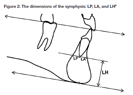
These lateral cephalometric radiographs were then traced, using Handelman's criteria.6 The dimensions of the mandibular symphysis were measured in millimetres, using a ruler. In order to assess the mandibular symphysis dimensions, two reference lines were used, i.e. the occlusal plane line and the tangent line parallel to the occlusal plane passing through the apices of the lower incisors (refer to Figure 2). The following mandibular symphysis dimensions were measured on the tangent line that runs parallel to the occlusal plane: the dimension of the bone labial to the root apex of the lower incisor apex (LA), the shortest distance from the root apex to the outer surface of the labial cortical plate; the dimension of the bone lingual to the root apex of the lower incisor (LP), the shortest distance from the root apex to the outer surface of the lingual cortical plate; and the dimension of the bone from the lower incisor apex to the lowest point of the mandibular symphysis (LH), that is the shortest distance from the root apex to the inferior part of the mandibular symphysis (see Figure 2). These measured variables were recorded on the data collection form, and entered into an Excel spreadsheet.
The traced lateral cephalograms were grouped into three classes, based on their skeletal relationship. The ANB20, facial plane angle21, convexity22 and Wits analysis23 were used to confirm each patient's skeletal relationship. ANB was used to classify skeletal jaw relationship (ANB =50 normal, ANB >50 Class II and ANB <50 Class III). The control group comprised 60 skeletal Class I lateral cephalometric radiographs. The test group comprised 120 lateral cephalometric radiographs, divided equally into a group of 60 skeletal Class II and a group of 60 skeletal Class III of black South African adult patients. There were equal numbers of males and females in each skeletal Class.
Lateral cephalometric landmarks and measurements Measurements according to skeletal relationship
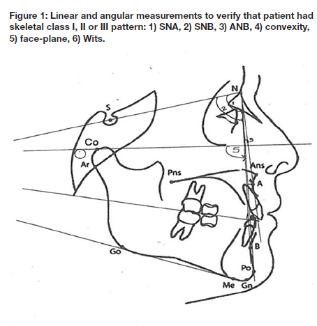
The classification and verification of the patient's skeletal relationship was achieved by using the following cephalometric angular linear measurements (refer to Figure 1):
• SNA angle: angle formed where the lines connecting nasion and point A to S-N plane intersect.20
• SNB angle: angle formed where the lines connecting the nasion plane and point B to S-N plane intersect.20
• ANB angle: subtraction of SNB angle value from SNA angle value.20
• Wits appraisal: a perpendicular line from point A of the maxilla and from point B of the mandible onto the Occlusal plane (OP).23
• Convexity: a linear measurement of the distance from Point A to the nasionpogonion line.22
• Face plane: angle formed between the Frankfort horizontal plane and the nasionpogonion line.21
Measurements of the mandibular symphysis dimensions
The following linear measurements were used in this study (Figure 2), following Handelman's criteria6.
• LP - bone lingual to mandibular incisor apex: a line drawn through the apex of the mandibular central incisors to the lingual cortex, parallel to the occlusal plane.
• LA - bone labial to mandibular incisors apex: a line drawn through the apex of the mandibular central incisors to the labial cortex, parallel to the occlusal plane.
• LH - bone inferior to mandibular incisor apex: the shortest distance from the apex of the mandibular incisors to the lowest point on the mandibular symphysis, that is crossed by a line parallel to the occlusal plane.
Eighteen radiographs were selected for intra-examiner reliability to evaluate and assess the accuracy of the single investigator. The measurements were repeated a month later. Inter-examiner reliability was established in order to evaluate and assess the accuracy of the measurements of the first and second investigator.
Statistical analysis system (SAS) 9.4 computer software program was used to determine all the continuous variables. Student's t-test was administered to compare the mean values of the mandibular symphysis dimensions of males and females, and to check for any major differences.
The value p<0.05 signifies the level of confidence. Oneway ANOVA test was administered to compare the mean values of the mandibular symphysis dimensions of different skeletal types and facial types, and to check for any major differences. The value p<0.05 signifies the level of confidence. All the p values equal to or greater than 0.05 were considered statistically significant. The magnitude of association between the original and second measurements of the mandibular symphysis dimensions were analysed using the Pearson Correlation Coefficient.
RESULTS
The intra-examiner and inter-examiner reliability was tested by randomly selecting and re-measuring 10% of the total sample and the results were analysed using the Pearson correlation Coefficient. Results showed strong correlations between the repeated and the original measurements for all measured variables of the mandibular symphysis dimensions. These findings are consistent with those of a study by Alhadlaq24 who mentioned that the method of measurement of the manual cephalometric tracing was reliable and reproducible. The results are summarized in tables 1 to 3.
Comparison of measured variables between female skeletal Class I and Skeletal Class III
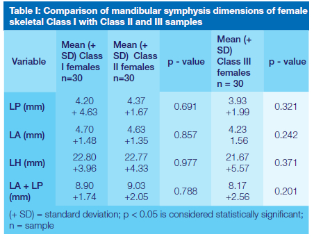
The study found no statistically significant differences between skeletal Class I and skeletal Class III.
Comparison between male skeletal Class I and Skeletal Class II
There were no statistically significant differences between the skeletal Class I and skeletal Class II male sample, except for the LA (bone labial to lower incisor apices). This was significantly larger in skeletal Class I as compared to the skeletal Class II.
Comparison between male of skeletal Class I and Skeletal Class III
No statistically significant differences were found between the skeletal Class I and skeletal Class III male sample for any of the measured variables.
Gender comparisons
Comparison between male and female skeletal Class I
The bone inferior to the lower incisor apices was significantly greater in females than in males with a skeletal Class I pattern.
Comparison between male and female skeletal Class II
The bone labial to the lower incisor apices was significantly larger in females than in males with a skeletal Class II pattern.
Comparison between male and female skeletal Class III
The bone inferior to the lower incisor apex was significantly greater in males than in females with a skeletal Class III pattern.
Comparisons of the mandibular symphysis dimensions in average (control group) and different facial type
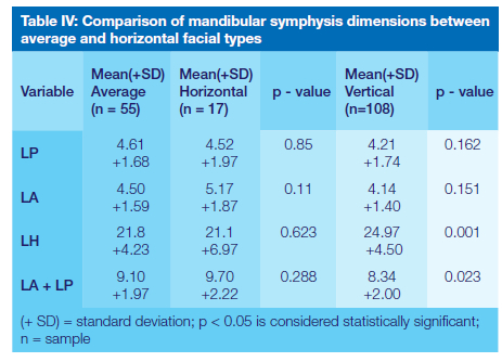
Comparison between average and horizontal facial type
No statistically significant differences were found between the horizontal and average facial types, indicating that none of the mean values for the mandibular symphysis dimensions differed significantly. A trend emerged in the average sample of the increased bone labial to lower incisor apices, increased bone inferior to lower incisor apices and increased bone lingual to the lower incisor apices. In the horizontal sample, there was a tendency for the total width of the mandibular symphysis to be greater than that of the average sample but this was not significant.
Comparison between average and vertical facial type
Two of the measured mandibular symphysis dimensions demonstrated statistically significant differences. The mean values of the mandibular symphysis dimensions of (the bone inferior to the lower incisor apices and the total width of the mandibular symphysis) of the vertical facial type were significantly greater than those of the average group sample.
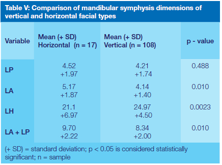
Comparison between vertical and horizontal facial types
Three of the mandibular symphysis dimensions demonstrated statistically significant differences. The mean values of the mandibular symphysis dimensions (the bone labial to the lower incisor apices, the bone inferior to the lower incisor apices and the total width of the mandibular symphysis) of the horizontal facial type were significantly larger than those of the vertical group.
DISCUSSION
In this study, the cephalometric norms of the mandibular symphysis dimensions of black South African adult patients were established (LA 4.7mm, LP 4.27mm, LH 24.2mm and LA+LP 8,8mm) and compared across different skeletal patterns. The sample was grouped according to skeletal relationship, gender and facial types.
This study found that values of LA (the bone labial to the lower incisor apices) were greater in males with a skeletal Class I pattern (control) than in males with a skeletal Class II pattern. It is possible that males with a skeletal Class I pattern had more proclined lower incisors than males with a skeletal Class II pattern. Proclined lower incisors in subjects with a skeletal Class I pattern are associated with bimaxillary protrusion. However, in this study Skeletal Class I patients presented with features of Bimax one (BM1) that present with a balanced profile and efficient lips which is considered an ideal profile in most south African black population, but these features are considered protrusive when compared to Caucasian population.25-27 This type of bimaxillary protrusion (BM1) does not require extraction or treatment at all. Treatment of patients with bimaxillary protrusion two (BM2) and bimaxillary protrusion three (BM3) may require the extraction of premolars, followed by retraction of the incisors as well as surgery in some cases. It is therefore important to know the boundaries of the mandibular symphysis dimensions before retracting the lower incisors, as moving teeth beyond these boundaries may cause damage to the periodontal tissues, resulting in root resorption, dehiscence and fenestrations.4
The findings of this study correspond with those of Alhadlaq28 who reported an increased LA (bone labial to the lower incisor apices) in males and females with a skeletal Class I pattern when they were compared to males and females with a skeletal Class II pattern. The findings of this study are also consistent with those of Molina-Berlanga29 who reported that increased LA (bone labial to the lower incisor apices) was associated with protruded lower incisors; when the lower incisors are protruded their root apices tend to rotate towards the lingual side, causing the thickness of LA (the bone labial to the lower incisor apices) to increase.
No statistically significant differences were found in this study in any of the measured variables when comparisons were made between males with a skeletal Class III pattern and males with a skeletal Class I pattern (control group), as well as between females with a skeletal Class III pattern and females with a skeletal Class I pattern (control group). These findings are in contrast to those of Alhadlaq25 who found significant differences between males and females with a skeletal Class I pattern when compared to males and females with a skeletal Class III pattern. Alhadlaq29 found that males with a skeletal Class I pattern had an increased LA (bone labial to the lower incisor apices) than males with a skeletal Class III pattern. An increased thickness of LA (the bone labial to the lower incisor apices) is associated with proclined lower incisors where the root apex rotates towards the lingual side causing the LA (bone labial to the lower incisor apices) to increase.29 Retraction of the lower incisors in patients with a skeletal Class I pattern should be carefully planned to avoid perforation of the lingual cortical plate, which may cause root resorption.6
The findings of this study are similar with those of Molina-Berlanga29 who found no significant differences of measured variables of the mandibular symphysis dimensions between subjects with a skeletal Class I pattern and subjects with a skeletal Class III pattern, although their study did not account for gender.
There was a trend towards increased bone inferior to the lower incisor apices in males and females with a skeletal Class III pattern than in males and females with a skeletal Class I pattern (control group), although this was not significant. This finding corresponds to that of Alhadlaq28 who further explained that increased bone inferior to the lower incisor apices in subjects with a skeletal Class III pattern demonstrated a dentoalveolar compensation caused by over-eruption of the lower incisors to approximate the upper incisors. Over-eruption of the lower incisors leads to a narrow mandibular symphysis dimension. Handelman6 reported similar findings, although his sample was not stratified according to gender. Patients with narrow mandibular symphysis of less than normal 8.8mm, should involve tipping movements of the lower incisors rather than bodily movement, to prevent damage to the periodontal tissues.30
Treatment of patients with bimaxillary protrusion depend on the severity of the case. Patients with mild protrusion classified as Bimax one (BM1), usually accept their facial profile and do not request treatment. Those with moderate Bimax two (BM2) and severe protrusion Bimax three (Bmx3), request treatment to have their teeth retracted.27 During retraction of anterior teeth, reciprocal movement of teeth may be allowed in Bimax2 patients, however, Bimax3 patients will required devices to prevent anchorage loss so that the space created through extraction of two upper first premolars and two lower first premolars is largely utilized for retraction of anterior teeth. In extreme cases of bimaxillary protrusion treatment may include both orthodontic and surgical treatment e.g. four first premolars extraction followed by segmental alveolar osteotomies to close the extraction sites to reduce the dental protrusion6 When comparisons were made between males and females with a skeletal Class I pattern in this study, no statistically significant differences were found between any of the measured variables, except in the case of the bone inferior to the lower incisor apex. In this regard, there was a more significant increase of the bone inferior to the lower incisor apices in males with a skeletal Class I pattern than in females. This indicated that males with a skeletal Class I pattern demonstrated a more vertical growth pattern than their female counterparts.13 Individuals with a vertical growth pattern present with increased bone inferior to the lower incisor apices as a result of extruded lower incisors and elongated mandibular symphysis dimensions.
This finding corresponds to that of Alhadlaq28 who found that males with a skeletal Class I pattern had increased bone inferior the lower incisor apices when compared to their female counterparts. This finding is also similar to that of the previous studies7,31 both of whom found that the height of the mandibular symphysis dimensions was greater in males than in females. Clinicians should know that patients with increased bone inferior of the lower incisor apices have a narrow (less than 8.8mm of the total width), mandibular symphysis which means that the movement of the lower incisors is limited. Such patients should be treated with a combination of orthodontic treatment and surgery in order to avoid iatrogenic sequelae.32
There were no statistically significant differences found between male and female subjects with a skeletal Class II pattern, other than the bone labial to lower incisor apices. A significant increase of the bone labial to the lower incisor apices was found in females with a skeletal Class II pattern when compared to male counterparts. This may have been the result of extruded and proclined lower incisors causing the bone labial to lower incisor apex to increase, as the root apex of the lower incisor rotates towards the lingual side.3,29 In other words, the labial movement of the lower incisors in females with a skeletal Class II pattern should be avoided since the roots of the lower incisors may make contact with the lingual cortical plate and suffer root resorption.6 These findings correspond in part with those of Alhadlaq28 who found significant differences between the males and females with a skeletal Class II pattern in all variables.
In this study, comparisons of males and females with a skeletal Class III pattern showed no significant differences in any of the measured variables except for the bone inferior the lower incisor apices. In this regard, there was a significant increase of the bone inferior to the lower incisor apices in the males when compared to their female counterparts. Males with a skeletal Class III pattern demonstrated a more vertical growth pattern, resulting in extruded lower incisors and narrow mandibular symphysis dimensions.13 When treating patients with narrow symphyses, the lower incisors should be moved by tipping rather than bodily.30 Similarly, Alhadlaq28 found that males had an increased bone inferior to the lower incisor apices than their female counterparts. In this study, the males showed greater mean values than their female counterparts, which correlated with other such studies.33,31 This study found no statistically significant differences between subjects with a horizontal facial type and those with an average facial type. The findings of this study are in contrast with those of Handelman6 who reported that the LP and the LA + LP were significantly thicker in subjects with horizontal facial types compared to subjects with average facial type. These patients with horizontal facial types present with less proclined or normal position of lower incisors.29
In this study an increased LH (bone inferior to the lower incisor apices) and a decrease LA+LP (total width of the mandibular symphysis) were noted in subjects with a vertical facial type when these were compared to subjects with an average facial type. These results correspond to those of Handelman6 who found subjects with a vertical facial type had a narrow LA+LP (total width of the mandibular symphysis) and increased LH (bone inferior to the lower incisor apices) when compared to subjects with a horizontal facial type and those with average facial type. Handelman6 explained that an increased LH (bone inferior to lower incisor apices) and decreased LA+LP (total width of the mandibular symphysis) are associated with over-erupted lower incisors and a thinning of the mandibular symphysis dimensions. Such thinning results in the labial movement of the lower incisors as well as their retraction of the lower incisors causing damage to the periodontal tissues.6
In the present study, subjects with a horizontal facial type were found to have significantly increased LA (bone labial to lower incisor apices), and increased LA+LP (total width of the mandibular symphysis dimension), as well as decreased LH (bone inferior to the lower incisor apices) when compared to subjects with a vertical facial type. Findings were similar to those of a study by Ponraj et al.34 who reported that decreased LH (bone inferior to lower incisor apices) in subjects with a horizontal facial type could be the result of the mandible rotating forward, in the absence of vertical mandibular symphysis remodeling with increased growth of the ramus. These patients thus presented with a deep bite and a reduced lower facial height. Ponraj et al.34 further reported that increased LA (bone labial to lower incisor apices) and increased LA+LP (total width of the mandibular symphysis dimensions) in the subjects with a horizontal facial type might be attributable to extreme muscle activity, since the masseter activity is significantly longer in subjects with a horizontal facial type. The distance between the root apex of the lower incisor and the LA (bone labial to lower incisor apex) was shown to be greater in subjects with a horizontal facial type than in subjects with a vertical facial type.35 Ponraj et al.34 reported that the thick anterior alveolus in subjects with a horizontal facial type allows the clinician to move lower incisors freely, without any fear of adverse effects. A decrease in the LA (bone labial to lower incisor apex) and in the LA+LP (total width of to the mandibular symphysis dimension) in subjects with a vertical facial type demonstrated that subjects with a vertical facial type had narrow mandibular symphysis dimensions. The increase in the LH (bone inferior to the lower incisor apices) may be due to over-eruption of lower incisors, causing the mandibular symphysis to elongate. These results correspond with those of previous studies34,29 that reported narrower mandibular symphysis dimensions in subjects with vertical facial type than in subjects with horizontal facial type. Further retraction of lower incisors in subjects with vertical facial types could be achieved without any loss of torque. During the retraction stage, careful mechanics must be used to ensure that teeth are positioned within the cancellous bone to prevent iatrogenic side effects. According to Ponraj 34 corticotomy might be beneficial in subjects with a vertical facial types since the procedure is less traumatizing to the teeth and to the alveolar bone. Corticotomy will allow 4mm en masse retraction of anterior teeth along with alveolar housing thereby preventing iatrogenic effects such as root resorption and dehiscence.36
CONCLUSION
Significant differences in mandibular symphysis dimensions were not found when variables of skeletal Class I subjects (control group) were compared to those of skeletal class II and III patterns except LA (the bone labial to the lower incisor apices) that was significantly larger in skeletal Class I sample as compared to skeletal Class II sample. When the mandibular symphysis dimensions were compared according to gender, no statistically significant differences were found except LH (the bone inferior to lower incisor apices) was significantly larger in males than in female sample of skeletal Class I pattern.
With regard to facial type, significant differences in mandibular symphysis dimensions were observed, particularly when the average faced subjects were compared with the vertical faced subjects, and when the vertical faced subjects were compared to the horizontal faced subjects. No statistically significant differences were found when the average faced subjects were compared to the horizontal faced subjects.
Clinicians should take into consideration the mandibular symphysis dimensions when treating patients with a vertical facial pattern as these patients are more likely to have narrow (less than 8.8mm of the total width) mandibular symphysis dimensions and may require surgery or corticotomy.
REFERENCES
1. Jacobson A, Evans, WG, Preston, CB, & Sadowsky PL. Mandibular prognathism. American Journal of Orthodontics. 1974 Aug 1;66 (2):140- 71. [ Links ]
2. Bibby RE. Incisor relationships in different skeletofacial patterns. The Angle Orthodontist. 1980 Jan;50(1):41-4. [ Links ]
3. Maniyar M, Kalia A, Hegde A, Gautam RG, Mirdehghan N. Lower incisor dentoalveolar compensation and symphysis dimensions in class II and class III patients. International Journal of Dental and Medical Specialty. 2014;1(2):20-4. [ Links ]
4. Mulie RM. The limitations of tooth movement within the symphysis, studied with laminagraphy and standardized occlusal Alms. J clin Orthod. 1976;10:882-93. [ Links ]
5. Aasen TO, Espeland L. An approach to maintain orthodontic alignment of lower incisors without the use of retainers. The European Journal of Orthodontics. 2005 Jun 1;27(3):209-14. [ Links ]
6. Handelman CS. The anterior alveolus: its importance in limiting orthodontic treatment and its influence on the occurrence of iatrogenic sequelae. The Angle Orthodontist. 1996 Apr 1;66(2):95-110. [ Links ]
7. Arruda KE, Valladares Neto J, Almeida GD. Assessment of the mandibular symphysis of Caucasian Brazilian adults with well-balanced faces and normal occlusion: the influence of gender and facial type. Dental Press Journal of Orthodontics. 2012 Jun;17(3):40-50. [ Links ]
8. Buschang PH, Julien K, Sachdeva R, Demirjian A. Childhood and pubertal growth changes of the human symphysis. The Angle Orthodontist. 1992 Sep;62(3):203-10. [ Links ]
9. Hoenig JF. Sliding osteotomy genioplasty for facial aesthetic balance: 10 years of experience. Aesthetic plastic surgery. 2007 Aug 1;31(4):384-91. [ Links ]
10. Gould SJ. The exaptive excellence of spandrels as a term and prototype. Proceedings of the National Academy of Sciences. 1997 Sep 30;94(20):10750-5. [ Links ]
11. Gould SJ. The structure of evolutionary theory. Harvard University Press; 2002 Mar 21. [ Links ]
12. Sherwood RJ, Hlusko LJ, Duren DL, Emch VC, Walker A. Mandibular symphysis of large-bodied hominoids. Human biology. 2005 Dec 1:735-59. [ Links ]
13. Björk A. Prediction of mandibular growth rotation. American journal of orthodontics. 1969 Jun 1;55(6):585-99. [ Links ]
14. Swasty D, Lee J, Huang JC, Maki K, Gansky SA, Hatcher D, Miller AJ. Cross-sectional human mandibular morphology as assessed in vivo by cone-beam computed tomography in patients with different vertical facial dimensions. American Journal of Orthodontics and Dentofacial Orthopedics. 2011 Apr 1;139(4): e377-89. [ Links ]
15. Nojima K, Nakakawaji K, Sakamoto T, Isshiki Y. Relationships between mandibular symphysis morphology and lower incisor inclination in skeletal class III malocclusion requiring orthognathic surgery. The Bulletin of Tokyo Dental College. 1998 Aug;39(3):175-81. [ Links ]
16. Yamada C, Kitai N, Kakimoto N, Murakami S, Furukawa S, Takada K. Spatial relationships between the mandibular central incisor and associated alveolar bone in adults with mandibular prognathism. The Angle Orthodontist. 2007 Sep;77(5):766-72. [ Links ]
17. Yu Q, Pan XG, Ji GP, Shen G. The association between lower incisal inclination and morphology of the supporting alveolar Bone-A coneBbeam CT study. International journal of oral science. 2009 Dec;1(4):217-23. [ Links ]
18. Endo T, Ozoe R, Kojima K, Shimooka S. Congenitally missing mandibular incisors and mandibular symphysis morphology. The Angle Orthodontist. 2007 Nov;77(6):1079-84. [ Links ]
19. Tweed CH. The Frankfort-mandibular incisor angle (FMIA) in orthodontic diagnosis, treatment planning and prognosis. The Angle Orthodontist. 1954 Jul;24(3):121-69. [ Links ]
20. Steiner CC. Cephalometrics for you and me. American journal of orthodontics. 1953 Oct 1;39(10):729-55. [ Links ]
21. Downs WB. Variations in facial relationships: their significance in treatment and prognosis. American journal of orthodontics. 1948 Oct 1;34(10):812-40. [ Links ]
22. Ricketts RM, Bench RW, Gugino CF, Hilgers JJ, Schulhof RJ. Part 4, the use of superimposition areas to establish treatment design. Bioprogressive Therapy. 1979:55-69. [ Links ]
23. Jacobson A. The "Wits" appraisal of jaw disharmony. American Journal of Orthodontics and Dentofacial Orthopedics. 1975 Feb 1;67(2):125-38. [ Links ]
24. Alhadlaq AM. Association between anterior alveolar dimensions and vertical facial pattern among Saudi adults. The Saudi dental journal. 2016 Apr 1;28(2):70-5. [ Links ]
25. Beukes S, Dawjee SM, Hlongwa P. Soft tissue profile analysis in a sample of South African Blacks with bimaxillary protrusion: scientific. South African Dental Journal. 2007 Jun 1;62(5):206-12. [ Links ]
26. Dawjee SM, Hlongwa P, Becker PJ. Is orthodontics an option in the management of bimaxillary protrusion?: scientific. South African Dental Journal. 2010 Oct 1;65(9):404-8. [ Links ]
27. Sethusa MP, Williams VA. A pilot study to establish a visual template for classifying bimaxillary protrusion profiles among Black South Africans: case study. South African Dental Journal. 2010 Nov 1;65(10):458-60. [ Links ]
28. AlHadlaq A. Anterior alveolar dimensions among different classifications of sagittal jaw relationship in Saudi subjects. The Saudi Dental Journal. 2010 Apr 1;22(2):69-75. [ Links ]
29. Molina-Berlanga N, Llopis-Perez J, Flores-Mir C, Puigdollers A. Lower incisor dentoalveolar compensation and symphysis dimensions among Class I and III malocclusion patients with different facial vertical skeletal patterns. The Angle Orthodontist. 2013 Nov;83(6):948-55. [ Links ]
30. Garib DG, Yatabe MS, Ozawa TO, Silva Filho OG. Alveolar bone morphology under the perspective of the computed tomography: defining the biological limits of tooth movement. Dental Press Journal of Orthodontics. 2010 Oct;15(5):192-205. [ Links ]
31. Al-Barakati SF, Alhadlaq AM. Anterior alveolar dimensions in Class I Saudi subjects. Journal of Pakistan Dental Association. 2007;(16):95-102 [ Links ]
32. Enhos S, Uysal T, Yagci A, Veli i, Ucar FI, Ozer T. Dehiscence and fenestration in patients with different vertical growth patterns assessed with cone-beam computed tomography. The Angle Orthodontist. 2012 Sep;82(5):868-74. [ Links ]
33. Hassan AH. Cephalometric norms for Saudi adults living in the western region of Saudi Arabia. The Angle Orthodontist. 2006 Jan;76(1):109-13. [ Links ]
34. Ponraj RR, KoRATH VA, Nagachandran DV, PARAMESwARAN RA, Raman P, Sunitha C, Khan N. Relationship of Anterior Alveolar Dimensions with Mandibular Divergence in Class I Malocclusion-A Cephalometric Study. Journal of clinical and diagnostic research: JCDR. 2016 May;10(5): ZC29. [ Links ]
35. Gracco A, Luca L, Bongiorno MC, Siciliani G. Computed tomography evaluation of mandibular incisor bony support in untreated patients. American Journal of Orthodontics and Dentofacial Orthopedics. 2010 Aug 1;138(2):179-87. [ Links ]
36. Ferguson DJ, Wilcko WM, Wilcko MT. Selective alveolar decortication for rapid surgical-orthodontic of skeletal malocclusion treatment. Bell WE, Guerrero C. Distraction osteogenesis of the facial skeleton. Hamilton, ON: BC Decker, Inc. 2007:199-203. [ Links ]
 Correspondence:
Dikeledi Maureen Gininda
Telephone no.: 0731711158
Email: dikeledigininda@yahoo.com
Author contributions:
1 . Dikeledi Maureen Gininda: 60%
3.Mohamed Imran Khan: 40%

