


Services on Demand
Article
 English (pdf)
 Article in xml format
 Article references
Indicators

Related links
-
 Cited by Google
 -
 Similars in Google
Share

 Permalink
South African Dental Journal
On-line version ISSN 0375-1562
Print version ISSN 0011-8516
S. Afr. dent. j. vol.77 n.1 Johannesburg Feb. 2022
http://dx.doi.org/10.17159/2519-0105/2022/v77no1a7
RADIOLOGY CASE
C SmitI; A UysII
IBChD, MSc (Maxillofacial and Oral Radiology). Department of Oral Pathology and Oral Biology, Faculty of Health Sciences, University of Pretoria. ORCID: 0000-0003-4047-6356
IIBSc, BChD, PGDipDent (Endodontics), PGDipDent (Aesthetic Dentistry), MSc (Maxillofacial Radiology), PhD (Anatomy). Department of Anatomy, Faculty of Health Sciences, University of Pretoria. ORCID: 0000-0001-8250-7662
HYPERPNEUMATISATION OF THE SKULL BONES
CASE
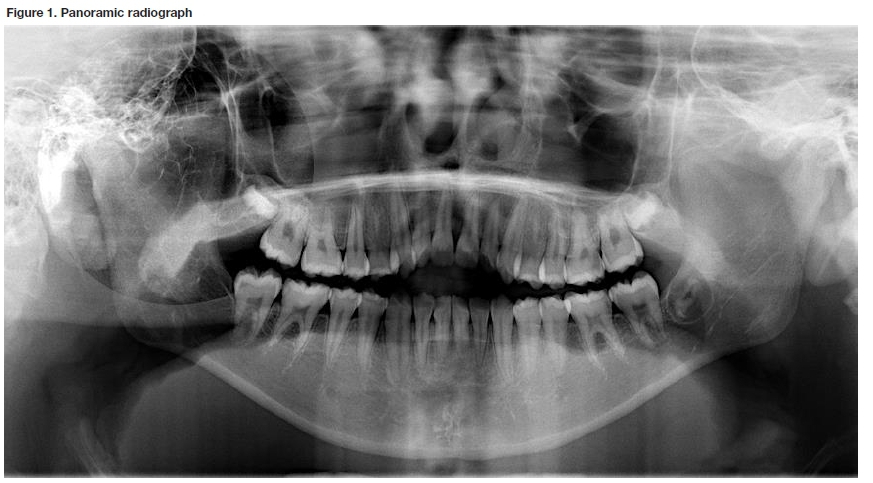
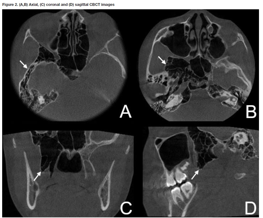
A healthy,12-year-old female patient, presented with an anterior open bite requesting orthodontic treatment. A panoramic radiograph was requested for treatment planning (Figure 1). An incidental finding of well-defined multilocu-lar radiolucencies were detected superimposed over the right ramus and middle cranial fossa region. The patient was asymptomatic with no clinical signs of facial asymmetry. A cone-beam computerised tomography (CBCT) scan was requested to exclude any occult pathology (Figure 2).
INTERPRETATION
The CBCT images showed multiple air-filled spaces that are continuous with the mastoid air cells, affecting the temporal and sphenoidal bones (Figure 2 A-D). The airspaces were limited to the right side and were associated with areas of mild expansion, best visualised in the lateral and medial pterygoid plates (Figure 2C). Pneumatisation is the process of airspace formation within the bones of the cranium. Pneumatisation or enlargement of the paranasal sinuses, as well as the extension of mastoid air cells anteriorly to involve the temporomandibular joint complex, are commonly reported.1 However hyperpneumatisation involving the temporal, occipital and parietal bones are rare with a reported prevalence of 0.0003%.2,3 The aetiology is due to a combination of congenital and environmental factors, including increased pressure within the middle ear.3
These entities are often asymptomatic with infrequent reports of tinnitus or headaches. Radiologically they present with a multilocular/ honeycomb appearance with minimal expansion. Recognition of this entity is important as the structural integrity of the bone is affected with an increased risk of fracture after minor trauma.2 Additionally, it should be differentiated from other pathological entities such as aneurysmal bone cysts or intrabony vascular malformations.
Valsalva manoeuvres in an attempt to relieve the tinnitus are discouraged in these patients as this can lead to damage to the tympanic membrane.3 Placement of pressure-equalisation tubes can assist in the alleviation of symptoms.3
Cone beam computed tomography is an accessible modality which can be used for detailed assessment and measurement of air space volumes and surgical planning in these cases.
Authors declaration
Funding: This research did not receive any specific grant from funding agencies in the public, commercial, or not-for-profit sectors.
Conflict of Interest: The authors declare that they have no conflict of interest.
Ethics approval: This study was approved by the University of Pretoria, Faculty of Health Sciences Research Ethics Committee (Reference no.: 423/2021). All procedures followed the ethical standards of the Helsinki Declaration of 1975, as revised in 2008.
REFERENCES
1. Şalli GA, Özcan i, Pekiner FN. Prevalence of pneumatization of the articular eminence and glenoid fossa viewed on cone-beam computed tomography examinations in a Turkish sample. Oral Radiol. 2020;36(1):40-46. doi:10.1007/s11282-019-00378-1 [ Links ]
2. AbdulAzeez M, Huber P-Z, Alsaadi S, Vladimir C-N, Salazar LM, Hoz S. Cranio-cervical bone hyperpneumatization: An overview and illustrative case. J Acute Dis. 2018;7(4):145. doi:10.4103/2221-6189.241007 [ Links ]
3. Tomblinson CM, Deep NL, Weindling SM, et al. Craniocervical pneumatization: Estimation of prevalence and imaging of treatment response. Otol Neurotol. 2016;37(6):708-712. doi:10.1097/MAO.0000000000001024 [ Links ]
 Correspondence:
Chané Smit
Department of Oral Pathology and Oral Biology
Faculty of Health Sciences.
Tel: +27 (0)12 319 2311
Email: chane.smit@up.ac.za
Authors contribution:
C Smit: 70%
A Uys: 30%

