


Services on Demand
Article
 English (pdf)
 Article in xml format
 Article references
Indicators

Related links
-
 Cited by Google
 -
 Similars in Google
Share

 Permalink
South African Dental Journal
On-line version ISSN 0375-1562
Print version ISSN 0011-8516
S. Afr. dent. j. vol.76 n.8 Johannesburg Sep. 2021
http://dx.doi.org/10.17159/2519-0105/2021/v76no8a9
RADIOLOGY CASE
Maxillofacial Radiology 193 Bridging analogue and digital imaging in occlusal radiography
C. NelI; Z. YakoobII
IBChD, MSc (Maxillofacial and Oral Radiology), Department of Oral Pathology and Oral Biology, University of Pretoria, Pretoria, South Africa. ORCID Number: 00000003-4047-6356
IIBChD, PDD (Maxillofacial and Oral Radiology), MSc (Maxillofacial and Oral Radiology). Department of Oral Pathology and Oral Biology, University of Pretoria, Pretoria, South Africa. ORCID: 0000-0003-1966-5574
CASES
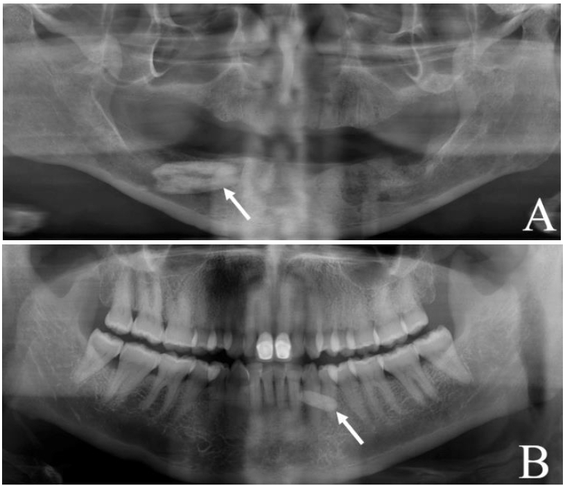
Two patients presented with incidental findings of well-defined radiopacities located in the mandible (Figure 1A & B).
INTERPRETATION
When radiopacities are detected in the mandible it is imperative to establish whether they are centrally (within or attached to the bone) or peripherally (in the soft tissues) located. The establishment of locality aids in the development a differential diagnosis.
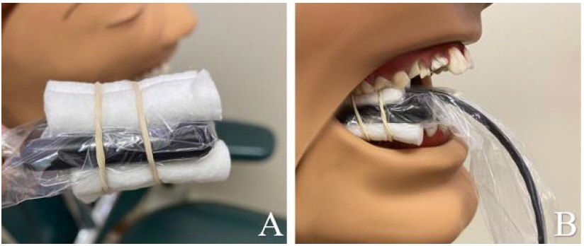
Two low-dose intraoral methods can be used in this regard, namely the buccal object-rule and mandibular occlusal radiographs. In the golden age of film and the more modern photostimulable phosphor plates, the process of acquiring an occlusal radiograph is typically straightforward. However, this process is difficult with the more commonly used charged coupled device (CCD) or complementary metal-oxide semiconductor (CMOS) digital sensors. An alternative method may be used to secure the sensor with two dental disposable cotton rolls at the top and bottom, held in place with an elastic band.
The exposure side of the sensor is then positioned against the segment of the jaw in the region of interest (Figure 2A & B).
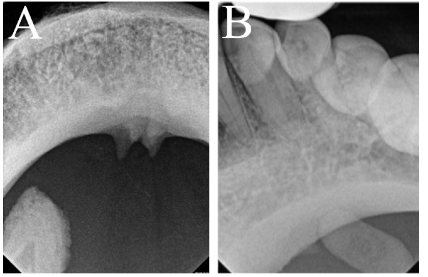
Using this method it was established that the radiopacities of both cases were located peripherally in the soft tissues, supporting a diagnosis of sialoliths (Figure 3A & B).
Salivary gland stones, better termed sialoliths, occur due to deposition of calcium salts around a central nidus of bacteria or debris. Their formation can be enhanced by xerostomia or chronic sialadenitis. Sialoliths most frequently occur in the submandibular gland followed by the parotid. The predilection for the submandibular gland is due to the thicker mucinous secretions produced by the gland and the long torturous path of Wharton's duct.1 Clinically, patients may experience swelling and intermittent pain around mealtimes. Panoramic radiography usually highlights oval or elongated radiopacities superimposed over the mandible.2 Imaging from another angle is needed for final confirmation of the diagnostic hypothesis. The treatment consists of surgical removal of the sialolith with repair of the associated duct.1
References
1. Baurmash HD. Submandibular Salivary Stones: Current Management Modalities. J Oral Maxillofac Surg. 2004;62(3):369-378. doi:10.1016/j.joms.2003.05.011 [ Links ]
2. Ribeiro A, Keat R, Khalid S, et al. Prevalence of calcifications in soft tissues visible on a dental pantomogram: A retrospective analysis. J Stomatol Oral Maxillofac Surg. 2018;119(5):369-374. doi:10.1016/j.jormas.2018.04.014 [ Links ]
 Correspondence:
Chané Nel
Department of Oral Pathology and Oral Biology, University of Pretoria
Pretoria, South Africa
Email: chane.nel@up.ac.za
Author contributions:
1 . Chané Nel: Primary author - 70%
2 . Zarah Yakoob: Second author - 30%

