


Services on Demand
Article
 English (pdf)
 Article in xml format
 Article references
Indicators

Related links
-
 Cited by Google
 -
 Similars in Google
Share

 Permalink
South African Dental Journal
On-line version ISSN 0375-1562
Print version ISSN 0011-8516
S. Afr. dent. j. vol.76 n.3 Johannesburg Apr. 2021
http://dx.doi.org/10.17159/2519-0105/2021/v76no3a8
RADIOLOGY CASE
CJ NortjéI; J WaltersII
IBChD, PhD, ABOMR, DSc. Faculty of Dentistry, University of the Western Cape. ORCID Number: 0000-0002-9717-5514
IIBChD PDD (MFR) PGD (OS) MSc (MFR), Department of Oral and Maxillofacial Radiology, Faculty of Dentistry, University of the Western Cape, Tygerberg Oral Health Centre, South Africa. ORCID Number: 0000-0002-0593-6890
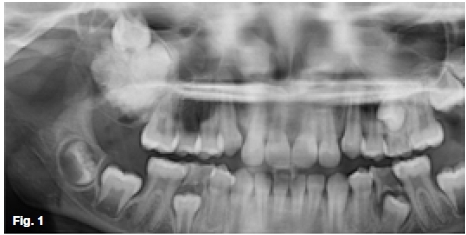
This 10-year-old boy presented with a main complaint of a carious painful primary molar in the third quadrant. A pantomograph revealed an incidental mass in the right posterior maxilla (Figure 1). No other symptoms were reported. What are the most important radiological features and what is your provisional diagnosis?
INTERPRETATION
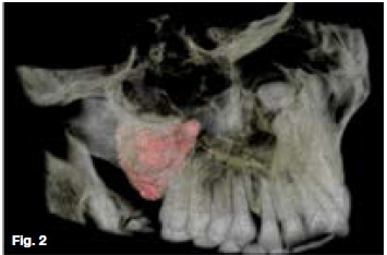
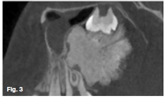
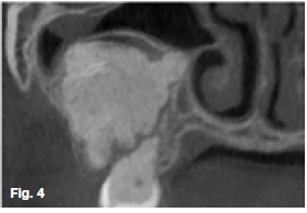
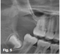
Appearing as a solitary radiopaque mass encompassed within a well-defined corticated cystic-like capsule and inclusion of the third molar. Expansion, thinning, and interruption of the cortex with protrusion into the maxillary sinus was discernible. Root resorption at the 16 with impaction and displacement of the third molar was apparent. 3D volume rendering (Figure 2) demonstrate the extensions. Sagittal (Figure 3) and coronal (Figure 4) CBCT slices depict a lobulated mass appearing as a tooth-like predominantly intermediate-density accompanied with specs of high-densities throughout and a missing second molar. Histopathological confirmation of an ameloblastic fibro-odontoma (AFO) was made. A follow-up cropped pantomograph (Figure 5) indicated no recurrence. A member of the mixed odontogenic tumours, demonstrating features of an ameloblastic fibroma and an odontoma.
The WHO notes it to be an immature representation of the latter. It is a benign neoplasm consisting of odon-togenic epithelium, ectomesenchyme and dental hard tissue formation. Compared to the ameloblastic fibroma and ameloblastic fibrodentinoma. The AFO's inductive changes are more advanced with enamel and dentine present. Frequency ranges from 0.3% to 3.7%. With 98.9% of cases observed before the age of twenty and a mean of 9-years-old. Similarly, odontomas also develop during the tooth-forming years. Therefore, meticulous radiographic interpretation can facilitate provisional diagnosis. Radio-graphically lesions appear unilocular or multilocular with internal content ranging from multiple specs of calcifications to solid odontoma-like masses. Diagnostic features include a fine cortical outline, a thick lucent rim, and the ability to cause significant tooth displacement when compared to similar appearing lesions of the same size. There is a slight male predominance with a ratio of 1.4:1. Predilection has been shown for the posterior mandible, though all regions of the jaws can be affected. Usually asymptomatic, slow-growing, and deemed solely as a central intraosseous lesion. Most are associated with an unerupted or impacted tooth where investigation leads to initial discovery. Treatment consists of surgical enucleation.
References
1. Langlais RP, Langland OE, Nortjé CJ. Diagnostic Imaging of the Jaws. Williams & Wilkins. 1995. [ Links ]
2. Reichart P, Philipsen, HP. Odontogenic Tumors and Allied Lesions. Quintessence. 2004. [ Links ]
 Correspondence:
Jaco Walters
Department of Oral and Maxillofacial Radiology, Faculty of Dentistry
University of the Western Cape, Tygerberg Oral Health Centre
South Africa.
Email: jawalters@uwc.ac.za
Author contributions:
1 . Christoffel J Nortjé: Principal author - 50%
2 . Jaco Walters: Second author - 50%

