


Serviços Personalizados
Artigo
 Inglês (pdf)
 Artigo em XML
 Referências do artigo
Indicadores

Links relacionados
-
 Citado por Google
 -
 Similares em Google
Compartilhar

 Permalink
South African Dental Journal
versão On-line ISSN 0375-1562
versão impressa ISSN 0011-8516
S. Afr. dent. j. vol.76 no.1 Johannesburg Fev. 2021
http://dx.doi.org/10.17159/2519-0105/2021/v76no1a3
REVIEW
A comparison of root canal transportation and centering ability between WaveOne® Gold and Protaper Next® files, using micro-computed tomography
A GajoumI; E PatelII; IE MunshiIII; S TootlaIV
IBDS, MScDent., Department of Conservative Dentistry and Endodontics, Faculty of Dentistry, Alasmarya University, Zliten, Libya. ORCID Number: 0000-0003-4061-6291
IIBDS, MScDent, Department of Paediatric & Restorative Dentistry, School of Oral Health Sciences, Faculty of Health Sciences, University of the Witwatersrand, South Africa. ORCID Number: 0000-0002-7683-4867
IIIBSc, BCHD, MDent, Department of Oral Rehabilitation, School of Oral, Health Sciences, Faculty of Health Sciences, University of the Witwatersrand, South Africa
IVBCHD, MScDent, Department of Paediatric & Restorative Dentistry, School of Oral Health Sciences, Faculty of Health Sciences, University of the Witwatersrand, South Africa. ORCID Number: 0000-0001-9014-9455
ABSTRACT
AIM: This study compared the transportation and centering ability of ProTaper Next (PTN) and WaveOne Gold (WOG) files in curved permanent teeth using micro-computed tomography (μCΤ
METHODOLOGY: Twenty-four molar teeth with curved roots were divided randomly into two equal groups. The root canals of one group was prepared using PTN files, and the other using WOG files. Pre-instrumentation and post-instrumentation imaging were taken for all the teeth. The dentine thickness of the pre-and the post-instrumentation cross sections was measured at eight different points at three levels: 3, 5 and 7mm from the apex, by two dentists using image analysis software. The data were analysed using one-way ANOVA, at a 5% significance level
RESULTS: The transportation in both groups was within the range accepted in the literature. The WOG file exhibited significantly less root canal transportation compared with the PTN file (p=0.001). The WOG file showed a significantly (p<0.001) higher mean centering ratio of 0.4286 when compared to that of PTN at 0.2448
CONCLUSIONS: Using a novel technique to measure canal transportation, this study found that the WOG and PTN systems were both suitable for preparation of curved molar root canals, but the WOG showed significantly less canal transportation and better centering ability than the PTN system
Keywords: Centering ability, Protaper Next, root canal transportation, WaveOne Gold.
INTRODUCTION
Biomechanical preparation of the root canal should result in a tapered preparation that maintains the original path of the canal.1 This is particularly pertinent in the apical third of curved canals because of the propensity to straighten the canal and the development of complications like, ledge formation, zipping, perforation and root canal transportation.2,3
Root canal transportation and centering ability measurements have been used to compare filing systems and techniques.4-6 Root canal transportation is defined as "Removal of canal wall structure from the outside curve in the apical half of the root canal due to the tendency of files to restore themselves to their original linear shape during canal preparation; may lead to ledge formation and possible perforation".7
A centred root canal preparation is another way to express an ideal root canal preparation without transportation. However, it must be kept in mind that the sole use of this method is flawed as it does not account for total circumferential transportation but only transportation in certain directions. The centering ability of a file is its ability to keep centred in the canal during instrumentation and is important for ideal root canal enlargement and to avoid weakening of root canal structure.8 While root canal transportation is usually measured in millimetres or micrometres, centering ability is measured using a ratio of 0 to 1. A centering ratio of 1 indicates perfect centering ability whereas, a ratio closer to zero indicates that the root canal wall was unequally prepared.
Over the years, manufacturers of nickel-titanium (NiTi) file systems have introduced various changes to the metallic structure and the designs of the files in order to improve their performance. Different cross sectional and longitudinal designs were produced to minimize apical transportation and to achieve a faster and more predictable canal preparations. These improvements include changes in design, metallurgy, and even the motion which the file is driven with.
ProTaper Next (PTN) file system (Dentsply Tulsa Dental Specialties, USA) is made from M-Wire; a thermo-mechanically treated NiTi metal. PTN was introduced in 2013 and is a continuous rotation system. It contains three crystalline phases: martensite, R-phase, and austenite9 and has shown improved cyclic fatigue resistance in comparison with conventional NiTi alloys.10 The main characteristic of PTN files is that the centre of the file mass is offset, which is claimed to provide a number of advantages, not least of which is the ability to prepare a size of canal that would otherwise require larger and stiffer files.11,12 PTN files showed competitive results when compared to other file systems in some studies.13,14
The WaveOne Gold (WOG) reciprocating file system (Dentsply Maillefer, Switzerland) was launched in 2015. After having established an effective glide path, a single file is required to shape the entire canal (in most cases, according to the manufacturer). The files have an off-centred parallelogram cross-section similar to PTN. WOG files have significantly greater flexibility and resistance to torsional stress compared to Reciproc (VDW, Germany) and Twisted File Adaptive (Kerr Endodontics, Glendora, Orange, CA, USA).15
Recently, methods to measure root canal transportation involving micro-computed tomography (μCT) and cone-beam computed tomography (CBCT) have become popular.13,16-19 Whilst CBCT produces 3-D images, the spatial resolution is considered to be inferior to pCT which conserves specimens and provides 3-D high resolution images.20,21
The aim of this study was to compare the root canal transportation and centering ability produced by the WOG reciprocating file system and PTN filing system in root canal treatment using pCT scans. The study uses a novel eight points measurement technique modified from Gambill et al.4 to measure the amount of canal transportation at three different levels along the length of the root canal.
Material and methods
24 extracted maxillary and mandibular first molars with complete root apices were randomly divided into two groups: PTN and WOG. The mesiobuccal and distobuc-cal roots of maxillary first molars, and the mesiobuccal and mesiolingual root canals of mandibular first molars were used, if they had roots with separate root canals and root canal curvatures of between 20 and 40 degrees.13 Excluded were calcified root canals, resorbed roots/root canals, root canals which did not allow a size #8 K-file to be inserted to the major foramen and those that allowed the passive placement of a #15 K-file to within 1 mm of the major foramen. Teeth with fractured roots were also excluded as were those with any previous attempts of endodontic treatment. Ethical clearance for the use of freshly extracted teeth (which were placed immediately after extraction in a 37% formalin solution) was granted by the Human Research Ethics Committee of the University of the Witwatersrand, South Africa; certificate number M160262.
Teeth were placed in a Protrain platform (Simit Dental, supplied by Dentsply, South Africa) and access cavities of sufficient size were prepared using round diamond burs and the Endo Z bur (Dentsply Maillefer, Switzerland). The canals of selected teeth were explored with a size #10 K-file (Dentsply Maillefer, Switzerland), which was advanced passively into the canal until the tip reached the apical foramen. The working length was established while using magnification (Zeiss OPMI Pico dental microscope, 31.25X magnification), from a standardised coronal point to the anatomical apical foramen minus 1 mm. The occlusal surfaces of the teeth were ground down so that the working length of all teeth was 17mm.
Schneider's method was used to measure the curvature of the root canal, while Pruett's method was used to measure radius of root canal curvature.22,23 The angle and the radius of curvature was calculated using the Digimizer 4 image analysis software (MedCalc Software®). There-after the teeth were embedded in an acrylic resin block 25 mm depth x 15 mm x 15mm. Base plate wax was used to prevent the acrylic resin from entering the apical foramen. Pre-instrumentation images were taken using a Nikon Metrology XTH 225/320 LC Micro-CT scanner at a voxel size of 15μm, 80KV and 95μA.
For all teeth, the glide path was prepared using the ProGlider file (Dentsply Maillefer, Switzerland), according to manufacturer instructions. The X-Smart™ Plus micromotor (Dentsply Maillefer, South Africa) was used to drive the files and adjusted for the two file systems. One group was instrumented using the primary size (25/.07) WOG file according to manufacturer instructions and the other group instrumented using a PTN file X1 (017/0.04) followed by the X2 (025/0.06) file. A #10 K-file was used in both groups between every step to maintain canal patency. 10 to 12mg of 17% EDTA (RC-Prep, Premier Dental, USA) was loaded on every rotary file to lubricate the root canal.
The individual canals were irrigated with 3 ml of 2.5% sodium hypochlorite between each rotary file. Each instrument was discarded after use in four canals, but any instrument that deformed was discarded immediately. A final irrigation with saline was applied to each root canal and a post-instrumentation μΟΤ image taken.
Two calibrated assessors compared the pre-and post-instrumentation images using VGSTUDIO MAX 3.0 software (Volume Graphics GmbH, Germany) by measuring the canal cross-sections. When the value between the two raters was different, the mean value of the readings was taken as a final value.
The technique used to measure canal transportation was modified from Gambill et al.4 Instead of two measurements, this study used eight measurements (Fig. 1) after superimposition of pre-and post-instrumentation images. The root canal transportation and the centering ability ratio were measured in four directions as the axes A-B, C-D, E-F and G-H.

All readings were taken at each of the three levels from the apex (3, 5 and 7mm from the root apex). The measurements along each axis were recorded and the values of opposing distances along an axis were subtracted from each other. A result of zero for the equation (e.g. (A1-A2) - (B1-B2)) was interpreted as no transportation. A positive value meant that the direction of canal transportation is in the direction of the first part of the equation and a negative value meant the direction of the second part of the equation.
A result of up to 0.15 mm was considered acceptable root canal transportation as most rotary NiTi instruments produce 0.15 mm or less.24 A result of >0.30 mm at the apical end was unacceptable transportation25 and values between 0.15 and 0.30 mm were considered borderline. In order to determine the centering ability, the ratios of the measurements along each axis were calculated, and transposed if necessary to reach a value of between 0 and 1. For example if the result of  was more than 1 that meant
  must be used instead to obtain a value between 0 and 1. Hence the formula was a variation as necessary of the following:
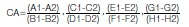
A result of 1 means optimal centering ability while a result of zero means no centering ability.
RESULTS
Three X2 PTN files separated during the study, with no separation recorded for the WOG files. Since separation occurred after the full working length had been reached, the three affected teeth were included in the analysis. The average curvature of the canals was 26.43° and 26.54° in the PTN group and the WOG group, respectively. The average radius of curvature in the PTN group was 5.49mm, and in the WOG group was 5.52mm.
Root canal transportation and centering ability results were normally distributed and were analysed using a one-way ANOVA, using Stata Version 13.1 (Stata Corp LP, Lakeway Drive, College Station, Texas, USA). Where there was a statistically significant difference, Tukey's multiple comparison post-hoc test was performed to show the site of the difference. A p value of <0.05 was considered statistically significant.
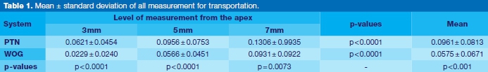
Canal transportation
At all levels of measurement, as well as the overall mean of all measurements, the PTN system was statistically significantly greater than the WOG system (Table 1).
The frequency and direction of root canal transportation at each measurement level is shown in Table 2. At the 3mm and 5mm levels, both systems caused greater canal transportation toward the outside, as compared with the inside of canal curvature. However, the opposite was true at the 7mm sections. The PTN system had a lower range and frequency of samples with no canal transportation (4.2 to 8.3%) than the WOG filing system (8.3 to 33.3%).
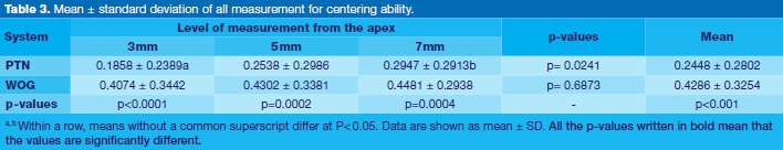
Centering ability
The centering ability was significantly different (p<0.0001) between the two systems, with the WOG system performing significantly better overall, and at each of the three levels (Table 3). The WOG also showed consistency between the different levels, whereas the PTN system had significant differences between the 3mm and 7mm levels.
DISCUSSION
The aim of this study was to compare the canal transportation and centering ability produced by a rotary file system, PTN, and a reciprocal file system, WOG, in freshly extracted permanent molars with severe canal curvature. In the present study, freshly extracted teeth were used, since they more accurately mimic the clinical situation.26 μCT scans were used to measure canal transportation and centering ability at three points along the length of the canal, representing the apical, middle and coronal thirds of the canal.
Root canal transportation
The technique that was used to measure root canal transportation was modified from the technique developed by Gambill et al.4 which was limited to using two measurements along a single plane. Measurement in only one direction may not be able to adequately show the geometric changes in three dimensions (3D) along the length of the canal. This study modified Gambill's technique to enable an eight points circumferential analysis that allowed for an enhanced assessment of the mechanical action of the file systems.
Table 2 shows that transportation was observed to have occurred circumferentially, and not just in one direction. This affirms the use of the eight-point measurement technique for assessing canal transportation.
Apical canal transportation of up to 0.15 mm is acceptable and should not be greater than 0.30 mm24 as it negatively affects apical sealing.25 Although the results of this study show that the PTN system produced more canal transportation and poorer centering ability compared to the WOG system, the canal transportation of PTN was within the accepted range and was similar to that obtained by Silva et al.27 (0.061 to 0.144mm), Zhao et al.13 (0.62mm), and Zanesco et al.14 (0.055 to 0.081 mm). The results for the WOG system in this study were superior to those reported by van der Vyfer et al.28
This study also confirms the findings from previous studies that the direction of canal transportation in the 3mm and 5mm sections under both systems was predominantly toward the outside curvature, while in 7mm section it was toward the inside of the curvature.29,30
Centering ability
Centering ability, which indicates whether or not the dentine removal over the prepared area is spread evenly by the instrument. The ability of an instrument to remain centred within the natural canal path during preparation is essential for adequate enlargement without weakening the root structure.8 Good centering ability reduces the risk of transportation, zipping, elbow formation and other preparation errors.
The results of this study concur with the findings by Tambe et al.31 who demonstrated that the WaveOne (reciprocation) file (Dentsply Maillefer, Switzerland) remained better centred in the canal than the ProTaper. However, this is in contrast with McRay et al.17 who showed no significant difference between WaveOne (reciprocation) file and Pro-Taper Universal (continuous rotation) (Dentsply Maillefer, Switzerland). This could be because WaveOne and Pro-Taper Universal files have similar design, taper and size and made from the same form of NiTi alloy.
There are several possible explanations for the findings of this study. Zhao et al.13 recognised that the centering ability is influenced both by the design features of the instruments (size, taper, flexibility, and type of alloy) as well as the anatomy of the root canal.
The WOG filing system had superior centering ability compared with the PTN filing system, in all three sections along the length of the root canal. This may be attributed to the following factors:
1. The thermal treatment of the WOG file gives it greater flexibility, allowing the file to follow the root canal anatomy without considerable resistance, and confirms the findings of Elsaka et al.15 thatthe WOG file had significantly greater flexibility and resistance to torsional stress compared to Reciproc and Twisted File Adaptive.
2. The use of a single file requires the gradual introduction of the file to the working length, whereby the file reaches the full working length after the coronal part is partially prepared.
3. The kinematics of the instruments was originally thought to play a role in canal transportation, and reciprocal motion was thought to make the file more centred.32 However, a review of the instrumentation kinematics of engine driven NiTi instruments showed conflicting results on the effect of reciprocating instruments on canal transportation.33 They attribute the conflicting results to different instruments and methodologies of the respective studies.
This study is in agreement with previous studies that the Primary size of WOG file has a high cyclic fatigue resistance compared to other Ni-Ti files,34,35 and recommends the use of the WOG Primary or smaller size file in severely curved root canals to attain superior shaping and centering ability, and to avoid file fracture.
A limitation of this study was that larger sizes of the WOG files were not tested. Caution must be taken when extrapolating these results to the larger file sizes, as they would differ in flexibility. Further studies are needed to evaluate the root shaping ability of the larger sizes of WOG files due to their differing flexibility.
CONCLUSIONS
The WOG and PTN systems were both suitable for preparation of molar root canals with severe curvature, but the WOG showed significantly less canal transportation and better centering ability than the PTN system. This study recommends the use of a novel technique to measure root canal transportation. This technique enables an improved eight point circumferential analysis of the prepared canals.
ACRONYMS
NiTi: Nickel Titanium
μΟΤ: Micro-Computed Tomography
CBCT: Cone-Beam Computed Tomography
PTN: ProTaper Next
WOG: WaveOne Gold
KV: Kilovolt
μA: Micro Amber
EDTA: Ethylenediaminetetraacetic Acid
References
1. Schilder H. Cleaning and shaping the root canal. Dent Clin North Am. 1974; 18: 269-96. [ Links ]
2. Sathorn C, Palamara JE, Messer HH. A comparison of the effects of two canal preparation techniques on root fracture susceptibility and fracture pattern. J Endod. 2005; 31(4): 283-7. [ Links ]
3. Hargreaves KM, Berman LH. Cohen's pathways of the pulp expert consult. Elsevier Health Sciences. 2015; Oct 2. [ Links ]
4. Gambill JM, Alder M, Carlos E. Comparison of nickel-titanium and stainless steel hand-file instrumentation using computed tomography. J Endod. 1996;22(7):369-75. [ Links ]
5. Short JA, Morgan LA, Baumgartner JC. A comparison of canal centering ability of four instrumentation techniques. J Endod. 1997; 23(8): 503-7. [ Links ]
6. Gluskin AH, Brown DC, Buchanan LS. A reconstructed computerized tomographic comparison of Ni-Ti rotary GT™ files versus traditional instruments in canals shaped by novice operators. Int Endod J. 2001; 34(6): 476-84. [ Links ]
7. Glossary of Endodontic Terms. American Association of Endodontists 9th Edn. Chicago, American Association of Endodontists. 2016. Available at https://www.aae.org/specialty/clinical-resources/glossary-endodontic-terms/. [ Links ]
8. Kandaswamy D, Venkateshbabu N, Porkodi I, Pradeep G. Canal-centering ability: An endodontic challenge. J Conserv Dent. 2009; 12(1): 3. [ Links ]
9. Alapati SB, Brantley WA, lijima M, et al. Metallurgical characterization of a new nickel-titanium wire for rotary endodontic instruments. J Endod. 2009; 35(11): 1589-93. [ Links ]
10. Johnson E, Lloyd A, Kuttler S, Namerow K. Comparison between a novel nickel-titanium alloy and 508 nitinol on the cyclic fatigue life of ProFile 25/. 04 rotary instruments. J Endod. 2008; 34(11): 1406-9. [ Links ]
11. Haapasalo M, Shen Y. Evolution of nickel-titanium instruments: from past to future. Endod Topics. 2013; 29(1): 3-17. [ Links ]
12. Saber SE, Nagy MM, Schäfer E. Comparative evaluation of the shaping ability of ProTaper Next, iRaC e and Hyflex CM rotary NiTi files in severely curved root canals. Int Endod J. 2015; 48(2): 131-6. [ Links ]
13. Zhao D, Shen Y, Peng B, Haapasalo M. Root canal preparation of mandibular molars with 3 nickel-titanium rotary instruments: a micro-computed tomographic study. J Endod. 2014; 40(11): 1860-4. [ Links ]
14. Zanesco C, Só MV, Schmidt S, Fontanella VR, Grazziotin-Soares R, Barletta FB. Apical transportation, centering ratio, and volume increase after manual, rotary, and reciprocating instrumentation in curved root canals: analysis by micro-computed tomographic and digital subtraction radiography. J Endod. 2017; 43(3): 486-90. [ Links ]
15. Elsaka SE, Elnaghy AM, Badr AE. Torsional and bending resistance of WaveOne Gold, Reciproc and twisted file adaptive instruments. Int Endod J. 2017; 50(11): 1077-83. [ Links ]
16. Marzouk AM, Ghoneim AG. Computed tomographic evaluation of canal shape instrumented by different kinematics rotary nickel-titanium systems. J Endod. 2013; 39(7): 906-9. [ Links ]
17. McRay B, Cox TC, Cohenca N, Johnson JD, Paranjpe B. A micro-computed tomography-based comparison of the canal transportation and centering ability of ProTaper Universal rotary and WaveOne reciprocating files. Quintessence Int. 2014; 45(2): 101-8. [ Links ]
18. Gergi R, Arbab-Chirani R, Osta N, Naaman A. Micro-computed tomographic evaluation of canal transportation instrumented by different kinematics rotary nickel-titanium instruments. J Endod. 2014; 40(8): 1223-7. [ Links ]
19. Baek JY, Yoo HM, Park DS, et al. Comparison of the shaping abilities of three nickel-titanium instrumentation systems using micro-computed tomography. JDS. 2014; 9(2): 111-7. [ Links ]
20. Freire LG, Gavini G, Cunha RS, Santos MD. Assessing apical transportation in curved canals: comparison between cross-sections and micro-computed tomography. Braz Oral Res. 2012; 26(3): 222-7. [ Links ]
21. Shen YA, Cheung GS. Methods and models to study nickeltitanium instruments. Endod Topics. 2013; 29(1): 18-41. [ Links ]
22. Schneider SW. A comparison of canal preparations in straight and curved root canals. Oral Surg Oral Med Oral Pathol. 1971; 32(2): 271-5. [ Links ]
23. Pruett JP, Clement DJ, Carnes Jr DL. Cyclic fatigue testing of nickel-titanium endodontic instruments. J Endod. 1997; 23(2): 77-85. [ Links ]
24. Peters OA. Current challenges and concepts in the preparation of root canal systems: a review. J Endod. 2004; 30(8): 559-67. [ Links ]
25. Wu MK, Fan B, Wesselink PR. Leakage along apical root fillings in curved root canals. Part I: effects of apical transportation on seal of root Allings. J Endod. 2000; 26(4): 210-6. [ Links ]
26. Nagaraja S, Murthy BS. CT evaluation of canal preparation using rotary and hand NI-TI instruments: An in vitro study. J Conserv Dent. 2010: 13(1): 16. [ Links ]
27. Silva EJ, Pacheco PT, Pires F, Belladonna FG, De-Deus G. Microcomputed tomographic evaluation of canal transportation and centring ability of ProTaper Next and Twisted File Adaptive systems. Int Endod J. 2017; 50(7): 694-9. [ Links ]
28. van der Vyver PJ, Paleker F, Vorster M, de Wet FA. Root Canal Shaping Using Nickel Titanium, M-Wire, and Gold Wire: A Micro-computed Tomographic Comparative Study of One Shape, ProTaper Next, and WaveOne Gold Instruments in Maxillary First Molars. J Endod. 2019; 45(1): 62-7. [ Links ]
29. Tasdemir T, Aydemir H, Inan U, Ünal O. Canal preparation with Hero 642 rotary Ni-Ti instruments compared with stainless steel hand K-file assessed using computed tomography. Int Endod J. 2005; 38(6): 402-8. [ Links ]
30. Pasternak-Júnior B, Sousa-Neto MD, Silva RG. Canal transportation and centring ability of RaCe rotary instruments. Int Endod J. 2009; 42(6): 499-506. [ Links ]
31. Tambe VH, Nagmode PS, Abraham S, Patait M, Lahoti PV Jaju N. Comparison of canal transportation and centering ability of rotary Protaper, One Shape system and Wave One system using cone beam computed tomography: An in vitro study. J Conserv Dent. 2014; 17(6): 561. [ Links ]
32. Roane JB, Sabala CL, Duncanson Jr MG. The "balanced force" concept for instrumentation of curved canals. J Endod. 1985; 11(5): 203-11. [ Links ]
33. Qapar ID, Arslan H. A review of instrumentation kinematics of engine-driven nickel-titanium instruments. Int Endod J. 2016; 49(2): 119-35. [ Links ]
34. Topguoglu HS, Düzgün S, Akti A, Topguoglu G. Laboratory comparison of cyclic fatigue resistance of WaveOne Gold, Re-ciproc and WaveOne files in canals with a double curvature. Int Endod J. 2017; 50(7): 713-7. [ Links ]
35. Özyürek T. Cyclic fatigue resistance of Reciproc, WaveOne, and WaveOne Gold nickel-titanium instruments. J Endod. 2016; 42(10): 1536-9. [ Links ]
 Correspondence:
Ebrahim Patel
Department of Paediatric & Restorative Dentistry, School of Oral Health Science
7 York Road, Parktown, Johannesburg, 2193, South Africa
Email: ebrahim.patel@wits.ac.za
Author contributions:
1. Abdulrzag Gajoum: Principal author - 55%
2. Ebrahim Patel: Co-author - 15%
3. Ismail E Munshi: Co-author - 15%
4. Saidah Tootla: Co-author - 15%

