


Serviços Personalizados
Artigo
 Inglês (pdf)
 Artigo em XML
 Referências do artigo
Indicadores

Links relacionados
-
 Citado por Google
 -
 Similares em Google
Compartilhar

 Permalink
South African Dental Journal
versão On-line ISSN 0375-1562
versão impressa ISSN 0011-8516
S. Afr. dent. j. vol.75 no.4 Johannesburg Mai. 2020
http://dx.doi.org/10.17159/2519-0105/2020/v75no4a7
CASE RFPORT
Lichenoid Granulomatous Stomatitis - an oral medicine case book
L RobinsonI; AW van ZylII; WFP van HeerdenIII
IBChD, PDD (Maxillofacial Radiology), PDD (Forensic Odontology), Department of Oral Pathology and Oral Biology, School of Dentistry, Faculty of Health Sciences, University of Pretoria. ORCID Number: 0000-0002-0549-7824
IIBChD, MChD, Private Practice Periodontist, Hermanus & Honorary Professor, Department of Oral Medicine and Periodontics, Faculty of Health Sciences, University of Witwatersrand
IIIBChD, MChD, FC Path (SA) Oral Path, PhD, DSc, Department of Oral Pathology and Oral Biology, School of Dentistry, Faculty of Health Sciences, University of Pretoria. ORCID Number: 0000-0003-2494-667X
CASE REPORT
A 72-year-old male patient presented with a painful area on the labial mucosa of the upper lip adjacent to tooth 12. He complained of a non-healing "bruise" on the inside of his lip that had been present for about 4-weeks. The patient was a non-smoker and reported taking anti-hypertensive medication (Lisinopril, hydrochlorothiazide) for 10-years. A prior colonoscopy 4 years ago revealed adenomas.
On examination, a swelling was noted with the overlying mucosa appearing erosive and erythematous with peripheral white striae, clinically similar to lichen planus. The lesion also involved the facial gingiva associated with teeth 12 & 22 (Figure 1). This red-white lesion was excised as well as minor salivary glands that extruded during the biopsy procedure. No additional pharma-cotherapeutic agents were administered.

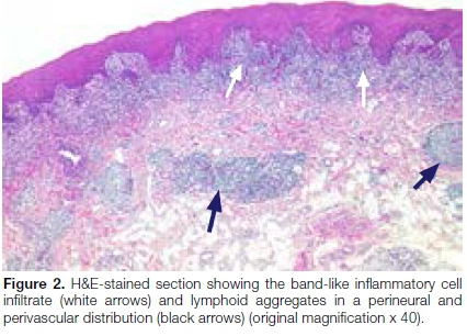
The specimen submitted from the lip lesion consisted of a mucosa-covered tissue fragment measuring 12x5x4mm. Histological evaluation confirmed the presence of a tissue fragment surfaced by stratified squamous epithelium with areas of hyperparakeratosis, as well as vacuolar degeneration of the basal cell layer with associated apoptotic bodies. A band-like lymphohistiocytic infiltrate was seen in the underlying superficial lamina propria. Secondarily, varying degrees of granulomatous inflammation within the superficial lichenoid inflammatory infiltrate was noted. These poorly formed granulomas were composed of epithelioid macrophages, however no giant cells or central necrosis could be appreciated. Additionally, lymphoid follicles were seen, with a striking perineural and perivascular distribution (Figures 2 & 3). No foreign material was noted under polarised light. Periodic acid-Schiff (PAS) and Ziehl-Neelsen histoche-mical stains failed to highlight any fungal elements or acid-fast bacilli respectively.

In conclusion, a final diagnosis of lichenoid granulomatous stomatitis was made.
The patient was followed-up one month after total excision of the lesion to reassess for further treatment. He reported that healing was uneventful, and all symptoms had disappeared after the excision biopsy. Intra-oral examination showed an absence of any clinical signs in the original area (Figure 4).

The patient will be followed-up for routine examination every few months and was instructed to immediately report back should symptoms reappear.
DISCUSSION
The presence of lichenoid inflammation with concomitant granulomatous inflammation is an uncommon observation within the oral cavity. Many diseases are typified by either lichenoid or granulomatous inflammation.1 However when both patterns occur simultaneously, problems arise in determining which pattern represents the primary disease process, or whether the coexistence of both patterns represents a distinctive disease entity.
The term lichenoid granulomatous stomatitis (LGS) was first described in literature by Robinson et al. in 2006.1 Lichenoid inflammation may render the oral mucosa susceptible to the ingress of foreign material, resulting in granuloma formation. LGS has been reported in cases of foreign body gingivitis. In a series of 61 foreign body gingivitis cases, investigators reported the presence of both patterns of inflammation in 26% of biopsies studied.2 In the present case, no foreign material could be identi-fled under polarised light.
To date the largest review by Hakeem et al.3 in 2019 identified 47 patients with LGS. In this study, patient demographics showed a female predilection of 1.9:1 with a mean age of 59 years. Seventy-nine percent of patients were older than 55 years. Patients commonly presented with a solitary lesion, with most cases occurring on the attached gingiva followed by the buccal mucosa and vestibule. With regards to clinical description, 38% were described as erythroleukoplakia, 36% as leukoplakia, and 26% as purely erythematous lesions.
There was an equal incidence of presentation amongst patients regarding painful or non-painful lesions. The clinical impressions for all cases in this study (for which multiple were listed in some instances) included lichen planus (17 cases), dysplasia/carcinoma in sifu/squamous cell carcinoma (11 cases), vesiculobullous lesions (9 cases), trauma-associated (5 cases), leukoplakia (5 cases), allergy (2 cases) and other differentials (4 cases). No clinical diagnosis was reported in 7 cases.3
Histologically, LGS consists of three distinctive components. First, is the presence of lichenoid inflammation, characterised by hyperkeratosis, basal cell degeneration with associated apoptotic bodies and a band-like lymphohistiocytic inflammatory cell infiltrate. Secondly, variable degrees of granulomatous inflammation can be seen throughout the corium. Importantly, all granulomas consist of epithelioid macrophages without giant cells or areas of necrosis. Thirdly, lymphoid follicles are present in the corium showing a prominent perineural distribution.1
Additional studies ruling out infective agents and foreign material should be performed in suspected cases. A study of six cases of LGS by Robinson et al.1 found that presence of fungal hyphae was not associated with a lichenoid inflammatory reaction.1 Secondly, granulomatous inflammation is typical of deep mycoses and not superficial candidosis.
Patients taking certain medications may develop LGS, which may ultimately resolve with discontinuation of the medication.3 Additionally, the case review by Robinson et al., reported two patients known to be on medications that have an association with lichenoid eruptions, namely Naproxen (Non-steroidal anti-inflammatory drug), Atenolol (β-adrenoceptor blocker), and Ramipril (Angio-tensin-converting enzyme inhibitor).4 Furthermore, these groups of drugs have also been implicated in both lichenoid and granulomatous dermatitis.
Equally rare is the presence of both patterns of inflammation in dermatological conditions. Lichenoid granulomatous dermatitis (LGD) was first described by Gonzalez in 1986.5 A study by Magro and Crowson6 reported a series of 40 patients with skin lesions showing lichenoid dermatitis with a granulomatous component. The majority of these cases had confounding medical problems associated with the disease, however one-fifth of the cases were considered idiopathic. Furthermore, in 12 cases an infective cause was implicated. The agent was either a or bacterial infection and not fungal in origin.6 Both inflammatory patterns have also been reported to coexist in a rare skin condition, lupus erythematosus profundus.7
In total approximately 57 cases have been previously reported as LGD. The gender ratio reported in the prior cases showed a slight female predilection of 1.3:1 with mean age of 48 years. The trunk, arms, and legs were the most common location. Dermatologic lesions mostly presented as erythematous or as maculopapular entities.6,8-9
Although many similarities were found when comparing histological features of LGS and LGD, some important differences were noted. Cases from the oral mucosa did not show an interstitial array between collagen fibers surrounded by palisaded histiocytes, granuloma annu-lare-like appearance, focal Langhans giant cells or granulomatous vasculitis. Additionally, a prominent perivascular inflammatory infiltrate, as seen in LGS cases, was not emphasised in descriptions of lesions involving the skin.3
Literature is sparse regarding the treatment of LGS, however, it appears to respond well to similar regimens used in treating conventional lichen planus.3
References
1. Max Robinson C, Oxley JD, Weir J, Eveson JW. Lichenoid and granulomatous stomatitis: An entity or a non-specific inflammatory process? J Oral Pathol Med. 2006; 35(5): 262-7. [ Links ]
2. Gordon SC, Daley TD. Foreign body gingivitis: Clinical and microscopic features of 61 cases. Oral Surg Oral Med Oral Pathol Oral Radiol Endod. 1997; 83(5): 562-70. [ Links ]
3. Hakeem A, Bhattacharyya I, Aljabri M, Bindakhil M, Pachigar K, Islam MN, et al. Lichenoid reaction with granulomatous stomatitis: A retrospective histologic study of 47 patients. J Oral Pathol Med. 2019; 48(9): 846-54. [ Links ]
4. Scully C, Bagan JV. Adverse drug reactions in the orofacial region. Crit Rev Oral Biol Med. 2004; 15(4): 221-39. [ Links ]
5. Gonzalez JG, Marcus MD, Cruz DJ. Giant cell lichenoid dermatitis. J Am Acad Dermatol. 1986; 15(1): 87-92. [ Links ]
6. Magro CM, Crowson AN. Lichenoid and granulomatous dermatitis. Int J Dermatol. 2000; 39(2): 126-33. [ Links ]
7. Crowson AN, Magro C. The cutaneous pathology of lupus erythematosus: A review. J Cutan Pathol. 2001; 28(1): 1-23. [ Links ]
8. Bulur I, Gokalp H, Erdem O, Gurer M. A rare form of liche-noid tissue reaction: Lichenoid granulomatous dermatitis. J Med Cases. 2015; 6: 95-7. [ Links ]
9. Ghasemibasir H, Khezrian L, Sobhan MR. Lichenoid and granulomatous dermatitis: Report of two cases from Iran. Iran J Derm. 2015; 18(3): 136-9. [ Links ]
 Correspondence:
Willie FP van Heerden
Department of Oral Pathology and Oral Biology, University of Pretoria
South Africa
Email: willie.vanheerden@up.ac.za
Author contributions:
1 . Liam Robinson: Principle author - 50%
2 . André W van Zyl: Clinical case, treatment and follow-up - 25%
3 . Willie FP van Heerden: Diagnosis, histological images and advisor - 25%

