


Services on Demand
Article
 English (pdf)
 Article in xml format
 Article references
Indicators

Related links
-
 Cited by Google
 -
 Similars in Google
Share

 Permalink
South African Dental Journal
On-line version ISSN 0375-1562
Print version ISSN 0011-8516
S. Afr. dent. j. vol.74 n.10 Johannesburg Nov. 2019
http://dx.doi.org/10.17159/2519-0105/2019/v74no10a12
RADIOLOGY CASE
CJ Nortjé
BChD, PhD, ABOMR, DSc. Faculty of Dentistry, University of the Western Cape. ORCID Number: 0000-0002-9717-5514 Email: cnortje@uwc.ac.za
Below are three very rare and unusual lesions that may present in the jaws. What are the important radiological features and what are your differential diagnoses?
INTERPRETATION
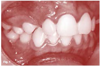
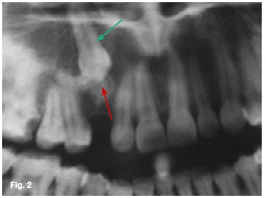
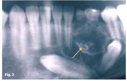
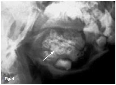
Figure 1 is of a patient with the complaint that her upper right canine has not erupted. The cropped pantomo-graph (Figure 2) shows an impacted canine with an irregular low density lesion at the incisal edge, (red arrow) is very similar to the dentin of the impacted canine (green arrow). A diagnosis of a dentinoma was made. This is an extremely rare tumour of odontogenic origin and occurs predominantly in the mandible and is frequently associated with an impacted tooth. The radio-graphic appearance is not specific, but usually there is a radiolucent area containing a large, solitary opaque mass or smaller masses of calcified material. Figure 3 is a cropped pantomograph of a fourteen year old female presenting with a swelling in the 42-35 region. A mixed radiolucent/radiopacity lesion causing displacement of left mandibular canine and first premolar is discernible. A histological diagnosis of ameloblastic fibro-dentinoma (AFD) was made. The AFD is a rare mixed odontogenic tumour composed of odontogenic epithelium, immature connective tissue and characterized by the formation of dysplastic dentin (ye ow). It is slow growing, is often an asymptomatic lesion with a predilection for males. Radiologically, it shows unilocular or multilocular radiolu-cency with or without radio-opaque areas. Histologically, it is similar to ameloblastic fibroma but also shows dentin formation. Figure 4 is an oblique lateral radiograph of a Ave year old male with a slow growing swelling in the premolar/molar region of the left mandible causing expansion and disturbance of eruption of the teeth in the region. Radiographically the lesion is characterized by a well-defined radiolucency containing several small, irregular fragments of tooth material (white arrow). A histological diagnosis of an ameloblastic fibro-odon-toma (AFO) was made. The lesion is very similar to the AFD, and consists of soft tissue, odontogenic epithelium, enamel and dentin. Occasionally the tumour is discovered during routine dental radiographic examination
Reference
1. Pindborg JJ, Hjorting-Hanson E: Atlas of Diseases of the Jaws, WB Saunders, 1974; 94-6. [ Links ]
2. Farman AG, Nortje CJ, Wood RE: Oral and Maxillofacial Imaging, 1st Ed, Mosby. St. Louis, Missouri 1993; 250-2. [ Links ]

