


Serviços Personalizados
Artigo
 Inglês (pdf)
 Artigo em XML
 Referências do artigo
Indicadores

Links relacionados
-
 Citado por Google
 -
 Similares em Google
Compartilhar

 Permalink
South African Dental Journal
versão On-line ISSN 0375-1562
versão impressa ISSN 0011-8516
S. Afr. dent. j. vol.74 no.6 Johannesburg Jul. 2019
http://dx.doi.org/10.17159/2519-0105/2019/v74no6a7
CASE REPORT
Age estimation of the skeletal remains of a child
VM Phillips
BDS; MChD; Dip Max-Facial Radiology; FC Path SA (Oral Path); PhD; DSC, Emeritus Professor, Department of Oral and Maxillo-Facial Pathology and Forensic Science, Faculty of Health Sciences, University of the Western Cape, Cape Town, South Africa. ORCID Number: 0000-0003-1432-6274
CASE REPORT
I was requested by the Forensic Science Laboratory in Plattekloof, Cape Town, to examine the skull of juvenile skeletal remains which had been recovered in Graaf Reinet, Eastern Cape. The skull was delivered to me at the Oral Health Centre of the University of the Western Cape and receipt was signed to maintain the chain of evidence.
The skull and lower jaw of the child were skeletonized with no residual soft tissues. The mandible was fractured in the mid-line. There was a depressed fracture of the posterior skull (Figure 1). The mandibular segments were cemented with cyanoacrylate cement (Super-glue) to facilitate dental radiography (Figure 2).


The maxilla showed the loss of the deciduous incisors and canines with underlying and erupting permanent teeth. Both the mandible and maxilla had partially erupted first permanent molars present, apart from the 36. The lower lateral incisors were starting to erupt lingually in the dental arch. There was a fracture of the anterior mandible in the region of right first incisor tooth and this tooth was missing (Fig. 2).
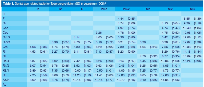
Periapical dental radiographs were taken of all the teeth in the upper and lower jaws (Fig. 3). These radio-graphic images were used in the determination of the dental developmental ages of the permanent mandi-bular teeth and thereby the estimated age of the child, using the Age Related Tables for South African children and juveniles (Table 1) (Tygerberg sample, Phillips 2009).1
The table shows the mean age at which the particular stage of calcification is visible, with the standard deviation in brackets. The Ac stage is omitted for age estimation. Only teeth that have not fully developed are used to estimate the age of the individual.
RESULTS
The calculation of the dental age is shown in the Table 2.
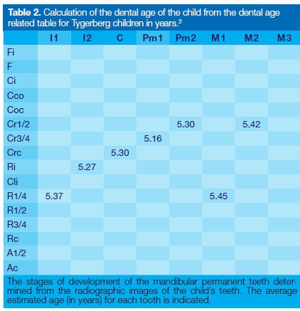
The estimated dental age of the child was calculated from the averages of the developmental ages of the teeth in the mandible.
Age calculation
The sum of the average developmental ages for seven permanent teeth in the mandible was calculated and the mean determined:

CONCLUSION
The skull and mandible of the child were assessed as being of mixed ethnic origin (mainly Negroid).
The dental age estimation of the child was 5.32 years (5 years 4 months) [SD = 6 months].
References
1. Phillips VM. Dental age related tables for children of various ethnic groups in South Africa. The Journal of Forensic Odon-to-Stomatology 2009; 27(2): 29-44. [ Links ]
2. Phillips VM, van Wyk Kotze TJ. Testing standard methods of age estimation by Moorrees, Fanning & Hunt and Demirjian, Goldstein & Tanner on three South African children samples. The Journal of Forensic Odonto-Stomatology 2009; 27(2); 20-28. [ Links ]
 Correspondence:
Vincent M Phillips
Department of Oral and Maxillo-Facial Pathology and Forensic Science
Faculty of Health Sciences, University of the Western Cape
Cape Town, South Africa
Email: vmphillips@uwc.ac.za

