


Serviços Personalizados
Artigo
 Inglês (pdf)
 Artigo em XML
 Referências do artigo
Indicadores

Links relacionados
-
 Citado por Google
 -
 Similares em Google
Compartilhar

 Permalink
South African Dental Journal
versão On-line ISSN 0375-1562
versão impressa ISSN 0011-8516
S. Afr. dent. j. vol.74 no.5 Johannesburg Jun. 2019
http://dx.doi.org/10.17159/2519-0105/2019/v74no5a8
RADIOLOGY CASE
CJ Nortjé
BChD, PhD, ABOMR, DSc. Faculty of Dentistry, University of the Western Cape. Email: cnortje@uwc.ac.za
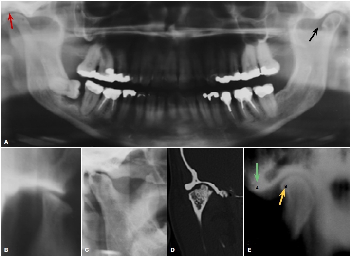
A 40 year old female complains of pain and limitation of movement of her right temporomandibular joint (TMJ). Clinical examination of the joint reveals palpable crepitus and limited movement. What are the important radiological findings and what is your diagnosis?
INTERPRETATION
Symptoms may derive from the impacted and infected 48, osteoarthritis of the TMJ's, joint space narrowed, (red arrow) or an osteophyte (black arrow). Osteoarthritis (OA), the most common type of arthritis (National Institute of Arthritis and Musculoskeletal and Skin Diseases), is caused by normal wear and tear of any joint. The cartilage deteriorates and may even disappear completely, the bone-to-bone contact causing pain, stiffness, and sometimes swelling. OA of the TMJ may include loss of articular cartilage and narrowing of the joint "space" (red arrow), condylar and anterior articular eminence flattening (Fig. B), formation of subcortical cysts and erosion (Fig. C). Axial CT scan (Fig. D) shows OA with a large lateral osteophyte on the superior condylar surface. Conventional tomogram (Fig. E) shows an anterior osteo-phyte (yellow arrow, dense cortical bone). The green arrow demonstrates eburnation of the eminence (ivorylike reaction of exposed bone). Previously thought non-inflammatory, osteoarthrosis was, an inadequate misnomer. Now accepted as a low grade inflammatory condition involving all components of the joint, a major cause of pain and disability in adults. About 10% of men and 18% of women over 60 years have symptomatic osteoarthritis, commonly of the knee, hip and hand joints, but also the TMJ. Diagnostic imaging (MRI) may be necessary to confirm diagnosis. Disc displacement and osteoarthritis may be shown, since both soft tissue and bone abnormalities can be assessed. However, for bone details CT is generally accepted as superior to MRI. CBCT is increasingly used to assess the TMJ, offering diagnostic accuracy and lower radiation dose, is advisable to ascertain that no other underlying pathology is present.
References
1. Rozylo-Kalinowska, I, Orhan, K: Imaging of the Temporomandibular Joint, Springer Nature Switzerland 2019 p264. [ Links ]
2. Farman AG, Nortje CJ, Wood RE: Oral and Maxillofacial Imaging, 1st Ed, Mosby. St. Louis, Missouri 1993 pp 366-9. [ Links ]

