


Serviços Personalizados
Artigo
 Inglês (pdf)
 Artigo em XML
 Referências do artigo
Indicadores

Links relacionados
-
 Citado por Google
 -
 Similares em Google
Compartilhar

 Permalink
South African Dental Journal
versão On-line ISSN 0375-1562
versão impressa ISSN 0011-8516
S. Afr. dent. j. vol.74 no.3 Johannesburg Abr. 2019
http://dx.doi.org/10.17159/2519-0105/2019/v74no3a9
RADIOLOGY CASE
CJ Nortjé
BChD, PhD, ABOMR, DSc. Faculty of Dentistry, University of the Western Cape. E-mail: cnortje@uwc.ac.za
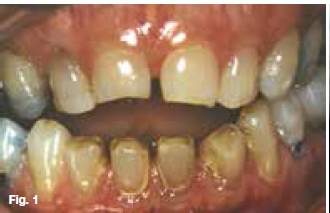
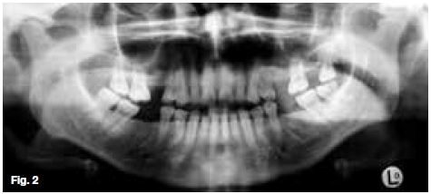
A sixty five year old female patient (Figure 1 & 2) presented at the Oral Health Centre with the main complaint of pain, swelling and stiffness of the right and left temporomandibular joints. Clinical examination revealed an anterior open bite and attrition of the posterior teeth. List your observations and give a differential diagnosis.
INTERPRETATION
The most important radiological observations are resorption of the articulating surfaces of the head of the condyle and articular eminence; sclerosis of the osseous components and a decrease in the size of the "joint space", an anterior open bite if one takes note that the posterior teeth are in close contact with each other and show signs of attrition. A provisional radiological diagnosis of Rheumatoid arthritis was made. Rheumatoid arthritis and related joint diseases consist of a number of inflammatory disorders that are characterized by prominent inflammation of the synovial membrane. It is a systemic disease with unknown aetiology and usually occurs within the 20-50 years age group but may occur at other ages. When it starts before the age of fifteen it is called the juvenile type of rheumatoid arthritis. Women are affected two to three times more often than men. In earlier reports the percentage flgures of involvement of the temporomandibular joint were rather low, but later studies utilizing improved radiographic techniques have found involvement of the joint in from 40 to 80 per cent of cases.
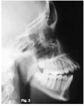
Clinically, the symptoms are usually pain and tenderness of the affected joint and stiffness or reduced mobility of the mandible. In the juvenile type, especially when the disease starts very early in life, marked growth changes may take place due to alterations in the condylar growth area. This results in the reduction of the forward and downward movement of the mandible, disto-occlusion and an anterior open bite (Figure 3). The classical clinical appearance of a bird-face may be the result of the growth changes. Radiographically, the condylar head may reveal flattening (Figure 2) or may be absent. The generalized osteoporosis may also involve the condyle, and subcondylar cysts may be formed Figure 4 (green arrow). The radiological joint changes are significantly related to the duration of the disease. Rheumatoid arthritis can affect any joint in the body, including those in the hands and Angers causing destruction of the joints Figure 5 (red arrows). The patient may experience hand pain, finger pain, swelling, and stiffness.


Reference
1. Farman AG, Nortjé CJ & Wood R E: Oral and Maxillofacial Imaging, 1st Ed, Mosby. St. Louis, Missouri 1993 pp. 368-9. [ Links ]

