


Serviços Personalizados
Artigo
 Inglês (pdf)
 Artigo em XML
 Referências do artigo
Indicadores

Links relacionados
-
 Citado por Google
 -
 Similares em Google
Compartilhar

 Permalink
South African Dental Journal
versão On-line ISSN 0375-1562
versão impressa ISSN 0011-8516
S. Afr. dent. j. vol.74 no.3 Johannesburg Abr. 2019
http://dx.doi.org/10.17159/2519-0105/2019/v74no3a3
CASE REPORT
Radiographic assessment of multiple pathoses and dental malocclusion: a case study
V SoniI; B BuchII; D KotsaneIII
IBChD (Pret), MSc (Dent)(Wits), Department of General Dental Practice, School of Oral Health Sciences, University of the Witwatersrand, Johannesburg
IIBSc (Hons) (Natal); SED (UCT); BDS (Wits); MSc (Dent) (Wits), Emeritus Professor-University of Pretoria, Visiting Professor and Part-time Consultant: Division of Maxillo-Facial and Oral Radiology, Department of General Dental Practice, School of Oral Health Sciences, university of the Witwatersrand, Johannesburg
IIIBDT, BDS (Medunsa), PDHE (Pret), MBA (RSB), Head, Dept. of General Dental Practice, School of Oral Health Sci-ences,University of the Witwatersrand, Johannesburg, South Africa
ABSTRACT
Odontomas are rare pathological abnormalities which may occur at any age. It is most unusual to observe an odontoma together with supernumerary teeth, more particularly if this is found in conjunction with an osteoma. This report emphasizes the underlying pathology of a case co-incidentally encountered in a 14 year old patient whose main complaint was of a painful 85 tooth.
An incisional biopsy of the area revealed the presence of mineralized tooth-related tissue resembling a complex odontoma. In conjunction with the radiographic evidence, which confirmed the presence of complex and compound odontomas as well as a number of small osteomas, a provisional diagnosis of Gardner's syndrome was made. The syndrome is a rare autosomal disease that is difficult to treat. If a dentist suspects a patient may present with Gardner's syndrome, the patient should be referred immediately for further medical management.
INTRODUCTION
Benign odontogenic tumours include ameloblastoma, ameloblastic fibroma, ameloblastic fibrodontoma, complex and compound odontomas.1 Of these, the most commonly encountered in patients less than 20 years of age are the complex and compound odontomas.2 These tumours are asymptomatic, slow- growing and may lead to malformation of adjacent developing teeth and malocclusion.
The complexity of odontomas has been studied and analyzed for many years. One example is that of supernumerary teeth developing largely within the maxillary permanent dentition and in conjunction with odontomas.3 These are usually asymptomatic, but may be associated with pathologic implications. One such manifestation is that of Gardener's Syndrome if the odontoma is also associated with the presence of osteomas.4-10 This is a life-threatening condition and it is essential that the presenting signs are not missed.
CASE REPORT
A 14 year-old female patient presented at the Wits Oral Health Centre complaining of a painful primary tooth (85) which appeared to have prevented eruption of the permanent successor, causing a severe malocclusion.
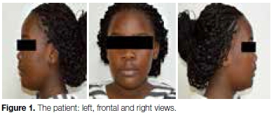
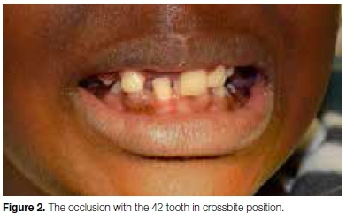
No medical problems were reported. Extra-oral findings revealed no abnormalities. Facial asymmetry was not detected (Fig 1). Intra-oral examination revealed missing premolars together with carious and painful mandibular primary second molars (Figs 1-5). An anterior cross bite involving tooth 42 was noted together with a deep overbite (Fig 2). The gingiva surrounding the painful tooth (85) was swollen with a buccal exudate (Fig 4).

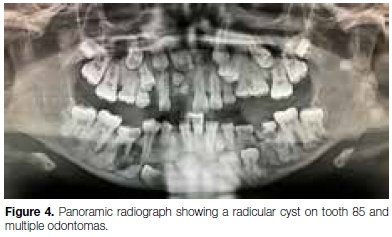
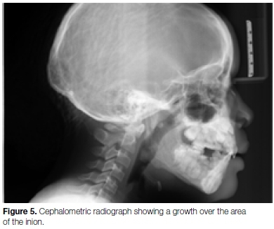
The panoramic radiograph revealed the presence of numerous unerupted and retained deciduous teeth, compound and complex odontomas (bi-mandibular) and a well circumscribed radiolucent lesion resembling a radicular cyst at the apex of the right mandibular deciduous second molar tooth. Several of the unerupt-ed teeth were supernumeraries. The angles of the mandible were deformed by presence number of ectopic teeth and small osteomas (Fig 4). The lateral cephalo-gram revealed a growth on the inion of the occipital bone (Fig 5).
The patient was classified as having a normal skeletal and dental class 1 profile (Fig 5). However, the bone deformities together with the presence of unerupted supernumeraries, ectopic teeth, osteomas and compound and complex odontomas suggested a diagnosis of Gardner’s syndrome, despite the apparent absence of visible epidermoid cysts of the skin.
The CBCT scan confirmed the presence of carious lesions of the teeth, together with multiple radio-opacities on the left angle and ramus of the mandible (Fig 6).
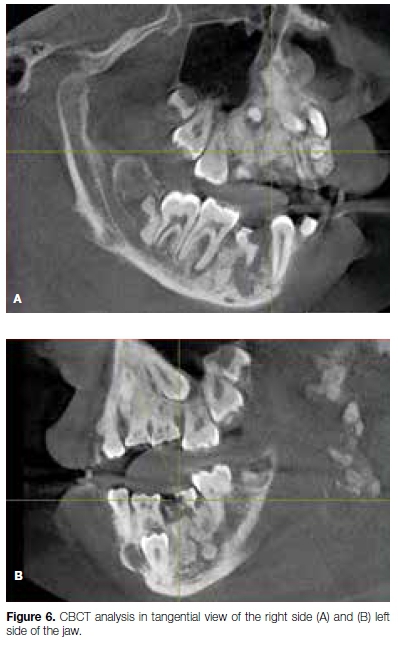
These displayed the appearance of osteomas, thereby confirming the provisional diagnosis of Gardner's syndrome.
An incisional biopsy was done on the right mandible at which the affected tooth and several fragments of bone were removed for microscopic examination. The latter revealed mineralized tissue, trabeculae of woven and lamellar bone together with fragments of enamel.
DISCUSSION AND CONCLUSION
The large number of supernumerary and ectopic teeth together with mixed odontomas and osteomas3-11 may be regarded as a rare occurrence.
Based on the clinical, radiological and biopsy findings, a diagnosis of Gardner's syndrome is justified. This is arare autosomal disease characterised by intestinal polyposis, connective tissue tumours and multiple osteomatosis.3,5-9,12 The malignant potential of the intestinal polyps, the presence of which the patient is totally unaware, makes it essential for a dentist to be aware of the signs for early detection of the condition.12
Treatment entailed surgical excision of the odontomas and enucleation of the cyst for aesthetic reasons. Thereafter fixed orthodontic appliance treatment would be considered when complete healing occurred.
Referral for colonoscopy was essential for final confirmation of Gardener's Syndrome and the surgical removal of colonic polyps. Recurrence of the odontomas and osteomas is not uncommon and would require possible further surgical intervention. Since there is no cure for Gardner's Syndrome, prevention of cancer is the primary aim in management.12
Acknowledgement
Sincere thanks to Professor E Rikhotso and his team from the Maxillo-facial and Oral Surgery Department for sharing the biopsy information with us. The authors would also like to thank Professor Dandajena, Dr Moshaoa and Professor Evans of the Orthodontic Department for their opinion on the orthodontic treatment approach of this case.
References
1. Gyulai-Gaál S, Takács D, Barabás J, Tarján I, Martonffy K, Szabó G, Suba Z. Mixed odontogenic tumor in children and adolescents. Fogorv Sz. 2007; 100(2):65-9. [ Links ]
2. De Oliveira BH, Campos V, Marçal S. Compound odontoma-diagnosis and treatment: three case reports. Ped Dent. 2001;23:151-7. [ Links ]
3. Pippi R. Odontomas and supernumerary teeth: is there a common origin? Int J Med Sci. 2014;11:1282. [ Links ]
4. Gaynor WN. Bilateral radicular cysts of mandibular deciduous teeth: a case report. New Zealand Dent J. 2012; 108:106-9. [ Links ]
5. Oner AY, Pocan S. Gardner's syndrome: A case report. BDJ.2006; 200; 666-7. [ Links ]
6. Panjwani S, Bagewadi A, Keluskar V, Arora S. Gardner's Syndrome. J Clinic Imaging Sci. 2011; 4: 1-4. [ Links ]
7. Seehra J, Patel S, Bryant C. Gardner's Syndrome revisited: clinical case and overview of the literature. J Orthodontics. 2016:43; 59-64. [ Links ]
8. Fonseca LC, Kodama NK, Nunes FC, Maciel PH, Fonseca FA, Roitberg M, de Oliveira JX, Cavalcanti MG. Radiographic assessment of Gardner's syndrome. Dentomaxillofacial Rad. 2007; 36: 121-4. [ Links ]
9. Koh KJ, Park HN, Kim KA. Gardner syndrome associated with multiple osteomas, intestinal polyposis, and epidermoid cysts. Imag. Sci Dent. 2016; 46:267-72. [ Links ]
10. Chung EH, Sun H, Yang YI, Yun SH, Jeon MK. Peripheral osteoma in the mandibular angle. Arch Plastic Surg. 2015;42:798. [ Links ]
11. Astekar M, Manjunatha BS, Kaur P, Singh J. Histopathological insight of complex odontoma associated with a dentigerous cyst. BMJ case reports; 2014:bcr2013200316. [ Links ]
12. Adisen MZ, Okkesim A, Misirlioglu M. The importance of early diagnosis of Gardner's syndrome in dental examination. Nige J Clinic Prac. 2018;21:114-6. [ Links ]
 Correspondence:
Vishani Soni
Department of General Dental Practice, School of Oral Health Sciences
University of the Witwatersrand
Johannesburg, South Africa
Email: vishani.soni@wits.ac.za

