


Serviços Personalizados
Artigo
 Inglês (pdf)
 Artigo em XML
 Referências do artigo
Indicadores

Links relacionados
-
 Citado por Google
 -
 Similares em Google
Compartilhar

 Permalink
South African Dental Journal
versão On-line ISSN 0375-1562
versão impressa ISSN 0011-8516
S. Afr. dent. j. vol.73 no.6 Johannesburg Jul. 2018
http://dx.doi.org/10.17159/2519-0105/2018/v73no6a6
RADIOLOGY CASE
Maxillofacial radiology case 162
C.J. Nortje
BChD, PhD, ABOMR, DSc. Faculty of Dentistry, University of the Western Cape. E-mail: cnortje@uwc.ac.za
Below are images of Peripheral Nerve Sheath Tumours (PNST) that presented in lower jaws of various patients that presented or referred to the Oral Health Science Centre of the Faculty of Dentistry, University of the Western Cape over the past 30 years. What are the important radiological features and what is your diagnosis?
INTERPRETATION
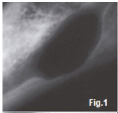
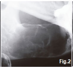
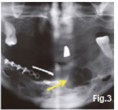
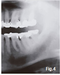
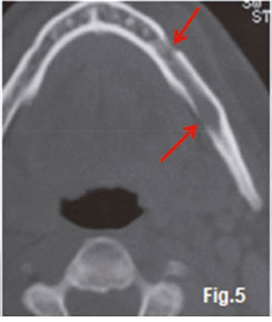
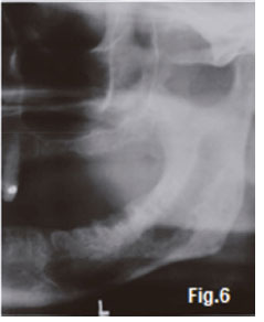
Figure 1 is a cropped pantomograph of a 25 year old female who presented with a history of pain and a slight asymmetry at the angle of the lower right mandible. Radiographically a well-defined corticated radiolucency is discernible in the 48 region. A histological diagnosis of a neurofibroma was made. A solitary neurofibroma occurs in an individual who does not have hereditary neurofibromatosis. The tumour is most often seen in the body of the mandible, sometimes in connection with the mental foramen. It is slow growing but may expand the cortical plate. Figure 2 is a cropped oblique lateral radiograph of a 51 year old male who presented with a slow growing painless lesion in the body of the left mandible. The lesion has a multilocular radiolucent appearance causing expansion of the mandible. A histological diagnosis of a benign Schwannoma was made. Most of the cases of Schwannoma or neurilemmomas present in the alveolar canal. They can occur at any age but are most common between 30 and 50 years. It is a benign tumour arising from and consisting solely of Schwann cells. Comparisons between Schwannomas and neurofibromas are frequent, but these tumours can be readily separated clinically and usually histologically. Schwannomas occur most commonly in the head and neck area where the tongue and floor of mouth are the most common sites. Because Schwannomas are painless and slow growing, they often reach sizes in excess of 5 cm x 5 cm before treatment is sought. Figure 3 is a cropped pantomograph of a 42 year old male presenting with pain in the 33 region (yellow arrow), with a history of being assaulted six months previously. The pantomograph shows a multilocular radiolucency with well demarcated borders. A histological diagnosis of a traumatic neuroma was made. Most intrabony traumatic neuromas are seen in the mandible as a result of injury to the inferior alveolar nerve caused by a fracture, extraction of an impacted tooth or by orthognathic surgery. On excision of a traumatic neuroma, healing is usually uneventful. Figures 4, 5 & 6 are images of a 51 year old male patient with a history of pain in the left mandible, problems on opening his jaw and also paraesthesia of the left lower jaw. Figure 4 shows enlargement of the inferior alveolar canal and the axial CT image (Fig. 5) shows perforation of the cortex in two places (red arrows). Figure 6, taken a few months after extraction of the posterior teeth and commencement of radiation therapy, shows further enlargement of the canal with irregular ragged borders and destruction of the cortex. A histological diagnosis of a malignant Schwannoma was made. Malignant Schwannoma is an uncommon neoplasm, consisting predominantly of spindle cell growth. The tumour is extremely rare in the maxillofacial region. The diagnosis of the tumour is difficult, because the histopathologic appearance of a Schwannoma is often quite similar to that of other types of sarcomas showing spindle cell growth, such as fibrosarcoma and leiomyosarcoma.
References
1. Farman AG, Nortjé CJ & Wood R E: Oral and Maxillofacial Imaging, 1st Ed, Mosby. St. Louis, Missouri 1993 pp. 342-344. [ Links ]
2. Langlais RP, Langland OE & Nortje CJ: Diagnostic Imaging of the Jaws 1st Edition, Williams & Wilkins, 1995, pp. 451-454. [ Links ]
3. Garn SM: The Earlier Gain and the Later Loss of Cortical Bone in Nutritional Perspective. Springfield, IL: Charles C Thomas, 1970. P. 231. [ Links ]

