


Servicios Personalizados
Articulo
 Inglés (pdf)
 Articulo en XML
 Referencias del artículo
Indicadores

Links relacionados
-
 Citado por Google
 -
 Similares en Google
Compartir

 Permalink
South African Dental Journal
versión On-line ISSN 0375-1562
versión impresa ISSN 0011-8516
S. Afr. dent. j. vol.73 no.4 Johannesburg may. 2018
RADIOLOGY
An alternative extra-oral digital technique for bitewing radiography
Crombie KI; Shaikh AII; Harnekar SYIII
INational Diploma in Diagnostic Radiography, Higher Education Diploma (UNISA), MSc Dent (UWC) Chief Radiographer I Lecturer Department of Diagnostics and Radiology, Faculty of Dentistry, University of the Western Cape, South Africa
IIBChD (UWC), MSc Dent (UWC), MChD (Orthodontics), Associate Professor Senior Specialist Department of Orthodontics, Faculty of Dentistry, University of the Western Cape, South Africa
IIIBChD, MSc Dent (UWC), PDD (Interceptive Orthodontics) (UWC), Senior Lecturer University of the Western Cape, South Africa
ABSTRACT
INTRODUCTION: The intraoral technique, currently the standard method of taking bitewing radiographs, is challenging, especially in children, and in patients with limited mouth opening.
OBJECTIVES: To assess an alternative, extra-oral, digital technique for bitewing radiographs.
METHODS
26 patients requiring bitewing radiographs were selected seriatim. A split mouth technique was used, taking an analogue intraoral bitewing radiograph on one side, and a modified, digital, posterior, segmental pantomogram of the contralateral side. Two calibrated observers evaluated the bitewing images, using a viewing box, and assessed the digital modified posterior segmental radiographic Images, using a computer monitor.
RESULTS: No statistically significant association was shown between the operators and the techniques used, i.e. it is not technique or operator sensitive.
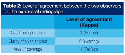
The extra-oral technique recorded perfect agreement (k=1) between the two observers for the categories of overlapping of teeth and area of coverage. For clarity of the alveolar crest there was strong agreement (k=0.8).
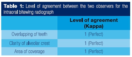
There was perfect agreement (k=1) between the two observers for all three categories examined on the intraoral bitewing radiographs.
CONCLUSION: An alternative and diagnostically accurate bitewing radiograph can be produced by modifying the patient positioning when taking a digital posterior segmental pantomogram.
INTRODUCTION
The bitewing radiographic film is the most widely used intraoral radiographic technique.1 It is currently the standard method of taking bitewing radiographs for oral and dental diagnostic evaluation. Bitewing radiographs typically show the contact surfaces from the distal of the canines to the most distal molars and are usually taken bilaterally. The indications for bitewing radiographs include diagnosis of proximal caries, assessment of the extent of the caries, identification of secondary caries under existing restorations and the assessment of the periodontium.2,4
This technique can be challenging especially in patients who are resistant to the placement of a radiographic film within the oral cavity due to problems related to discomfort, pain and stimulation of the gag reflex. Patients may displace the film, reject the Rinn holder, or reposition the film after placement, resulting in the failure of, or an inadequate, radiographic image. Difficulties may be further compounded in paediatric patients, and those who are anxious and fearful, patients with special needs and those presenting with trismus.
The method is also technique sensitive and errors will occur if the principles of the technique are not applied. The most typical errors that can occur are in the placement of the film, the vertical and horizontal angulations and the centering of the central ray of the x-ray beam.
Various other alternatives to the conventional bitewing radiographs have been suggested in the literature, including, intraoral as well as extra-oral techniques. Modifications to the intraoral technique Include: adjustments of the film packet (softening the corners, bending occlusal film in half), adaptation of the film holder (tongue depressor and rubber bands), various devices for film holder and patient jaw (mouth props, helmet with chin strap, Velero strips), modifications of the films (reverse bitewing i.e. placed in buccal vestibule). The extra-oral techniques that have been recommended are the lateral oblique technique and the conventional pantomogram. However, these techniques prove to be inadequate as they show poor detail, excessive superimposition and distortion.5
Scarfe (1994)6 and his associates compared the diagnostic accuracy of orthogonally projected panoramic image with conventional panoramic radiograph for the detection of Interproximal caries, using the conventional intraoral bitewing radiograph as a benchmark. They concluded that the orthogonal projection did not improve the diagnostic accuracy as had been suggested.
Newman and Friedman (2003)7 devised an aiming procedure for an extra-oral radiographic technique using a modified locating device which proved to be well tolerated by patients. One of the major shortcomings of this procedure is that it still requires patient cooperation. Another shortcoming was the repeated cone cutting that was obtained when the device was tested subsequently by Chen et al (2007).8
As a result of these challenges, an alternative extra-oral digital technique was devised and tested.
The purpose of this study was to assess that technique for taking bitewing radiographs.
MATERIALS AND METHODS
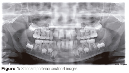
The Kodak 8000 Digital Panoramic System allows for sectional imaging of the panoramic view, of the two posterior segments extending from the condyle to the molars, and of an anterior segment extending from the canine to the canine (Fig 1).
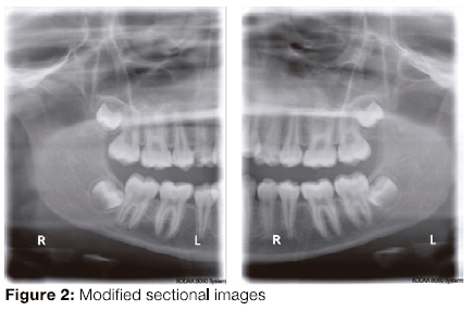
The posterior sectional function offered the potential for some modifications to be effected to optimize the view so as to obtain a view similar to that of a bitewing radiograph (Fig 2).
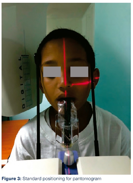
To obtain the modified sectional extra-oral digital bitewing radiograph the patient is repositioned more posteriorly so that the corner of the mouth laser beam coincides with that of the mid-sagittal laser beam (Fig 4).
This modified technique was first tested on a phantom head.
Modification to the Sectional technique
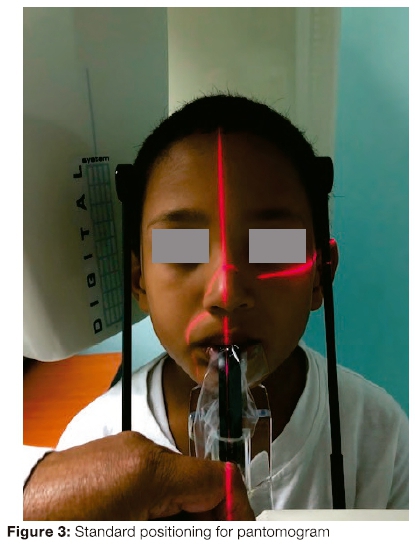
The patient position for the standard panoramic view is obtained with the aid of three reference laser beams: ala-tragus line, mid-sagittal plane and the comer of the mouth (Fig 3).
Patients presenting for bitewing radiographs were considered for the study sample. Adult patients with no overlapping, displaced or crowded teeth in the buccal segments were invited to participate in the study. The purpose of the study was explained to the patient. Each was reassured that the procedure involved no additional exposure or cost. Patients willing to participate were given written information and subsequently signed a consent form.
Participants were given the option to withdraw from the study at any stage without any consequences or compromise to further management.
A total of 26 patients were selected.
Materials and Methods
A split mouth technique was used, taking a standard intraoral bitewing radiograph on one side, and the modified extra-oral radiograph of the contralateral side.
A size 3 Kodak Ultra-speed film was placed in a bitewing Rinn holder which was positioned in the mouth, and the film exposed by a GE 1000 intraoral machine, with exposure factors of 70kV 10mA 0,8s. The accompanying radiograph of the contralateral side of the same patient was taken on a Kodak 8000 Digital Panoramic System, using the modified, extra-oral technique with exposure factors of 70kV 10mA 13s. All the radiographs were captured by the same operator.
The techniques were alternated between the left and right sides of each consecutive patient. The images obtained were numbered and randomized prior to evaluation. Two calibrated observers viewed and assessed the images.
A total of 26 pairs of radiographs (intraoral and extra-oral) were evaluated. The extra-oral (digital images) were viewed in a darkened room on a 39cm monitor with a resolution of 1024 χ 768 pixels. The intraoral bitewing (analogue) radiographs were all viewed on the same viewing light box in a darkened room. These images were independently assessed in random sequence. The variables assessed included crown overlap, clarity of the alveolar crest and area of coverage from the 1st premolar to the 3rd molar area using the following criteria:
1. Overlap of crowns (yes/no),
2. Clarity of alveolar crest (clear/unclear).
3. Area of coverage to include the 1st premolar to the most distal molar (yes/no)
The data were captured and analysed using the Microsoft Excel and the SPSS packages respectively.
Inter-examiner correlations were determined by means of the kappa test. A non-parametric test (Fisher exact test) was used to compare the data between the two techniques.
The following criteria were used to interpret the Κ value:

RESULTS
There was perfect agreement (k=1) between the two observers for all three categories examined on the intraoral bitewing radiographs (Table 1).
For the extra-oral technique there was perfect agreement (k=1) between the two observers for two of the categories examined, i.e., overlapping of teeth and area of coverage. For the clarity of the alveolar crest there was strong agreement (k=0.8) (table 2).
When a comparison was made between the two techniques with respect to the three criteria used, there was no association
between the operators and the techniques used, i.e. it is not technique or operator sensitive (Table 3).

This confirms that the proposed extra-oral technique can be considered to be an acceptable substitute for the traditional intra oral bitewing radiograph.
DISCUSSION
The modified technique was able to repeatedly produce diagnostically satisfactory digital bitewing radiographs. As observed, the use of the intraoral bitewing film and the Rinn holder can be uncomfortable, overwhelming, painful, and is even rejected by some patients, particularly patients with very small mouths as well as children. None of the patients showed any objection or hesitation in the taking of the extra-oral radiographs with the panoramic system.
CONCLUSION
This supports the hypothesis that an alternative, extra-oral, digital technique for taking bitewing radiographs may be clinically relevant among patients for whom the intraoral bitewing technique is particularly difficult to obtain, namely paediatric and other special needs patients.
Using the option of the posterior segmental program in the digital panoramic system, with modification of patient positioning produces an image comparable to the traditional intraoral bitewing radiograph.
Permission to reproduce the photographs was granted.
Acknowledgements
J Du Toit: SCftD(UWC), Dip Oral Surg. (WITS)
D Smit: BChD, MChD (Community Dentistry) (UWC)
References
1. Andlaw J, Rock WP. A Manual of Paediatric Dentistry. UK: Elsevier Health 1996. [ Links ]
2. Ghom A. Textbook of Oral Radiology. India: Elsevier 2008: Chapters 17&45. [ Links ]
3. Kaffe I, Moshe G, Laufer B, Littner MM. Detection of proximal carious lesions: Two-film versus four-film bitewing radiography. Oral Surg Oral Med Oral Path 1984;57:567-71. [ Links ]
4. Kidd EAM. Essentials of Dental Caries: The Disease and Its Management. UK: Oxford Medical Publications; 2005: Chapter 3, 42-58. [ Links ]
5. Casamassimo PS. Radiographic considerations for special patients-modifications, adjuncts, and alternatives. Pediatric Dentistry, 3 Special issue 21982; 448-54. [ Links ]
6. Scarfe WC, Langlais RP, Nummikoski P, Dove SB, McDavid WD, Deahl ST, Yuan CH. Oral Surgery, Oral Medicine, Oral Pathology 1994; 77:2:195-207.7. [ Links ]
7. Newman ME, Friedman S. Extraoral radiographic technique: an alternative approach. J Endod 2003; 29:419-21. [ Links ]
8. Chen C, Lin S, Chiu H, Lin Y, Chen Y, Lin L. An aiming device for an extraoral radiographic technique. J Endod 2007;33:758-S0. [ Links ]
 Correspondence:
Ms K Crombie
Tel 021 370 4482 021 9373112
Fax: 021 392 3250
Email:kcrombie@uwc.ac.za

