


Services on Demand
Article
 English (pdf)
 Article in xml format
 Article references
Indicators

Related links
-
 Cited by Google
 -
 Similars in Google
Share

 Permalink
South African Dental Journal
On-line version ISSN 0375-1562
Print version ISSN 0011-8516
S. Afr. dent. j. vol.73 n.4 Johannesburg May. 2018
RADIOLOGY
Maxillofacial radiology case 160
CJ Nortje
BChD, PhD, ABOMR, DSc, Faculty of Dentistry, University of the Western Cape. E-mail: cnortje@uwc.ac.za
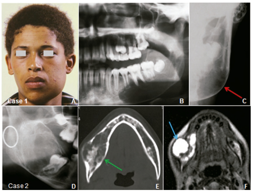
Below are two cases of a lesion that is not commonly seen in the jaws but has been found in most bones of the skeleton; however the majority occur in the long bones and in the spine. Discuss the radiological features and what is your diagnosis?
INTERPRETATION
Case 1 is a sixteen year old male (Fig.A) who presented with a history of a rapidly growing painful swelling in the lower left jaw causing displacement of the 37,38 (Fig.B) and ballooning expansion (red arrow) of the of the cortex (Fig.C). Case 2 is a fifteen year old female complaining of a fast growing swelling displacing and loosening of teeth in the lower right jaw. The cropped pantomograph show a multilocular lesion (Fig.D). An Axial CT view of the lesion show an expansive ballooned-out appearance with a thin cortex outline and presence of internal trabeculation (Fig.E). Figure F is an Axial MR T2 image showing a hyper-intense lesion with septae and buccal and lingual expansion. A histological diagnosis of an aneurysmal bone cyst was made. The aneurysmal bone cyst is an uncommon lesion and is a very rare finding in the jaws. The true nature of the lesion remains uncertain, although most pathologists regard it as probably reactive. Although the lesion is characteristically cystic and blood filled, the term "aneurysmal bone cyst" was suggested by Jaffe and Lichtenstein (1942) who described the characteristic "blown out" contour of the bone seen in radiographs of the lesion. The aneurysmal bone cyst has a characteristic 'ballooning' growth pattern which results in a radiolucent area with elevation of the periosteum to produce an ovoid or fusiform expansion of bone with the typical cortical expansion (Fig.C). The cyst constitutes only 1% of all nonodontogenic, non-epithelial cysts of the jaws. They most commonly occur in the long bones and the spine. Almost all lesions affecting the jaws present in the mandible, especially the posterior body and vertical ramus. They are found predominantly in young patients, rarely manifesting after the third decade of life with no gender predilection. Perforation of the cortex, pain and tenderness to palpation has been reported. Treatment of choice consists of enucleation with thorough curettage.
References
1. Jaffe HL & Lichtenstein L: Solitary unicameral bone cyst with an emphasis on the roentgen picture, the pathologic appearance, and the pathogenesis. Archives of Surgery 44,1942, 1004-1025. [ Links ]
2. Farman AG, Nortjé CJ & Wood R E: Oral and Maxillofacial Imaging, 1st Ed, Mosby. St. Louis, Missouri 1993 p. 228-231. [ Links ]

