


Services on Demand
Article
 English (pdf)
 Article in xml format
 Article references
Indicators

Related links
-
 Cited by Google
 -
 Similars in Google
Share

 Permalink
South African Dental Journal
On-line version ISSN 0375-1562
Print version ISSN 0011-8516
S. Afr. dent. j. vol.73 n.3 Johannesburg Apr. 2018
CASE STUDY
Bilateral ectopic eruption of permanent maxillary canines into the incisive fossa, evaluated using Micro Focus X-ray Computed Tomography: A Case study and brief literature review
Nyirenda TI; Bacci NII; Nchabeleng EIII; Billings BKIV; Ndou RV; Mazengenya PVI
ITrust Nyirenda: BBSc, BBSc Hons; MSccandidate; School of Anatomical Sciences, Faculty of Health Science, University of the Witwatersrand, Johannesburg, Gauteng, South Africa
IINicholas Bacci: BSc, BBSc Hons MSc; PhDCandidate, Assistant Lecturer; School of Anatomical Sciences, Faculty of Health Science, University of the Witwatersrand, Johannesburg, Gauteng, South Africa
IIIElsie Nchabeleng: BSc, BSc Hons; MSccandidate; School of Anatomical Sciences, Faculty of Health Science, University of the Witwatersrand, Johannesburg, Gauteng, South Africa
IVBrendon K Bilings: BSc, BSc Hons, MSc; PhiD candidate - Lecturer, Curator: Raymond A. Dart Collection of Human Skeletons; School of Anatomical Sciences, Faculty of Health Science, University of the Witwatersrand, Johannesburg, Gauteng, South Africa; School of Anatomical Sciences, Faculty of Health Science, University of the Witwatersrand, Johannesburg, Gauteng, South Africa
VRobert Ndou: BSc, BSc Med Hons, MSc(Medd), PhiD- Senior Lecturer; School of Anatomical Sciences, Faculty of Health Science, University of the Witwatersrand, Johannesburg, Gauteng, South Africa
VIPedzisai Mazengenya: BSc Hons, MSc, PhD- Lecturer; School of Anatomical Sciences, Faculty of Health Science, University of the Witwatersrand, Johannesburg, Gauteng, South Africa
ABSTRACT
Tooth development is a complex process whereby various genetic and environmental variables interact to achieve the final morphology and destination. Disruptions in the process lead to impaction and or ectopic eruption. Bilateral ectopic eruption of maxillary canine teeth into the incisive fossa is a rare phenomenon. This report describes bilateral permanent maxillary canine teeth erupting into the incisive fossa of the skull of an adult male African. The skull specimen was first examined physically, followed by Micro Focus X-ray Computed Tomography μCT) to determine the morphology and trajectory of the impacted and ectopically erupting teeth. Physical examination of the skull revealed a portion of the right maxillary canine tooth in the incisive fossa. μCT revealed the presence of right and left permanent maxillary canines within the palatine bone with cusps projecting into the incisive fossa. Both teeth were mature with well-developed root, root canal and crowns with distinct cusps. The root of the right impacted canine tooth was deflected at its apex. Tooth impaction is caused by mechanical disturbance in the path of the developing tooth. This information is vital to practicing maxillofacial surgeons during interpretation of the radiographs and surgical correction of disorders of the oral cavity.
Key words: Bilateral ectopic eruption, maxillary canines, incisive foramen, Microfocus Computed Tomography
INTRODUCTION
Tooth development is a continuous and complex process by which various biological, genetic and environmental variables interact to achieve the destination and final morphology of the tooth.1,2 Disruptions in tooth development may lead to impaction and or ectopic eruption,3,4 a frequently encountered clinical problem affecting the maxillary canine teeth. Impaction of these teeth is estimated to have a prevalence of 1-2 % in the general population, with 8% of all these cases being bilateral. Location of impacted ectopic tooth eruption may be palatal, labial and or buccal, but the former is more prevalent.4 Various other sites of ectopic tooth eruption have been reported including the maxillary sinus, mandibular condyle, coronoid process, orbit, nasal cavity and through the facial skin.1,5 However, the aetiology of tooth impaction and or ectopic eruption remains elusive. Several theories have been suggested including trauma, infection, pathologic conditions, dental crowding, genetic and developmental anomalies.2,6,7 Ectopic teeth may be asymptomatic, or may cause a variety of signs and symptoms, including facial pain, nasal obstruction, headache, epistaxis, foul-smelling rhinorrhea, external nasal deformities and nasolacrimal duct obstruction.8,9 They can be associated with abnormalities such as initiscaseosa with septal perforation, with aspergillosis, the formation of a rhinolith and with naso-oral fistula.10 The present study describes a rare case of bilateral ectopic eruption of maxillary canine teeth into the incisive fossa of an adult male dry skull.
CASE REPORT
During a routine inventory of the Raymond A. Dart Collection of Human Skeletons housed in the School of Anatomical Sciences at the University of the Witwatersrand, an erupted ectopic tooth in the incisive fossa of the hard palate was observed on a dry skull specimen of a 25 year old black African (Xhosa) male (Fig. 1A). The reported cause of death was suggestive of complications relating to lung carcinoma. External examination of the maxillary arch of the skull revealed intact premolar and molar tooth sets on both the left and right sides of the dental arch. Bilaterally, the central and lateral incisors were absent with evident signs of bone resorption at the sockets which had previously accommodated these teeth (Fig. 1A). All teeth were present on the mandibular arch except for the central and lateral incisors which were lost postmortem. There were no further tooth or bone abnormalities evident upon external observation.

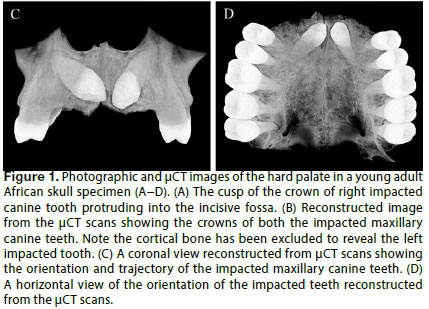
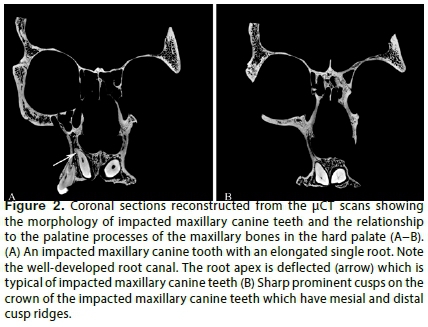
The skull was then scanned using Micro Focus X-ray Computed Tomography (μCT) and the scan analysed with Amira (5.4.5) software to determine the type and trajectory of the tooth based on the dental morphology. The permanent maxillary canines possess an elongated single root apex and a sharp prominent coronal cusp which has mesial and distal cusp ridges (Figs. 2A, B). Permanent maxillary canines are large in size and contain a root canal (Figs. 1C, D, 2A). The investigation revealed two permanent maxillary canines within the palatine processes of the maxilla, having their cusps projecting into the incisive foramen (Fig. 1B). Both teeth were seen to have infero-medial inclinations towards the midline (Figs. 1C, D). The root of the right impacted canine tooth was deflected at its apex (Fig. 2A).
DISCUSSION
General considerations on impacted maxillary canine teeth
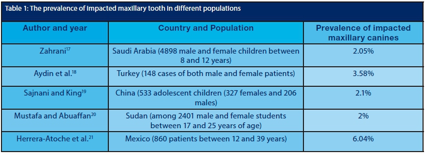
Canine teeth are the hallmark of a beautiful smile and of functional occlusion. They are also fundamental in successful dental arch development and disruptions in their development can lead to various tooth anomalies such as impaction and ectopic eruptions.3 Permanent maxillary canine teeth are the second most frequently impacted teeth after the third molar teeth.12,13 The general prevalence of impaction of permanent maxillary canine teeth is 1-2% in the general population with palatal impactions having the highest prevalence of 85%, the rest being displaced either buccally or labially.4,14 Permanent maxillary canine impaction occurs up to three times more frequently in females than in males.1 Several studies have been conducted to show variations in the prevalence of maxillary teeth impaction based on race and ethnic factors (Table 1). Morphologically, impacted maxillary canine teeth often present with deflected apical roots.16 The cause of apical root deflection is not clear, but the surrounding bone environment has been implicated.16
AETIOLOGY
While the actual causes of tooth impaction or ectopic eruption remain inconclusive, various theories have been linked to these conditions. Richardson and Russell4 suggested that genetics and mechanical guidance are critical in determining the impaction and subsequent ectopic eruption of the maxillary canine teeth. Maxillary canine teeth develop far away from their final destination and hence obstacles like supernumerary teeth, tumours, and displaced teeth may disrupt normal eruption.4,22 However, various researchers have concurred that the absence of mechanical guidance from the maxillary lateral incisor teeth, which erupt earlier than the canine teeth, and variations in their root morphology are central to the impaction and subsequent ectopic eruption of canine teeth.3,15,23 It appears that the presence of lateral incisors with an appropriate root length and developed at the exact time are critical in providing guidance to the migrating canine teeth.12
In addition, maxillary canine impaction can be as a result of either prolonged retention or early loss of deciduous canine teeth. In certain instances, tooth size-arch discrepancies can also lead to impaction or ectopic eruption of the canines.12 Regrettably, past medical history in the present case does not provide any information on how lateral incisors were lost and at what stage, hence it is not possible to reconcile the case with these theories. However, the observation of the absence of lateral incisors and signs of bone resorption on their healed sockets may suggest premature loss of the lateral incisors resulting in bilateral ectopic eruption of the maxillary canines.
Genetic theory describes palatally impacted maxillary canines as often presenting with other dental abnormalities in tooth size, shape, number and structure.22 Several abnormalities are believed to have a common hereditary link, manifested as a developmental disturbance during embryonic growth.24 It is reported that 33% of patients with palatally impacted canines also present with congenitally missing teeth and, in particular, patients with congenitally absent maxillary lateral incisors record a prevalence of palatally impacted canines of 2.4 times more than that of the general population.4,12 Besides, other congenital conditions such as anklylosis, cleft lip and cleft palate have been found in association with tooth impactions and ectopic eruptions.1,12
CLINICAL COMPLICATIONS ASSOCIATED WITH THE INCISIVE CANAL AND IMPACTED ECTOPIC CANINE TEETH
The maxillary incisive foramen is a funnel shaped opening that conveys the nasopalatine nerves and arteries from the nasopalatine canal to the anterior palate.25 During life, the maxillary incisive papilla overlays the incisive fossa. The maxillary incisive papilla is an important anatomical landmark in prosthetic dentistry particularly for arrangement and alignment of teeth to the midline.25 The incisive papilla also marks the site for the administration of local anaesthesia in the anterior palate.26,27 The maxillary incisive canal together with the incisive foramen connects the palate to the floor of the nasal cavity.28
Although no serious clinical complications have been reported as a result of surgical disturbance to the contents of the incisive canal,29 temporary sensory discomfort has been reported following surgical transection of the nasopalatine nerve during the first week after surgery.30 Conversely, complications arising from pressure on neurovascular structures due to prolonged use of dental implants have been reported leading to neurological dysfunction.25,31 The cusps of the ectopic maxillary canines in the present case projected into the incisive fossa, suggesting the possibility of compressing the nasopalatine nerve and vessels in the incisive foramen.
In conclusion, the current presentation may be valuable to dental and maxillofacial surgeons for it highlights and focusses attention on a possible altered path of tooth eruption of the maxillary canines and associated clinical sequelae.
Acknowledgements:
The work was supported by the Faculty of Health Sciences Staff individual research grants; Grant Number: 001254842110151211050 0000000000000004985
Author contributions: TN, EN and NB were involved in data collection/ inventory and preliminary writing of the paper. BKB, RN and PM designed and guided the manuscript development and review of the manuscript. PM is the principal investigator. All authors approved the manuscript.
Conflict of interest: None
ACRONYMS
μCT : Micro Focus X-ray Computed Tomography
References
1. Hattab FN, Yassin O, Rawashdeh M. Supernumerary teeth: report of three cases and review of the literature ASDc J Dent Child. 1994;6: 382-93. [ Links ]
2. Buyukkurt MC, Omezli M, Miloglu O. Dentigerous cyst associated with an ectopic tooth in the maxillary sinus: a report of three cases and review of the literature. Oral Surgery, Oral Medicine, Oral Pathology, Oral Radiology, and Endodontology 2010;109: 67-71. [ Links ]
3. Charles A, Duraiswamy S, Krishnaraj R, Jacob S. Surgical and orthodontic management of impacted maxillary canines. SRM Journal of Research in Dental Sciences 2012;3: 198-203. [ Links ]
4. Richardson G, Russell KA. A review of impacted permanent maxillary cuspids-diagnosis and prevention. Journal-Canadian Dental Association 2000;66: 497-502. [ Links ]
5. Yeung K, Lee K. Intranasal tooth in a patient with a cleft lip and alveolus. The Cleft palate-Craniofacial Journal 1996;33: 157-9. [ Links ]
6. Raghoebar G, Boering G, Vissink A, Stegenga B. Eruption disturbances of permanent molars: a review. Journal of Oral Pathology & Medicine. 1991;20: 159-66. [ Links ]
7. Baykul T, Dogru H, Yasan H, Aksoy MÇ. Clinical impact of ectopic teeth in the maxillary sinus. Auris Nasus Larynx 2006;33: 277-81. [ Links ]
8. Alexandrakis G, Hubbell RN, Aitken PA. Nasolacrimal duct obstruction secondary to ectopic teeth. Ophthalmology 2000;107: 189-92. [ Links ]
9. Chen A, Huang J-K, Cheng S-J, Sheu C-Y. Nasal teeth: report of three cases. American Journal of Neuroradiology 2002;23: 671-3. [ Links ]
10. El-Sayed Y. Sinonasal teeth. The Journal of Otolaryngology 1995;24: 180-3. [ Links ]
11. Rostami A, Geissbühler U, Schellenberger F, Zanolari P. Computed tomographic and radiographic examination of dental structures in South American camelid specimens of different ages. BMC Veterinary Research 2014;10: 4. [ Links ]
12. Bishara SE, Ortho D. Impacted maxillary canines: a review. American Journal of Orthodontics and Dentofacial Orthopedics 1992;101: 159-71. [ Links ]
13. Ngan P, Hornbrook R, Weaver B, editors. Early timely management of ectopically erupting maxillary canines. Seminars in Orthodontics; Elsevier 2005. [ Links ]
14. Yavuz M, Aras M, Büyükkurt M, Tozoglu S. Impacted mandibular canines. J Contemp Dent Pract 2007;8: 78-85. [ Links ]
15. Peck S, Peck L, Kataja M. The palatally displaced canine as a dental anomaly of genetic origin. The Angle Orthodontist 1994;64: 250-6. [ Links ]
16. Rohlin M, Rundquist L. Apical root anatomy of impacted maxillary canines: A clinical and radiographic study. Oral Surgery, Oral Medicine, Oral Pathology 1984;58: 141-7. [ Links ]
17. Zahrani A. Impacted cuspids in a Saudi population: prevalence, etiology and complications. Egyptian Dental Journal 1993;39: 367-74. [ Links ]
18. Aydin U, Yilmaz H, Yildirim D. Incidence of canine impaction and transmigration in a patient population. Dentomaxillofacial Radiology 2004;33: 164-9. [ Links ]
19. Sajnani AK, King NM. Prevalence and characteristics of impacted maxillary canines in Southern Chinese children and adolescents. Journal of Investigative and Clinical Dentistry 2014;5: 38-44. [ Links ]
20. Mustafa R, Abuaffan A. Prevalence of impacted canines among Sudanese university students. Brazilian Dental Science 2014;17: 27-33. [ Links ]
21. Herrera-Atoche JR, Agüayo-de-Pau MdR, Escoffié-Ramírez M, Aguilar-Ayala FJ, Carrillo-Ávila BA, Rejón-Peraza ME. Impacted maxillary canine prevalence and its association with other dental anomalies in a Mexican population. International Journal of Dentistry 2017; 2017; 7326061. [ Links ]
22. Baccetti T. A controlled study of associated dental anomalies. The Angle Orthodontist 1998;68: 267-74. [ Links ]
23. Becker A. Orthodontic treatment of impacted teeth. American Journal of Orthodontics and Dentofacial Orthopedics 1998;113:368. [ Links ]
24. Bjerklin K, Kurol J. Ectopic eruption of the maxillary first permanent molar: etiologic factors. American Journal of Orthodontics 1983;84: 147-55. [ Links ]
25. Salemi F, Atarbashi Moghadam F, Shakibai Z, Farhadian M. Three-dimensional assessment of the nasopalatine canal and the surrounding bone using cone-beam computed tomography. Journal of Periodontology & Implant Dentistry 2016;8:1-7. [ Links ]
26. Scheid RC, Weiss G. Woelfel's Dental Anatomy: its relevance to Dentistry. Lippincott Williams Wilkins 2012. [ Links ]
27. Al-Amery SM, Nambiar P, Jamaludin M, John J, Ngeow WC. Cone beam computed tomography assessment of the maxillary incisive canal and foramen: considerations of anatomical variations when placing immediate implants. PloS One 2015;10: e0117251. [ Links ]
28. Ennis L, Berry H, Phillips J. Normal anatomical landmarks of the teeth as seen in roentgenogram. Dental Roentgenology, 6th edn Lea and Febiger, Philadelphia 1967:pp 334-407. [ Links ]
29. Asaumi R, Kawai T, Sato I, Yoshida S, Yosue T. Three-dimensional observations of the incisive canal and the surrounding bone using cone-beam computed tomography. Oral Radiology 2010;26: 20-8. [ Links ]
30. Filippi A, Pohl Y, Tekin U. Sensory disorders after separation of the nasopalatine nerve during removal of palatal displaced canines: prospective investigation. British Journal of Oral and Maxillofacial Surgery 1999;37: 134-6. [ Links ]
31. Gupta J, Ali SP. Cone beam computed tomography in oral implants. National Journal of Maxillofacial Surgery 2013;4:2-6. [ Links ]
 Correspondence::
Dr. Pedzisai Mazengenya
Tel.: +27-11-717-2204, Fax: +27-11-717-2422.
Email: pedzisai.mazengenya@wits.ac.za

