


Serviços Personalizados
Artigo
 Inglês (pdf)
 Artigo em XML
 Referências do artigo
Indicadores

Links relacionados
-
 Citado por Google
 -
 Similares em Google
Compartilhar

 Permalink
South African Dental Journal
versão On-line ISSN 0375-1562
versão impressa ISSN 0011-8516
S. Afr. dent. j. vol.73 no.2 Johannesburg Mar. 2018
RADIOLOGY CASE
Maxillofacial Radiology Case 158
CJ Nortjé
BChiD, PhiD, ABOMR, DSc. Faculty of Dentistry, University of the Western Cape. E-mail: cnortje@uwc.ac.za
Below are photos of various stages of a lesion that may present in the jaws. Studies published in the literature found this lesion to be most common in black middle-aged (+-40yrs old) women. Discuss the radiological features. What is the most important clinical test that you will perform on the patient before making a final diagnosis?
INTERPRETATION
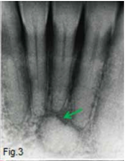
In the first stage (Fig. 1) the radiograph shows well defined apical radiolucencies apical to the teeth. The teeth are intact, as are the periodontal ligament spaces. In the second or intermediate stage (Fig. 2) the lesion is partly radiolucent and partly radiopaque. Usually the hard tissue formation is initiated centrally in the lesion. In the third stage (Fig. 3), also called the mature stage, the lesion is transformed into a mineralized, radiopaque mass which is surrounded by a narrow radiolucent zone (green arrow). A clinical diagnosis of periapical cemental dysplasia was made. Apical cemental dysplasias (apical cementomas) are benign lesions that contain cementum-like tissue and originate from cellular elements of the periodontal space. They are non-expansile radiolucencies in the early or mature stage. There may be single or multiple lesions. Bone expansion is absent, and pain is not a feature and no treatment is required. As the lesion matures, increasing amounts of cementum-like material are laid down in the lesion. In the mature stage, the radiographic appearance of a cementoma is a well-defined radiopaque lesion (Fig. 4) usually bordered by a thin radiolucent line or band (white arrow).


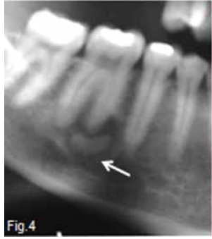
It is the radiographic feature of the radiolucent band separating the calcified mass from the bone that distinguishes the apical cementomas from osteosclerosis and condensing osteitis. As the lesion progresses, the fibroblasts differentiate into cementoblasts and the formation of cementum begins. Usually cementum is formed as cement-like structures (Fig. 5) which later coalesce to form solid masses (yellow arrow). Apical cemental dysplasia is not commonly seen in the upper jaw (Fig. 6). In the early radiolucent stages of a developing apical cemental dysplasia it is important to check the vitality of the involved tooth to exclude the possible presence of an apical infection.


References
1. Langlais RP, Langland OE & Nortje CJ: Diagnostic Imaging of the Jaws 1st Edition, Williams & Wilkins, 1995, pp. 268-72. [ Links ]
2. Farman AG, Nortjé CJ & Wood R E: Oral and Maxillofacial Imaging, 1st Ed, Mosby. St. Louis, Missouri 1993 pp. 319-24. [ Links ]

