


Serviços Personalizados
Artigo
 Inglês (pdf)
 Artigo em XML
 Referências do artigo
Indicadores

Links relacionados
-
 Citado por Google
 -
 Similares em Google
Compartilhar

 Permalink
South African Dental Journal
versão On-line ISSN 0375-1562
versão impressa ISSN 0011-8516
S. Afr. dent. j. vol.72 no.10 Johannesburg Nov. 2017
http://dx.doi.org/10.17159/2519-0105/2017/v72no10a2
RESEARCH
Accuracy of acetate overlays in bite mark comparison: How accurate is an ideal bite pattern?
N MohamedI; V M PhillipsII
IBChD (Stell), BSc Hons (Paed Dent) (Stell), MSc (Paed Dent) (Stell), PhD (Stell). Senior Lecturer, Department of Paediatric Dentistry, Faculty of Dentistry, University of the Western Cape
IIBDS (Wits), MChD (Stell), FC Path SA (Oral Path), Dip Max-Fac Radiology, PhD (UWC), DSc (UWC). Professor, Department of Oral Pathology and Forensic Sciences, Faculty of Dentistry, University of the Western Cape
ABSTRACT
Forensically, a bite mark on human skin is reliant on the matching of the alignment and position of the dentition of the perpetrator with the bruise pattern inflicted by the bite. If there is more than one suspect, the bite pattern of each suspect needs to be analysed. At least hypothetically, a bite delivered by a person who has had orthodontic treatment will result in a bruise pattern of an ideal arrangement of the teeth. If there are two suspects, both of whom have had orthodontic treatment, could that "ideal" alignment compromise identification of the perpetrator of the bite mark?
AIM: To determine the accuracy of an ideal bite pattern and whether an exact match could be obtained when comparing acetate overlays with bite patterns registered in wax of treated orthodontic cases.
METHOD: The biting patterns of upper and lower teeth of each of the study models were recorded in grey bite registration wax (Alminax®). Two examiners viewed the bite mark patterns and correlated them with the study models.
RESULT: In some cases an exact match between the teeth of the plaster model and the bite mark was not possible.
INTRODUCTION
General dental practitioners do not deal with forensic dentistry on a daily basis but their awareness should be raised regarding bite marks as these are often seen in cases of child and elder abuse. The dental practitioner should be able to make a clinical assessment of a suspected case of abuse and report the case to the police.
In many criminal cases the dentitions of suspects have been compared with bite marks left on the skin in order to determine whether the perpetrator in question could be held accountable for the crime.1,2 The accuracy of the bruise patterns when compared with the biting patterns of the upper and lower teeth of a suspect has been questioned. A degree of concordance should be demonstrable between the bite marks left on an impression surface (the skin) and the dentition of a suspect.3 There is, however, no consensus in the literature regarding the actual number of concordant features that are needed to implicate an individual as being the perpetrator.4 In principle as many concordant features as possible should be recorded when the comparisons are made.
It has been suggested that bite mark evidence should never be used to convict a suspect,3 despite the variations in caries experience, dental treatment received, environmental factors and wear-and-tear, that makes each the morphology of each dentition unique.2,5 Features such as crowding, asymmetry, missing or filled teeth, supernumerary teeth, diastemata and attrition as well as the combination of these features could result in a unique bite pattern.4
Despite that unique quality, how these features are recorded on the skin can produce bite marks that are so similar that one may be indistinguishable from another.2,5 Thus, inaccurate interpretation of a bite mark may lead to wrongful conviction of a suspect.2,6 At the very least, bite mark analysis could either exclude a suspect as the possible perpetrator or suggest that a degree of probability could exist that the suspect inflicted the bite mark.7
Cases with obvious irregularities, such as tooth rotations that are unique to an individual, have been used as evidence in the conviction of a criminal, but in numerous cases the bite mark evidence has not been convincing due to a lack of accuracy in the correlation between the bruise patterns and the teeth of the suspect. When comparing the dental features, the positions of the teeth, inter-canine distance, shape of the arches and tooth sizes should be taken into consideration.6 The area of the tooth biting surfaces, tooth rotation and width, centric position and other unique characteristics, including absent teeth, should also be noted.2,8 These distinct features are easily correlated, but a perfect row of teeth may not produce enough evidence for a match.
The objective of orthodontic treatment is to arrange the upper and lower dentition of a patient into a "normal" Class I occlusion for aesthetic as well as functional and health reasons. Young patients with malocclusions are subjected to long term mechanical adjustment of the dentition. Sometimes, extraction of premolar teeth is required to attain a Class I occlusion. The teeth are moved and rotated to attempt normal catenary alignment and thereby improve mastication, reduce interdental food retention and subsequent periodontal disease.
Dental study models of orthodontic patients at the completion of their treatment show an almost perfect catenary curve of the maxillary and mandibular teeth. Some minor rotations can persist, especially of the mandibular incisors. The maxillary and mandibular incisors also vary in size (mesio-distally) and the relationship between the maxillary central and lateral incisors can be sufficiently peculiar to be used for identification. The variable nature of bite marks on the skin makes identification of a positive match difficult. The question, however, is "If an ideal bite is recorded, is it possible to obtain a 100% match between the teeth of the plaster model and the bite mark"?
AIM
The aim of this study was to determine whether it is possible to accurately match the teeth of a sample of orthodontic plaster study models and an ideal bite mark registered in wax, using the acetate overlay technique.
MATERIALS AND METHODS
A cross-sectional, comparative study was carried out. Plaster of Paris study models of the upper and lower teeth of 26 dentate young adults who had completed their orthodontic treatment were used. The models were obtained by random selection from the database of the Orthodontic Department database at the Dental Faculty of the University of the Western Cape. All models had to have fully-erupted permanent teeth. This was purely a records-based (archival) study. No names or personal details of the patients were available. Models were identified only by means of a number (Figure 1). Patient confidentiality was therefore preserved.

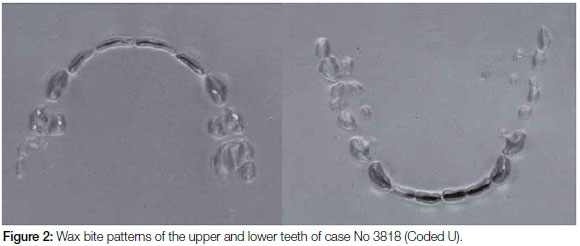
To create an ideal bite pattern for each individual, the biting patterns of the upper and lower teeth of each of the study models were recorded in grey bite registration wax (Alminax®) to create an accurate impression of the biting patterns of the upper and lower teeth. The wax was heated with a flame to soften it and placed on a firm flat surface; then the teeth of each study model were pressed into the wax to record the bite pattern (Figure 2).
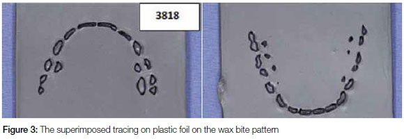
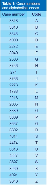
The method of bite mark comparison routinely used by author VMP is to trace the bite pattern of each jaw on plastic foil and to then superimpose the tracing over the actual bite mark. Thus the wax biting patterns of the upper and lower teeth of each of the cases were traced onto plastic transparent foil using a fine permanent marker pen (Figure 3). Alphabetical characters from A to Z were assigned to the tracings. The list of alphabetical labels and the correlating case numbers were kept separately so that blind comparisons could be made (Table 1).
Two examiners independently analysed the cases and tried to identify matched pairs of the transparency tracings and the wax bite patterns. This was undertaken in the following manner;
The first analysis was to match the tracings of both the upper and lower jaws simultaneously with the upper and lower wax bite patterns. The wax bite patterns for each case were arranged on a table surface. Tracings of the upper and lower bite patterns, A to Z, were severally superimposed on each wax pattern until a match was obtained. This matched pair was then eliminated from the analysis. The results obtained by each examiner were recorded.
The second analysis was to identify matches of the upper teeth only and then matches of the lower teeth only. A similar method of matching was used. The results of each examiner were recorded.
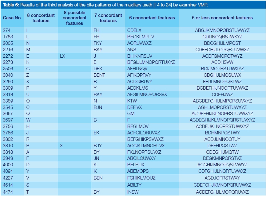
The third analysis (Tables 5 to 7) examined the section of the dental arch spanning from the first premolar on the left side to the first premolar on the right side in the upper and lower arches. (In many of the cases of bite marks on the skin the pattern of bruises is inflicted by the upper and lower anterior teeth and rarely extends beyond the 2nd premolars.)
This meant that a maximum of eight concordant features could be obtained for each of the upper and for each of the lower arches. Each researcher performed the matching process for the maxilla and mandible together and then for each arch separately. The number of concordant features for each jaw were recorded as follows:
•8 concordant features-definite match
•8 similar features but not a definite match
•7 concordant features-highly probable match
•6 concordant features-possible match
•5 concordant features-no match
Concordant features were noted if there was a match in the following between the transparency overlay and the wax bite pattern:
•the pattern of tooth distribution
•the spatial alignment of the teeth
•the shape of the arch-teeth had to fall within the dental arch
•the width of the incisal edges of the teeth
•angulation of teeth/ incisal edges of teeth
RESULTS
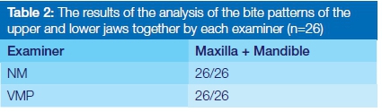
First analysis: When the upper and lower wax biting patterns were superimposed with the tracings of both dental arches, both examiners were able to match every case accurately i.e. 100% match (Table 2).
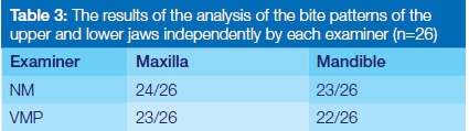
Second analysis: When each of the tracings were independently superimposed on the wax bite patterns of the mandibular and maxillary dentitions the degree of accuracy was found to be less accurate (Table 3).
Third analysis: Using the anterior 16 teeth (1st premolar to 1st premolar) of the upper and lower jaws separately, the tracings of each case were superimposed over these teeth to obtain a pattern match. The findings are reflected in Tables 4 to 7. In those Tables, the case numbers are shown in the first column. The tracings are labelled A to Z. The second column shows the exact match (eight concordant features) of the tracings with the bite patterns. The third column shows tracings where eight possible concordant features were matched. The fourth column shows those tracings where seven concordant features between the tracings and the bite patterns were obtained. The fifth column shows those cases where six concordant features were obtained. The sixth column shows those cases with five or less concordant features.
The first column in Table 4 demonstrates a high degree of accuracy in matching the cases. The third column shows two tracings (A & B) where eight possible concordant features were matched.
The third column in Table 5 shows four tracings (G, EG and G) where eight possible concordant features were matched.
The third column in Table 6 shows three tracings (LX, and X) where eight possible concordant features were matched.
The third column in Table 7 shows that for case No. 1783 tracing G has eight possible concordant features. Similarly for case 3766 the tracings G, L and M have eight possible concordant features. Case 3818 has eight possible concordant features with B, P and S; Case 3949 has eight possible concordant features with E, K and S; Case 4474 has eight possible concordant features with S and Case 4614 has eight possible concordant features with O and P.
DISCUSSION
The bite mark patterns recorded in the wax were ideal and accurate replications of the bite patterns of each of the study models were obtained. The tracings onto the plastic overlays of each of the biting patterns of the upper and lower teeth of the cases were systematically and sequentially superimposed over each wax bite pattern and the number of concordant features recorded.
It was clear from the results that when the mandible and maxilla were examined together as a single entity, the tracings could easily be matched to the wax bite patterns. This was repeated on more than one occasion with the same result. Both examiners scored a 100% match each time. When both arches were viewed together, these ideal bite patterns were thus unique enough to be able to identify an exact match, even when the teeth were perfectly aligned.
When the arches were examined independently of each other, the maxillary arches were more easily matched than were the mandibular arches, but it was more difficult to identify an exact match.
The variability between the examiners could be attributed to the fact that Examiner NM is a general dentist and Examiner VMP is a forensic pathologist. Taking the variability of bite marks into consideration, the pathologist was therefore more inclined to be more lenient in his assessment. Examiner NM tended to be stricter in assessing the possibility of a match. Despite this, it was clear that both examiners found that more than one tracing could be matched to a wax bite when the maxillary and mandibular arches were viewed independently of each other.
This study shows that even in the ideal situation where the bite mark patterns in the wax are a perfect replication of the dental arches of the maxilla and the mandible, there are several of the biting patterns that are so similar that an absolute match is not possible.
A bite mark on human skin is often seen as only bruises and analysis requires that the teeth of the perpetrator be matched with those bruises. Often there are imperfections in the bruise patterns due to abrasion of the skin during the infliction of the bite. The malleability and distortion of the human tissues also contribute to distorted representations and hence inaccuracies in matching with the perpetrator's teeth.
CONCLUSION
This study emphasized that even under ideal circumstances where the impression of each tooth was recorded accurately; an exact match between the acetate overlay and the teeth of the plaster model is not possible in some cases e.g. where more than one "perpetrator's" bite pattern was very similar. In clinical situations where the examination of a bite mark in human skin often takes place long after the infliction thereof, the appearance of bite marks are variable depending on the degree of force applied and the movement of the victim.
The bite mark on skin usually consists of a pattern of bruises or puncture wounds, and is far less accurate for identification purposes. The latest literature confirms the inaccuracy of bite marks and suggests that it cannot be used as primary identification data to implicate a perpetrator of a bite mark.
There were several duplicate matches where more than one set of models could have made the impression in the wax. The plaster of Paris study models of patients who had undergone orthodontic treatment had very similar dental arch morphology. This added to the argument that if a bite mark were inflicted by a person who had an ideal dental arch and there were two or more suspects who had undergone orthodontic treatment, it would be difficult to accurately match their bite patterns with the bite mark.
Caution should therefore be exercised when analysing bite marks especially where the alleged perpetrator has a "perfect set of teeth". There should be a move away from using this as a definitive means of identification of perpetrators of abuse, assault or murder.
References
1. Reddy, SS, Rakesh, N, Kaushik, A, Devaraju, D, Kumar, N. Evaluation of the accuracy, precision and validity of hydrophilic vinyl polysiloxane impression material for bite mark analysis. Experimental and Clinical Sciences Journal 2011;10: 55- 61. [ Links ]
2. Pretty, IA. The barriers to achieving an evidence base for bitemark analysis. Forensic Science International 2006; 159S: S110- S120. [ Links ]
3. Taroni, F, Mangin, P, Perrior, M. Identification concept and the use of probabilities in forensic odontology-an approach by philosophical discussion. Journal of Forensic Odontostomatology 2000; 18:15- 7. [ Links ]
4. Bernitz, H, van Heerden, W, Solheim, T, Owen, J. A technique to capture, analyse and quantify anterior tooth rotations for application in court cases involving tooth marks. Journal of Forensic Science 2006; 51: 624-9. [ Links ]
5. Blackwell, SA, Taylor, RV, Gordon, I, Ogleby, CL, Tanijiri, T, Yoshino, M, Donald, MR, Clement, JG. 3-D imaging and quantitative comparison of human dentitions and simulated bite marks. International Journal of Legal Medicine 2007; 121: 9- 17. [ Links ]
6. Bernitz, H, Owen, JH, van Heerden, WFP, Solheim, T. An integrated technique for the analysis of skin bite marks. Journal of Forensic Science 2008; 53 (1):194- 8. [ Links ]
7. Lessig, R, Wenzel, V, Weber, M. Bite mark analysis in forensic routine case work. Experimental and Clinical Sciences Journal 2006; 5: 93- 102. [ Links ]
8. Sweet, D, Bowers M. Accuracy of Bite mark overlays: a comparison of five common methods to produce exemplars from a suspect's dentition. Journal of Forensic Science 1998; 43 (2): 362- 7. [ Links ]
 Correspondence:
Nadia Mohamed
Department of Paediatric Dentistry, Faculty of Dentistry,
University of the Western Cape
Private Bag X1 , Tygerberg, 7505
South Africa.
Tel: 021 937 3073 or 937 3056/7 Cell nr: 083 2705 105.
E-mail: namohamed@uwc.ac.za

