


Serviços Personalizados
Artigo
 Inglês (pdf)
 Artigo em XML
 Referências do artigo
Indicadores

Links relacionados
-
 Citado por Google
 -
 Similares em Google
Compartilhar

 Permalink
South African Dental Journal
versão On-line ISSN 0375-1562
versão impressa ISSN 0011-8516
S. Afr. dent. j. vol.72 no.8 Johannesburg Set. 2017
http://dx.doi.org/10.17159/2519-0105/2017/v72no8a6
RADIOLOGY CASE
Maxillo-facial radiology case 154
CJ Nortjé
BChD, PhD, ABOMR, DSc. Faculty of Dentistry, University of the Western Cape. E-mail: cnortje@uwc.ac.za
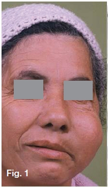
Below are two examples of the malignant tumour that most commonly may affect the maxillary sinuses. What are the important clinical and radiological features in these cases and what is your diagnosis? What is the importance of the maxillary sinuses for the dental practitioner?
INTERPRETATION
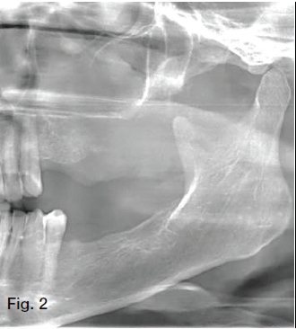
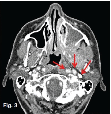
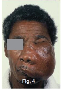
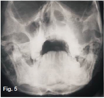
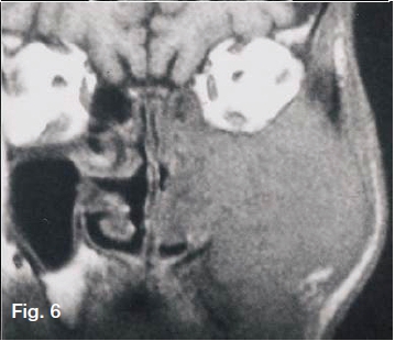
Figs. 1,2&3 present a clinical picture and radiographs of a fifty year old female patient with the main complaint of dental pain, loss of teeth, nasal obstruction and a numbness on the left side of the face.The cropped pantomograph (Fig.2) shows opacification and destruction of the floor and posterior border of the sinus. The axial CT shows a large destructive necrotic tumour originating from the left maxillary sinus with destruction of the medial and inferior floor of the sinus. The tumour infiltrated the left nasal passage destroying the hard palate and expanding into the roof of the mouth. The tumour also infiltrated the soft palate posteriorly, destroying the pterygoid plate on the left. Massive necrotic metastatic lymphadenopathy is clearly present in the neck on the left side Fig.3 (red arrows). The lower case (Fig.4) shows an extensive tumour affecting the left side of the face and extending into the orbit. The Water's view (Fig.5) shows destruction of the medial, lateral and orbital walls of the maxillary sinus with the tumour infiltrating the nasal cavity and orbit. A T1 weighted coronal MR image (Fig.6) show an expanded isointense homogeneous mass of the left antrum which has extended into the nasal cavity and ethmoid sinus. The palate is eroded and there is extension into the alveolar ridge. In both cases a histological diagnosis of a squamous carcinoma was made. Squamous carcinoma is the most common malignant neoplasm of nose and sinuses. Eighty percent of paranasal sinus carcinomas occur in the maxillary sinus. Men are affected twice as often as women. Facial or dental pains are very common early signs while nasal obstruction and epistaxis appear very late in the development of the lesion.
In conclusion, the importance of the maxillary sinus for the dental practioner is that the image of the sinuses appears almost consistently in radiographs taken on a daily basis in a normal dental practice. The radiograph often reveals associated pathology which is often overlooked or not identified, with serious consequences to the patient. Furthermore a pathological lesion can affect both the oral cavity and the sinus. Lesions in one of these regions may cause problems in the other region. An abnormal opening can also develop between these cavities and pain can be referred from one region to the other.
Reference
1. Farman AG, Nortjé CJ & Wood R E: Oral and Maxillofacial Imaging, 1st Ed, Mosby. St. Louis, Missouri 1993 p 395-8. [ Links ]

