


Serviços Personalizados
Artigo
 Inglês (pdf)
 Artigo em XML
 Referências do artigo
Indicadores

Links relacionados
-
 Citado por Google
 -
 Similares em Google
Compartilhar

 Permalink
South African Dental Journal
versão On-line ISSN 0375-1562
versão impressa ISSN 0011-8516
S. Afr. dent. j. vol.72 no.6 Johannesburg Jul. 2017
http://dx.doi.org/10.17159/2519-0105/2017/v72no6a6
RADIOLOGY CASE
Maxillofacial radiology case 152
CJ Nortjé
BChD, PhD, ABOMR, DSc. Faculty of Dentistry, University of the Western Cape. E-mail: cnortje@uwc.ac.za
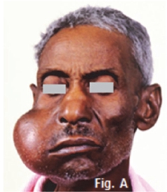
Below is a clinical picture and radiographic images of certain malignancies that may present in the salivary glands of the maxillofacial region. What are the important features and what are your diagnoses?
INTERPRETATION
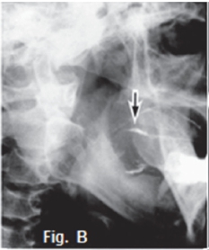
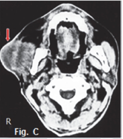
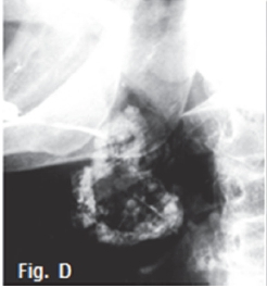
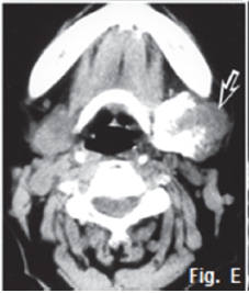
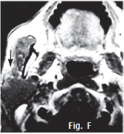
Fig. A is a photograph of a 65 year old male patient presenting with a slow growing swelling of the parotid gland on the right side of the face, which he noticed about eight months ago. Sialography of the parotid gland (Fig. B) shows that contrast medium uptake is prevented by tumour formation, resulting in a poorly imaged right parotid gland (arrow). Fig. C is an axial CT of the same patient demonstrating an ill-defined tumour with an infiltrative margin, indicating malignancy. The right masseter muscle also lacks definition because of infiltration by a malignant tumour (red arrow). A diagnosis of an epidermoid carcinoma was made. The tumour was termed mucoepidermoid by Stewart et al in 1945, who considered it to be divided into benign and malignant types. The WHO regards this tumour as a lesion with malignant potential but believes that, in a variety of clinical instances, it is inappropriate to call any of these tumours "carcinomas". The WHO consider this neoplasm to be intermediate between adenoma and carcinoma. Thirty percent of mucoepidermoid malignancies occur in the salivary glands and appear commonly in the 3rd to 4th decades. The sialogram in Fig. D shows irregular non filling defects suggestive of a malignant pleomorphic adenoma (malignant mixed tumour). The CT sialogram (Fig. E) of the same patient shows an ill-defined tumour mass (arrow). Pathologically, malignant pleomorphic adenoma results from the transformation of the epithelial tissue in a pleomorphic adenoma. These tumours occur predominantly in patients older than 50 years. The transverse T1 weighted MRI scan (Fig. F) of an adenocarcinoma of the right parotid gland shows a poorly defined, non-homogeneouus, infiltrating mass (arrow). Adenocarcinomas most commonly involve the minor salivary glands and submandibular glands, accounting for 5% to 10% of salivary gland tumours. A break in the duct and leakage of contrast medium are sialographic signs of malignancy, and are usually found in epidermoid carcinoma, adenocarcinoma, and high-grade mucoepidermoid carcinoma. However, most of the malignant tumours have a low rate of malignancy.
Reference
1. Farman AG, Nortje CJ & Wood R E: Oral and Maxillofacial Imaging, 1st Ed, Mosby. St. Louis, Missouri 1993 pp. 413, 423-4. [ Links ]

